
Krijgt de bodyguard van ons brein de ziekte van Parkinson klein?
We zijn allemaal wel al eens op een feestje geweest waar een dronken persoon de sfeer verpestte. Gelukkig gebeurt dit niet vaak, omdat aan de ingang van de meeste feestjes bodyguards staan die dronken personen de toegang weigeren en amokmakers uit de feestzaal verwijderen. Ook onze hersenen hebben een soortgelijke bodyguard, genaamd de choroid plexus. De studies uitgevoerd in kader van mijn masterthesis toonden aan dat de choroid plexus mogelijk een rol speelt in de verspreiding van Parkinson-gerelateerde eiwitten tijdens de ontwikkeling van de ziekte van Parkinson. Deze specifieke hersenstructuur kan op lange termijn een nieuw doelwit vormen voor een behandeling van de ziekte.
Amokmakers in de hersenen
De ziekte van Parkinson is een neurologische hersenaandoening die gekenmerkt wordt door bewegingsproblemen. De ziekte wordt gekarakteriseerd door een verlies van zenuwcellen in de substantia nigra. Deze hersenregio is verantwoordelijk voor het vlot verlopen van bewegingen. Het afsterven van neuronen in de substantia nigra veroorzaakt bijgevolg de typische bewegingsproblemen zoals beven, vertraagde bewegingen en spierstijfheid. Naast deze typische motorische symptomen wordt de ziekte van Parkinson ook gekenmerkt door niet-motorische problemen waaronder verlies van reukzin ten gevolge van schade aan de hersenreukregio. Opvallend is dat de reukproblemen zich vroeger manifesteren dan de bewegingsproblemen.
Tot op heden is het niet duidelijk waarom de zenuwcellen in de substantia nigra afsterven. Een mogelijke oorzaak is de ophoping van het eiwit alpha-synucleïne (αSyn). Dit eiwit komt in normale omstandigheden voor in de hersenzenuwcellen. De precieze functie en werking van αSyn zijn nog steeds niet gekend. Onderzoek toonde aan dat het eiwit bij Parkinsonpatiënten van structuur verandert. De misvormde αSyn-eiwitten klonteren samen en vormen eiwitophopingen. Deze ophopingen kunnen ook de normaal opgevouwen αSyn-eiwitten aanzetten om fout op te vouwen. Deze verspreiding is vergelijkbaar met het uit de hand lopen van een conflict. Eerst zijn slechts enkele amokmakers betrokken in een ruzie, maar al snel zetten ze omstaanders aan om mee te vechten en breidt het gevecht zich uit. Zo een sneeuwbaleffect zorgt ervoor dat de αSyn-eiwitten in steeds meer hersenregio’s een foute opvouwing krijgen en samenklonteren. De ophopingen beschadigen waarschijnlijk de zenuwcellen waardoor deze finaal afsterven. Waarom de eiwitten fout opvouwen en hoe de αSyn-eiwitten zich precies over de hersenen verspreiden, is tot heden niet volledig opgehelderd.
De bodyguard van ons brein
Een mogelijks belangrijke speler in de verspreiding van de ziekte van Parkinson is de choroid plexus. De choroid plexus is een sterk doorbloede structuur die de hersenen beschermt van de rest van ons lichaam. Afscherming is nodig omdat onze hersenen uiterst gevoelig zijn voor kleine omgevingsveranderingen. De choroid plexus is eigenlijk de bodyguard van ons brein en kan vergeleken worden met de portiers aan een discotheek. Een duidelijke gastenlijst beslist wie binnen mag en wie op de zwarte lijst komt. Op deze manier zijn onze hersenen afgeschermd van toxische stoffen. Een foute werking van deze hersenbarrière veroorzaakt schadelijke veranderingen in de hersenen. Ondanks de relevantie van deze structuur in de normale hersenwerking, is slechts weinig gekend over de rol van de choroid plexus in de ziekte van Parkinson.
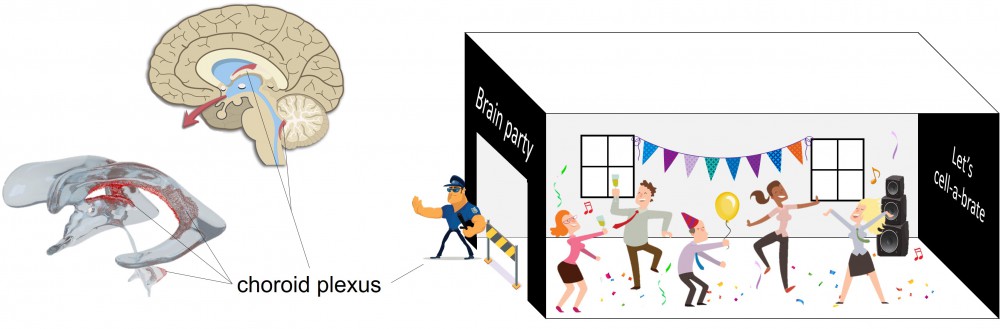
De rol van de choroid plexus in de ziekte van Parkinson
Om na te gaan of de choroid plexus een rol speelt in de verspreiding van de ziekte van Parkinson, werden de schadelijke, misvormde αSyn-eiwitten rechtstreeks geïnjecteerd dicht bij de choroid plexus in de hersenen van de proefdiermuizen. Injectie van αSyn-eiwitten is vergelijkbaar met het plaatsen van enkele amokmakers in de hersendiscotheek.
De experimenten besproken in mijn masterthesis toonden aan dat de geïnjecteerde αSyn-eiwitten grotendeels aan de choroid plexus bleven plakken. Het lijkt alsof de bodyguard van onze hersenen in staat is om de ‘amokmakereiwitten’ vast te nemen wanneer deze langs hem passeren. Hoe en waarom deze interactie gebeurt, blijft voorlopig nog een raadsel. Mogelijks kunnen de schadelijke eiwitten via de choroid plexus verwijderd worden uit de hersenen zodat het hersenfeestje zo weinig mogelijk wordt verstoord.
Er werd ook vastgesteld dat de choroid plexus tijdelijk zijn barrièrefunctie verliest na de injectie van de misvormde αSyn-eiwitten. Dit betekent dat de deuren van de hersendiscotheek wagenwijd open worden gezet zonder dat er nog controle is over wie binnen en buiten kan. Hierdoor is transport mogelijk van bepaalde stoffen naar de hersenen die in normale omstandigheden worden tegengehouden door de choroid plexus. Het is mogelijk dat de choroid plexus te druk bezig is met het verwijderen van de αSyn-eiwitten en hierdoor zijn barrièrefunctie tijdelijk verliest. Na enige tijd herstelde de barrièrefunctie zich opnieuw.
Daarnaast toonden de studies aan dat de αSyn-eiwitten een ontstekingsreactie in de choroid plexus en de geurhersenregio veroorzaakten. Dit is vergelijkbaar met een paniekreactie in de hersendiscotheek. Opmerkelijk was dat de ontstekingsreactie zich vroeger manifesteerde in de geurregio dan in de choroid plexus. Deze resultaten kunnen gelinkt worden aan het vroegtijdig optreden van reukproblemen bij Parkinsonpatiënten. Welke cellen en moleculen de ontstekingsreactie initiëren is niet precies duidelijk. Verder onderzoek is nodig om het totale ontstekingsproces in kaart te brengen.
Toekomstperspectief
Als verder onderzoek aantoont dat de choroid plexus in staat is om het schadelijk alpha-synucleïne te transporteren uit de hersenen naar het bloed, kan deze hersenstructuur een doelwit vormen voor therapie. Zo zou het mogelijk zijn om het transport in de choroid plexus te stimuleren. Meer onderzoek is echter nodig om de precieze rol van dit hersendeel in de ontwikkeling van de ziekte van Parkinson bloot te leggen.
Bibliografie
1. Kalia LV, Lang AE (2015) Parkinson's disease. Lancet 386: 896-912
2. Kim WS, Kagedal K, Halliday GM (2014) Alpha-synuclein biology in Lewy body diseases. Alzheimers Res Ther 6: 73
3. Ghosh D, Mehra S, Sahay S, Singh PK, Maji SK (2017) alpha-synuclein aggregation and its modulation. Int J Biol Macromol 100: 37-54
4. Burre J (2015) The Synaptic Function of alpha-Synuclein. J Parkinsons Dis 5: 699-713
5. Ghiglieri V, Calabrese V, Calabresi P (2018) Alpha-Synuclein: From Early Synaptic Dysfunction to Neurodegeneration. Front Neurol 9: 295
6. Miraglia F, Ricci A, Rota L, Colla E (2018) Subcellular localization of alpha-synuclein aggregates and their interaction with membranes. Neural Regen Res 13: 1136-1144
7. Roberts HL, Brown DR (2015) Seeking a mechanism for the toxicity of oligomeric alpha-synuclein. Biomolecules 5: 282-305
8. Braak H, Del Tredici K, Rub U, de Vos RA, Jansen Steur EN, Braak E (2003) Staging of brain pathology related to sporadic Parkinson's disease. Neurobiol Aging 24: 197-211
9. Rietdijk CD, Perez-Pardo P, Garssen J, van Wezel RJ, Kraneveld AD (2017) Exploring Braak's Hypothesis of Parkinson's Disease. Front Neurol 8: 37
10. Vargas JY, Grudina C, Zurzolo C (2019) The prion-like spreading of alpha-synuclein: From in vitro to in vivo models of Parkinson's disease. Ageing Res Rev 50: 89-101
11. Li JY, Englund E, Holton JL, Soulet D, Hagell P, Lees AJ, Lashley T, Quinn NP, Rehncrona S, Bjorklund A, et al. (2008) Lewy bodies in grafted neurons in subjects with Parkinson's disease suggest host-to-graft disease propagation. Nat Med 14: 501-3
12. Yamada K, Iwatsubo T (2018) Extracellular alpha-synuclein levels are regulated by neuronal activity. Mol Neurodegener 13: 9
13. Freundt EC, Maynard N, Clancy EK, Roy S, Bousset L, Sourigues Y, Covert M, Melki R, Kirkegaard K, Brahic M (2012) Neuron-to-neuron transmission of alpha-synuclein fibrils through axonal transport. Ann Neurol 72: 517-24
14. Rey NL, Petit GH, Bousset L, Melki R, Brundin P (2013) Transfer of human alpha-synuclein from the olfactory bulb to interconnected brain regions in mice. Acta Neuropathol 126: 555-73
15. Abounit S, Bousset L, Loria F, Zhu S, de Chaumont F, Pieri L, Olivo-Marin JC, Melki R, Zurzolo C (2016) Tunneling nanotubes spread fibrillar alpha-synuclein by intercellular trafficking of lysosomes. Embo j 35: 2120-2138
16. Steiner JA, Quansah E, Brundin P (2018) The concept of alpha-synuclein as a prion-like protein: ten years after. Cell Tissue Res 373: 161-173
17. Rostami J, Holmqvist S, Lindstrom V, Sigvardson J, Westermark GT, Ingelsson M, Bergstrom J, Roybon L, Erlandsson A (2017) Human Astrocytes Transfer Aggregated Alpha-Synuclein via Tunneling Nanotubes. J Neurosci 37: 11835-11853
18. Gelders G, Baekelandt V, Van der Perren A (2018) Linking Neuroinflammation and Neurodegeneration in Parkinson's Disease. J Immunol Res 2018: 4784268
19. Surmeier DJ (2018) Determinants of dopaminergic neuron loss in Parkinson's disease. Febs j 285: 3657-3668
20. Troncoso-Escudero P, Parra A, Nassif M, Vidal RL (2018) Outside in: Unraveling the Role of Neuroinflammation in the Progression of Parkinson's Disease. Front Neurol 9: 860
21. Theodore S, Cao S, McLean PJ, Standaert DG (2008) Targeted overexpression of human alpha-synuclein triggers microglial activation and an adaptive immune response in a mouse model of Parkinson disease. J Neuropathol Exp Neurol 67: 1149-58
22. Mogi M, Harada M, Narabayashi H, Inagaki H, Minami M, Nagatsu T (1996) Interleukin (IL)-1β, IL-2, IL-4, IL-6 and transforming growth factor-α levels are elevated in ventricular cerebrospinal fluid in juvenile parkinsonism and Parkinson's disease. Neurosci Lett 211: 13-16
23. Mogi M, Harada M, Kondo T, Riederer P, Inagaki H, Minami M, Nagatsu T (1994) Interleukin-1β, interleukin-6, epidermal growth factor and transforming growth factor-α are elevated in the brain from parkinsonian patients. Neurosci Lett 180: 147-150
24. Mogi M, Harada M, Riederer P, Narabayashi H, Fujita K, Nagatsu T (1994) Tumor necrosis factor-α (TNF-α) increases both in the brain and in the cerebrospinal fluid from parkinsonian patients. Neurosci Lett 165: 208-210
25. Sveinbjornsdottir S (2016) The clinical symptoms of Parkinson's disease. J Neurochem 139 Suppl 1: 318-324
26. Pringsheim T, Jette N, Frolkis A, Steeves TD (2014) The prevalence of Parkinson's disease: a systematic review and meta-analysis. Mov Disord 29: 1583-90
27. Abbas MM, Xu Z, Tan LCS (2018) Epidemiology of Parkinson's Disease-East Versus West. Mov Disord Clin Pract 5: 14-28
28. Papadopoulos VE, Nikolopoulou G, Antoniadou I, Karachaliou A, Arianoglou G, Emmanouilidou E, Sardi SP, Stefanis L, Vekrellis K (2018) Modulation of beta-glucocerebrosidase increases alpha-synuclein secretion and exosome release in mouse models of Parkinson's disease. Hum Mol Genet 27: 1696-1710
29. Youn J, Lee SB, Lee HS, Yang HO, Park J, Kim JS, Oh E, Park S, Jang W (2018) Cerebrospinal Fluid Levels of Autophagy-related Proteins Represent Potentially Novel Biomarkers of Early-Stage Parkinson's Disease. Sci Rep 8: 16866
30. De Bock M, Vandenbroucke RE, Decrock E, Culot M, Cecchelli R, Leybaert L (2014) A new angle on blood-CNS interfaces: a role for connexins? FEBS Lett 588: 1259-70
31. Johanson CE (2018) Choroid Plexus Blood-CSF Barrier: Major Player in Brain
Disease Modeling and Neuromedicine. J Neurol Neuromedicine
32. Simon MJ, Iliff JJ (2016) Regulation of cerebrospinal fluid (CSF) flow in neurodegenerative, neurovascular and neuroinflammatory disease. Biochim Biophys Acta 1862: 442-51
33. Mollenhauer B, Trautmann E, Otte B, Ng J, Spreer A, Lange P, Sixel-Doring F, Hakimi M, Vonsattel JP, Nussbaum R, et al. (2012) alpha-Synuclein in human cerebrospinal fluid is principally derived from neurons of the central nervous system. J Neural Transm (Vienna) 119: 739-46
34. Bates CA, Zheng W (2014) Brain disposition of alpha-Synuclein: roles of brain barrier systems and implications for Parkinson's disease. Fluids Barriers CNS 11: 17
35. Bates CA, Fu S, Ysselstein D, Rochet JC, Zheng W (2015) Expression and Transport of alpha-Synuclein at the Blood-Cerebrospinal Fluid Barrier and Effects of Manganese Exposure. Admet dmpk 3: 15-33
36. Shi M, Liu C, Cook TJ, Bullock KM, Zhao Y, Ginghina C, Li Y, Aro P, Dator R, He C, et al. (2014) Plasma exosomal alpha-synuclein is likely CNS-derived and increased in Parkinson's disease. Acta Neuropathol 128: 639-650
37. Pisani V, Stefani A, Pierantozzi M, Natoli S, Stanzione P, Franciotta D, Pisani A (2012) Increased blood-cerebrospinal fluid transfer of albumin in advanced Parkinson's disease. J Neuroinflammation 9: 188
38. Haussermann P, Kuhn W, Przuntek H, Muller T (2001) Integrity of the blood-cerebrospinal fluid barrier in early Parkinson's disease. Neurosci Lett 300: 182-4
39. Gray MT, Woulfe JM (2015) Striatal blood-brain barrier permeability in Parkinson's disease. J Cereb Blood Flow Metab 35: 747-50
40. Goldman JG, Andrews H, Amara A, Naito A, Alcalay RN, Shaw LM, Taylor P, Xie T, Tuite P, Henchcliffe C, et al. (2018) Cerebrospinal fluid, plasma, and saliva in the BioFIND study: Relationships among biomarkers and Parkinson's disease Features. Mov Disord 33: 282-288
41. Forland MG, Ohrfelt A, Dalen I, Tysnes OB, Blennow K, Zetterberg H, Pedersen KF, Alves G, Lange J (2018) Evolution of cerebrospinal fluid total alpha-synuclein in Parkinson's disease. Parkinsonism Relat Disord 49: 4-8
42. Tokuda T, Qureshi MM, Ardah MT, Varghese S, Shehab SAS, Kasai T, Ishigami N, Tamaoka A, Nakagawa M, El-Agnaf OMA (2010) Detection of elevated levels of α-synuclein oligomers in CSF from patients with Parkinson disease. Neurology 75: 1766-1770
43. van Niel G, D'Angelo G, Raposo G (2018) Shedding light on the cell biology of extracellular vesicles. Nat Rev Mol Cell Biol 19: 213-228
44. Caruso S, Poon IKH (2018) Apoptotic Cell-Derived Extracellular Vesicles: More Than Just Debris. Front Immunol 9: 1486
45. Zhang Y, Liu Y, Liu H, Tang WH (2019) Exosomes: biogenesis, biologic function and clinical potential. Cell Biosci 9: 19
46. Hessvik NP, Llorente A (2018) Current knowledge on exosome biogenesis and release. Cell Mol Life Sci 75: 193-208
47. Fussi N, Hollerhage M, Chakroun T, Nykanen NP, Rosler TW, Koeglsperger T, Wurst W, Behrends C, Hoglinger GU (2018) Exosomal secretion of alpha-synuclein as protective mechanism after upstream blockage of macroautophagy. Cell Death Dis 9: 757
48. Minakaki G, Menges S, Kittel A, Emmanouilidou E, Schaeffner I, Barkovits K, Bergmann A, Rockenstein E, Adame A, Marxreiter F, et al. (2018) Autophagy inhibition promotes SNCA/alpha-synuclein release and transfer via extracellular vesicles with a hybrid autophagosome-exosome-like phenotype. Autophagy 14: 98-119
49. Danzer KM, Kranich LR, Ruf WP, Cagsal-Getkin O, Winslow AR, Zhu L, Vanderburg CR, McLean PJ (2012) Exosomal cell-to-cell transmission of alpha synuclein oligomers. Mol Neurodegener 7: 42
50. Grapp M, Wrede A, Schweizer M, Huwel S, Galla HJ, Snaidero N, Simons M, Buckers J, Low PS, Urlaub H, et al. (2013) Choroid plexus transcytosis and exosome shuttling deliver folate into brain parenchyma. Nat Commun 4: 2123
51. Tietje A, Maron KN, Wei Y, Feliciano DM (2014) Cerebrospinal fluid extracellular vesicles undergo age dependent declines and contain known and novel non-coding RNAs. PLoS One 9: e113116
52. Thompson AG, Gray E, Mager I, Fischer R, Thezenas ML, Charles PD, Talbot K, El Andaloussi S, Kessler BM, Wood M, et al. (2018) UFLC-Derived CSF Extracellular Vesicle Origin and Proteome. Proteomics 18: e1800257
53. Balusu S, Van Wonterghem E, De Rycke R, Raemdonck K, Stremersch S, Gevaert K, Brkic M, Demeestere D, Vanhooren V, Hendrix A, et al. (2016) Identification of a novel mechanism of blood-brain communication during peripheral inflammation via choroid plexus-derived extracellular vesicles. EMBO Mol Med 8: 1162-1183
54. Coulter ME, Dorobantu CM, Lodewijk GA, Delalande F, Cianferani S, Ganesh VS, Smith RS, Lim ET, Xu CS, Pang S, et al. (2018) The ESCRT-III Protein CHMP1A Mediates Secretion of Sonic Hedgehog on a Distinctive Subtype of Extracellular Vesicles. Cell Rep 24: 973-986.e8
55. Emmanouilidou E, Melachroinou K, Roumeliotis T, Garbis SD, Ntzouni M, Margaritis LH, Stefanis L, Vekrellis K (2010) Cell-produced alpha-synuclein is secreted in a calcium-dependent manner by exosomes and impacts neuronal survival. J Neurosci 30: 6838-51
56. Delenclos M, Trendafilova T, Mahesh D, Baine AM, Moussaud S, Yan IK, Patel T, McLean PJ (2017) Investigation of Endocytic Pathways for the Internalization of Exosome-Associated Oligomeric Alpha-Synuclein. Front Neurosci 11: 172
57. Gustafsson G, Loov C, Persson E, Lazaro DF, Takeda S, Bergstrom J, Erlandsson A, Sehlin D, Balaj L, Gyorgy B, et al. (2018) Secretion and Uptake of alpha-Synuclein Via Extracellular Vesicles in Cultured Cells. Cell Mol Neurobiol
58. Grey M, Dunning CJ, Gaspar R, Grey C, Brundin P, Sparr E, Linse S (2015) Acceleration of alpha-synuclein aggregation by exosomes. J Biol Chem 290: 2969-82
59. Zhang S, Eitan E, Wu TY, Mattson MP (2018) Intercellular transfer of pathogenic alpha-synuclein by extracellular vesicles is induced by the lipid peroxidation product 4-hydroxynonenal. Neurobiol Aging 61: 52-65
60. Alvarez-Erviti L, Seow Y, Schapira AH, Gardiner C, Sargent IL, Wood MJ, Cooper JM (2011) Lysosomal dysfunction increases exosome-mediated alpha-synuclein release and transmission. Neurobiol Dis 42: 360-7
61. Stuendl A, Kunadt M, Kruse N, Bartels C, Moebius W, Danzer KM, Mollenhauer B, Schneider A (2016) Induction of alpha-synuclein aggregate formation by CSF exosomes from patients with Parkinson's disease and dementia with Lewy bodies. Brain 139: 481-94
62. Gui Y, Liu H, Zhang L, Lv W, Hu X (2015) Altered microRNA profiles in cerebrospinal fluid exosome in Parkinson disease and Alzheimer disease. Oncotarget 6: 37043-53
63. Lee Y, Dawson VL, Dawson TM (2012) Animal models of Parkinson's disease: vertebrate genetics. Cold Spring Harb Perspect Med 2
64. Visanji NP, Brotchie JM, Kalia LV, Koprich JB, Tandon A, Watts JC, Lang AE (2016) alpha-Synuclein-Based Animal Models of Parkinson's Disease: Challenges and Opportunities in a New Era. Trends Neurosci 39: 750-762
65. Jagmag SA, Tripathi N, Shukla SD, Maiti S, Khurana S (2015) Evaluation of Models of Parkinson's Disease. Front Neurosci 9: 503
66. Ko WKD, Bezard E (2017) Experimental animal models of Parkinson's disease: A transition from assessing symptomatology to alpha-synuclein targeted disease modification. Exp Neurol 298: 172-17967. Polinski NK, Volpicelli-Daley LA, Sortwell CE, Luk KC, Cremades N, Gottler LM, Froula J, Duffy MF, Lee VMY, Martinez TN, et al. (2018) Best Practices for Generating and Using Alpha-Synuclein Pre-Formed Fibrils to Model Parkinson's Disease in Rodents. J Parkinsons Dis 8: 303-322
68. Fortuna JTS, Gralle M, Beckman D, Neves FS, Diniz LP, Frost PS, Barros-Aragao F, Santos LE, Goncalves RA, Romao L, et al. (2017) Brain infusion of alpha-synuclein oligomers induces motor and non-motor Parkinson's disease-like symptoms in mice. Behav Brain Res 333: 150-160
69. Allen Brain Explorer. In
70. Volpicelli-Daley LA, Luk KC, Lee VM (2014) Addition of exogenous alpha-synuclein preformed fibrils to primary neuronal cultures to seed recruitment of endogenous alpha-synuclein to Lewy body and Lewy neurite-like aggregates. Nat Protoc 9: 2135-46
71. Bass JJ, Wilkinson DJ, Rankin D, Phillips BE, Szewczyk NJ, Smith K, Atherton PJ (2017) An overview of technical considerations for Western blotting applications to physiological research. Scand J Med Sci Sports 27: 4-2572. Stevens RW, Elmendorf D, Gourlay M, Stroebel E, Gaafar HA (1979) Application of fluoroimmunoassay to cerebrospinal fluid immunoglobulin G and albumin. J Clin Microbiol 10: 346-50
73. Collins MA, An J, Peller D, Bowser R (2015) Total protein is an effective loading control for cerebrospinal fluid western blots. J Neurosci Methods 251: 72-82
74. Barbour R, Kling K, Anderson JP, Banducci K, Cole T, Diep L, Fox M, Goldstein JM, Soriano F, Seubert P, et al. (2008) Red blood cells are the major source of alpha-synuclein in blood. Neurodegener Dis 5: 55-9
75. Haudenschild DR, Eldridge A, Lein PJ, Chromy BA (2014) High abundant protein removal from rodent blood for biomarker discovery. Biochem Biophys Res Commun 455: 84-9
76. Kaur C, Rathnasamy G, Ling EA (2016) The Choroid Plexus in Healthy and Diseased Brain. J Neuropathol Exp Neurol 75: 198-213
77. Yang DT, Lu X, Fan Y, Murphy RM (2014) Evaluation of Nanoparticle Tracking for Characterization of Fibrillar Protein Aggregates. AIChE J 60: 1236-1244
78. Lim NK, Moestrup V, Zhang X, Wang WA, Moller A, Huang FD (2018) An Improved Method for Collection of Cerebrospinal Fluid from Anesthetized Mice. J Vis Exp
79. Zhao H, Zhao J, Hou J, Wang S, Ding Y, Lu B, Wang J (2017) AlphaLISA detection of alpha-synuclein in the cerebrospinal fluid and its potential application in Parkinson's disease diagnosis. Protein Cell 8: 696-700
80. Mollenhauer B, Bowman FD, Drake D, Duong J, Blennow K, El-Agnaf O, Shaw LM, Masucci J, Taylor P, Umek RM, et al. (2019) Antibody-based methods for the measurement of alpha-synuclein concentration in human cerebrospinal fluid - method comparison and round robin study. J Neurochem 149: 126-138
81. Smith JS, Angel TE, Chavkin C, Orton DJ, Moore RJ, Smith RD (2014) Characterization of individual mouse cerebrospinal fluid proteomes. Proteomics 14: 1102-6
82. Sui YT, Bullock KM, Erickson MA, Zhang J, Banks WA (2014) Alpha synuclein is transported into and out of the brain by the blood-brain barrier. Peptides 62: 197-202
83. Iliff JJ, Wang M, Liao Y, Plogg BA, Peng W, Gundersen GA, Benveniste H, Vates GE, Deane R, Goldman SA, et al. (2012) A paravascular pathway facilitates CSF flow through the brain parenchyma and the clearance of interstitial solutes, including amyloid beta. Sci Transl Med 4: 147ra111
84. Hsu M, Rayasam A, Kijak JA, Choi YH, Harding JS, Marcus SA, Karpus WJ, Sandor M, Fabry Z (2019) Neuroinflammation-induced lymphangiogenesis near the cribriform plate contributes to drainage of CNS-derived antigens and immune cells. Nat Commun 10: 229
85. Plog BA, Nedergaard M (2018) The Glymphatic System in Central Nervous System Health and Disease: Past, Present, and Future. Annu Rev Pathol 13: 379-394
86. Rey NL, Petit GH, Bousset L, Melki R, Brundin P (2013) Transfer of human α-synuclein from the olfactory bulb to interconnected brain regions in mice. Acta Neuropathol 126: 555-573
87. Rodriguez L, Marano MM, Tandon A (2018) Import and Export of Misfolded alpha-Synuclein. Front Neurosci 12: 344
88. Masaracchia C, Hnida M, Gerhardt E, Lopes da Fonseca T, Villar-Pique A, Branco T, Stahlberg MA, Dean C, Fernández CO, Milosevic I, et al. (2018) Membrane binding, internalization, and sorting of alpha-synuclein in the cell. Acta Neuropathol Commun 6: 79-79
89. Lopes da Fonseca T, Villar-Piqué A, Outeiro TF (2015) The Interplay between Alpha-Synuclein Clearance and Spreading. Biomolecules 5: 435-471
90. Chavarria C, Rodriguez-Bottero S, Quijano C, Cassina P, Souza JM (2018) Impact of monomeric, oligomeric and fibrillar alpha-synuclein on astrocyte reactivity and toxicity to neurons. Biochem J 475: 3153-3169
91. Lee H-J, Suk J-E, Bae E-J, Lee S-J (2008) Clearance and deposition of extracellular α-synuclein aggregates in microglia. Biochem Biophys Res Commun 372: 423-428
92. Lalancette-Hebert M, Phaneuf D, Soucy G, Weng YC, Kriz J (2009) Live imaging of Toll-like receptor 2 response in cerebral ischaemia reveals a role of olfactory bulb microglia as modulators of inflammation. Brain 132: 940-54
93. Kim C, Ho DH, Suk JE, You S, Michael S, Kang J, Joong Lee S, Masliah E, Hwang D, Lee HJ, et al. (2013) Neuron-released oligomeric alpha-synuclein is an endogenous agonist of TLR2 for paracrine activation of microglia. Nat Commun 4: 1562
94. Xu J, Camfield R, Gorski SM (2018) The interplay between exosomes and autophagy – partners in crime. Journal of Cell Science 131: jcs215210
95. Back MJ, Ha HC, Fu Z, Choi JM, Piao Y, Won JH, Jang JM, Shin IC, Kim DK (2018) Activation of neutral sphingomyelinase 2 by starvation induces cell-protective autophagy via an increase in Golgi-localized ceramide. Cell Death Dis 9: 670
96. Pattingre S, Bauvy C, Levade T, Levine B, Codogno P (2009) Ceramide-induced autophagy: to junk or to protect cells? Autophagy 5: 558-60
97. Bussi C, Peralta Ramos JM, Arroyo DS, Gaviglio EA, Gallea JI, Wang JM, Celej MS, Iribarren P (2017) Autophagy down regulates pro-inflammatory mediators in BV2 microglial cells and rescues both LPS and alpha-synuclein induced neuronal cell death. Sci Rep 7: 43153
98. Gelb S, Stock AD, Anzi S, Putterman C, Ben-Zvi A (2018) Mechanisms of neuropsychiatric lupus: The relative roles of the blood-cerebrospinal fluid barrier versus blood-brain barrier. J Autoimmun 91: 34-44


