Bevriezen of vluchten: wat zou je doen?
Stel dat je een kleine, nieuwsgierige muis bent die op zoek is naar zijn avondeten. Plots gaat een donkere schaduw over je heen en voel je een roofdier boven je hoofd, een havik die misschien ook op zoek is naar eten. Je brein van 0,4 g ondergaat instinctief tientallen berekeningen in een fractie van een seconde. Valt de havik aan of komt hij langs? Moet je bevriezen om te voorkomen dat je wordt opgemerkt of proberen te ontsnappen?
Wat gebeurt er in de hersenen tijdens aangeboren gedrag?
Wanneer een muis instinctief verstart of wegrent voor een roofdier, lichten zijn hersenen op met activiteit die complexe, kronkelige netwerken overspant. Een primaire speler in deze netwerken is een structuur in de hersenen van zoogdieren (inclusief mensen) die de superieure colliculus wordt genoemd. De superieure colliculus combineert visuele, auditieve en tactiele informatie van sensorische organen en stuurt signalen door de hersenen om beweging naar of weg van stimuli te oriënteren en te sturen. Wat er gebeurt in de hersengebieden stroomafwaarts van de superieure colliculus is niet goed begrepen; dit project ontrafelt een stukje van dat mysterie.
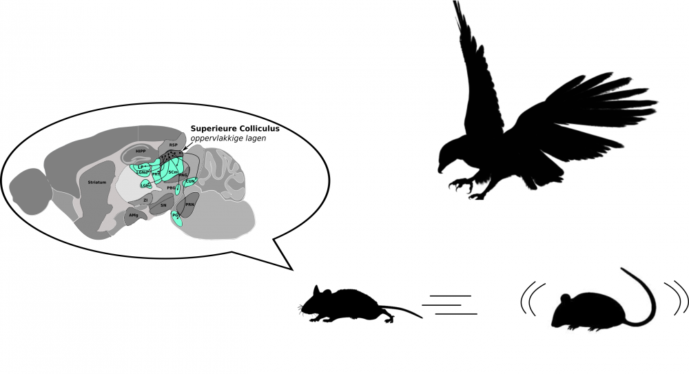
Figuur 1: Hersennetwerken beïnvloeden het aangeboren gedrag bij muizen
Is aangeboren gedrag bij muizen relevant voor ziekten bij de mens?
Ja, absoluut. Bij mensen spelen de netwerken van de superieure colliculus een belangrijke rol bij het verwerken van angst en zijn ze betrokken bij aandoeningen zoals autisme, angst en posttraumatische stressstoornis (PTSD). Begrijpen hoe de hersenen van muizen reageren op bedreigende prikkels is noodzakelijk voor het ontrafelen en uiteindelijk behandelen van ziekten bij de mens waarbij soortgelijke circuits betrokken zijn.
Onderzoek naar de bijdrage van een specifiek hersengebied aan aangeboren gedrag
Het Farrow-lab van Neuro-electronics Research Flanders (NERF) heeft onlangs ontdekt dat activatie van specifieke cellen, genaamd NTSR-neuronen, in de superieure colliculus muizen plotseling 1-2 seconden laat stoppen. Door deze snelle verschuiving in aandacht en de plotselinge locomotieve stilstand kan de muis stoppen en de omgeving onderzoeken. Het team gebruikte vervolgens functionele beeldvorming van het hele brein, waarbij hersenactiviteit wordt gemeten op basis van veranderingen in het bloedvolume, om na te gaan welke andere delen van de hersenen actief waren toen NTSR-cellen werden gestimuleerd. Ze waren verrast om activiteit te vinden in een hersengebied dat nooit is gekoppeld aan aangeboren gedragingen die worden aangedreven door de superieure colliculus. Dit kleine hersengebied, de posterieure paralaminaire kernen van de thalamus (PPnT), is minder intimiderend dan de naam klinkt! Eerder onderzoek heeft de PPnT in verband gebracht met auditieve angstconditionering, waarbij een knaagdier leert om een geluid te associëren met een onaangename ervaring zoals een voetschok. In dit project onderzoeken we de rol van de PPnT in aangeboren stopgedrag door de PPnT bij muizen uit te schakelen tijdens gelijktijdige activatie van NTSR-cellen. Verandert het stopgedrag van muizen wanneer de PPnT wordt gedempt? Dit was de vraag die we wilden beantwoorden.
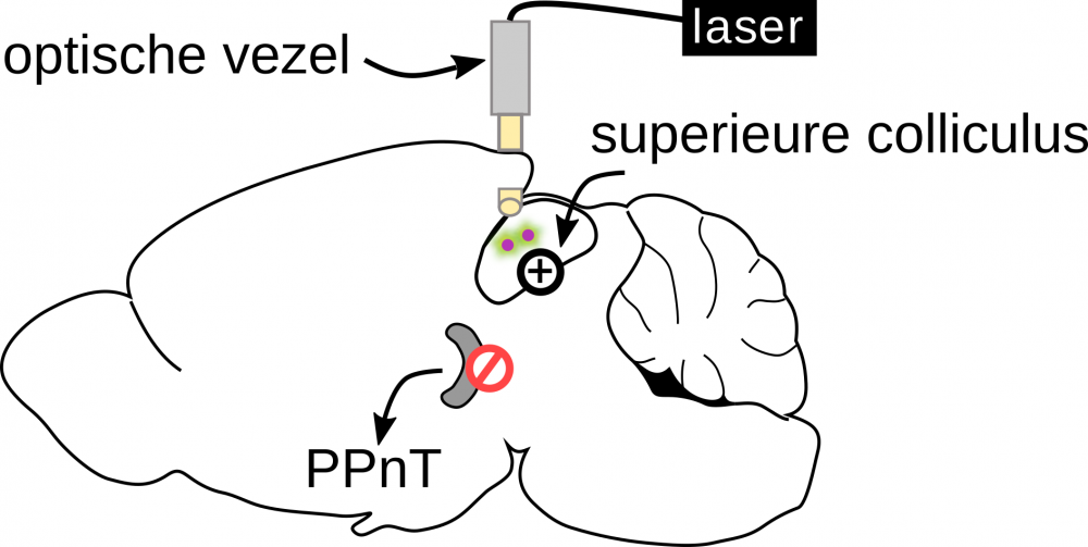
Figuur 2: Schematisch diagram van experimenteel ontwerp
Optogenetica en chemogenetica: krachtige tools om deze onderzoeksvraag te beantwoorden
Als optogenetica en chemogenetica klinken alsof ze rechtstreeks uit een sci-fi-roman komen, blijf dan rustig zitten en wacht tot je hoort hoe ze worden gebruikt om dierlijk gedrag nauwkeurig te kunnen manipuleren door lichte of kleine chemicaliën te gebruiken. Optogenetica is gebaseerd op genetische manipulatie van hersencellen, zodat ze reageren op een specifieke golflengte van licht. In ons geval hebben we chirurgisch een kleine optische vezel geïmplanteerd (ja, denk aan een minuscule streng van een LED lamp met lichtgevende vezels) net boven de superieure colliculus van muizen met NTSR-cellen die genetisch zijn gemanipuleerd om te worden geactiveerd door blauw laserlicht. De optische vezel in de hersenen van de muis was via een kabel aan de laser bevestigd, zodat bij handmatige activatie van de laser, een pulstrein van blauw licht van 1 seconde rechtstreeks naar de superieure colliculus werd gestuurd; dit activeerde NTSR-neuronen en resulteerde in plotseling stopgedrag.
Vervolgens hebben we chemogenetica gebruikt om de PPnT af te sluiten. We injecteerden een genetisch gemodificeerd virus rechtstreeks in de PPnT van muizen, waardoor de cellen daar tijdelijk werden gedeactiveerd. Dit gebeurde enkel als we een medicijn genaamd clozapine-N-oxide toedienden vóór de gedragstesten. Tijdens de test mochten muizen gedurende 20 minuten vrij rondlopen in een open veld, een houten kist. We hebben de laser geactiveerd toen de muis het centrum passeerde, ongeveer 15-20 keer, en we vergeleken het stopgedrag tussen muizen met een geremde PPnT en controlemuizen waarin de PPnT normaal functioneerde.
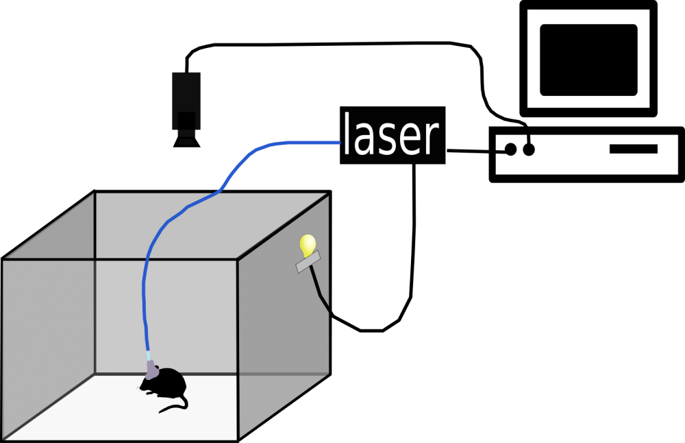
Figuur 3: Testopstelling in open veld
Het remmen van de PPnT verhoogt de gewenning aan activering van NTSR-neuronen
We ontdekten dat controlemuizen gedurende de hele test van 20 minuten consequent stopten bij elke laser activatie om NTSR-neuronen te activeren. Daarentegen waren muizen met een geremde PPnT gewend aan de laserstimulatie, wat betekent dat hun stopreactie geleidelijk afnam na herhaalde stimulaties. Dit betekende dat deze muizen geleidelijk hogere snelheden vertoonden tijdens laserstimulaties en significant lagere kansen om te stoppen in vergelijking met controles. Onze resultaten toonden aan dat het remmen van de PPnT de gewenning aan activering van NTSR-neuronen verhoogt.
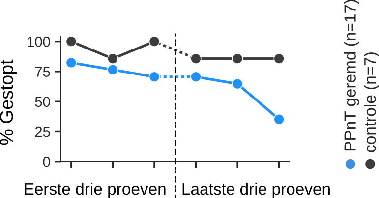
Figuur 4: Percentage muizen dat is gestopt vanwege laserstimulatie
Een nieuwe rol voor de PPnT: waarom is dit van belang?
Deze resultaten suggereren een nieuwe rol voor de PPnT; het bemiddelt aangeboren gedrag door gedragsaanpassing bij herhaalde stimulus te voorkomen. Evolutionair gezien is dit erg belangrijk voor een muis, aangezien het negeren van herhaalde maar relevante, mogelijk dreigende signalen, dodelijk kan zijn. De PPnT laat een verrassende prikkel niet onopgemerkt voorbijgaan. Deze resultaten kunnen met name relevant zijn voor de behandeling van PTSD, waarbij sprake is van overactiviteit in het angstreactiesysteem. Vooruitkijkend, zou het circuit dat we in onze studie hebben gemanipuleerd, erop kunnen worden gericht om patiënten met PTSD te helpen wennen aan niet-bedreigende stimuli.
Laten we een mentale sprong naar jou maken, onze kleine muis die over het veld rent met de havik boven ons. Mede dankzij de bijdrage van de PPnT aan de netwerken die aangeboren gedrag bepalen, stop je instinctief, vliegt de havik boven je hoofd en kun je in alle rust verder foerageren voor je diner.
Bibliografie
- Almeida, I., Soares, S.C., and Castelo-Branco, M. (2015). The Distinct Role of the Amygdala, Superior Colliculus and Pulvinar in Processing of Central and Peripheral Snakes. PLoS One10, e0129949.
- Armbruster, B. N., Li, X., Pausch, M. H., Herlitze, S., & Roth, B. L. (2007). Evolving the lock to fit the key to create a family of G protein-coupled receptors potently activated by an inert ligand. Proceedings of the National Academy of Sciences of the United States of America, 104(12), 5163–5168. https://doi.org/10.1073/pnas.0700293104.
- Arnault, P., and Roger, M. (1987). The connections of the peripeduncular area studied by retrograde and anterograde transport in the rat. J Comp Neurol258, 463–476.
- Arnault, P., and Roger, M. (1990). Ventral temporal cortex in the rat: connections of secondary auditory areas Te2 and Te3. J Comp Neurol302, 110–123.
- Asede, D., Bosch, D., Lüthi, A., Ferraguti, F., and Ehrlich, I. (2015). Sensory inputs to intercalated cells provide fear-learning modulated inhibition to the basolateral amygdala. Neuron86, 541–554.
- Assareh, N., Sarrami, M., Carrive, P., and McNally, G.P. (2016). The organization of defensive behavior elicited by optogenetic excitation of rat lateral or ventrolateral periaqueductal gray. Behav Neurosci130, 406–414.
- Bakker, R., Tiesinga, P., and Kötter, R. (2015). The Scalable Brain Atlas: Instant Web-Based Access to Public Brain Atlases and Related Content. Neuroinformatics13, 353–366.
- Barth, D.S., and MacDonald, K.D. (1996). Thalamic modulation of high-frequency oscillating potentials in auditory cortex. Nature383, 78–81.
- Basso, M.A., and May, P.J. (2017). Circuits for Action and Cognition: A View from the Superior Colliculus. Annu Rev Vis Sci3, 197–226.
- Beltramo, R., and Scanziani, M. (2019). A collicular visual cortex: Neocortical space for an ancient midbrain visual structure. Science363, 64–69.
- Benedek, G., Perény, J., Kovács, G., Fischer-Szátmári, L., and Katoh, Y.Y. (1997). Visual, somatosensory, auditory and nociceptive modality properties in the feline suprageniculate nucleus. Neuroscience78, 179–189.
- Bennett, C., Gale, S.D., Garrett, M.E., Newton, M.L., Callaway, E.M., Murphy, G.J., and Olsen, S.R. (2019). Higher-Order Thalamic Circuits Channel Parallel Streams of Visual Information in Mice. Neuron102, 477–492.e5.
- Bittencourt, A.S., Carobrez, A.P., Zamprogno, L.P., Tufik, S., and Schenberg, L.C. (2004). Organization of single components of defensive behaviors within distinct columns of periaqueductal gray matter of the rat: role of N-methyl-D-aspartic acid glutamate receptors. Neuroscience125, 71–89.
- Bordi, F., and LeDoux, J.E. (1994). Response properties of single units in areas of rat auditory thalamus that project to the amygdala. I. Acoustic discharge patterns and frequency receptive fields. Exp Brain Res98, 261–274.
- Bordi, F., and LeDoux, J.E. (1994). Response properties of single units in areas of rat auditory thalamus that project to the amygdala. II. Cells receiving convergent auditory and somatosensory inputs and cells antidromically activated by amygdala stimulation. Exp Brain Res98, 275–286.
- Campeau, S., and Davis, M. (1995). Involvement of the central nucleus and basolateral complex of the amygdala in fear conditioning measured with fear-potentiated startle in rats trained concurrently with auditory and visual conditioned stimuli. J Neurosci15, 2301–2311.
- Campeau, S., and Davis, M. (1995). Involvement of subcortical and cortical afferents to the lateral nucleus of the amygdala in fear conditioning measured with fear-potentiated startle in rats trained concurrently with auditory and visual conditioned stimuli. J Neurosci15, 2312–2327.
- Carrive, P. (1993). The periaqueductal gray and defensive behavior: functional representation and neuronal organization. Behav Brain Res58, 27–47.
- Chen, L., Wang, X., Ge, S., and Xiong, Q. (2019). Medial geniculate body and primary auditory cortex differentially contribute to striatal sound representations. Nat Commun10, 418.
- Comoli, E., Das, N.F.P., Vautrelle, N., Leriche, M., Overton, P.G., and Redgrave, P. (2012). Segregated anatomical input to sub-regions of the rodent superior colliculus associated with approach and defense. Front Neuroanat6, 9.
- Coolen, L.M., Veening, J.G., Petersen, D.W., and Shipley, M.T. (2003). Parvocellular subparafascicular thalamic nucleus in the rat: anatomical and functional compartmentalization. J Comp Neurol463, 117–131.
- Cynader, M., and Berman, N. (1972). Receptive-field organization of monkey superior colliculus. J Neurophysiol35, 187–201.
- De Franceschi G., Vivattanasarn, T., Saleem, A.B., and Solomon, S.G. (2016). Vision Guides Selection of Freeze or Flight Defense Strategies in Mice. Curr Biol26, 2150–2154.
- Dean, P., Mitchell, I.J., and Redgrave, P. (1988). Responses resembling defensive behaviour produced by microinjection of glutamate into superior colliculus of rats. Neuroscience24, 501–510.
- Dean, P., Redgrave, P., and Westby, G.W. (1989). Event or emergency? Two response systems in the mammalian superior colliculus. Trends Neurosci12, 137–147.
- Deng, H., Xiao, X., and Wang, Z. (2016). Periaqueductal Gray Neuronal Activities Underlie Different Aspects of Defensive Behaviors. J Neurosci36, 7580–7588.
- Doron, N.N., and Ledoux, J.E. (1999). Organization of projections to the lateral amygdala from auditory and visual areas of the thalamus in the rat. J Comp Neurol412, 383–409.
- Dräger, U.C., and Hubel, D.H. (1975). Responses to visual stimulation and relationship between visual, auditory, and somatosensory inputs in mouse superior colliculus. J Neurophysiol38, 690–713.
- Dräger, U.C., and Hubel, D.H. (1976). Topography of visual and somatosensory projections to mouse superior colliculus. J Neurophysiol39, 91–101.
- DuBois, R.M., and Cohen, M.S. (2000). Spatiotopic organization in human superior colliculus observed with fMRI. Neuroimage12, 63–70.
- Elsabbagh, M., Volein, A., Holmboe, K., Tucker, L., Csibra, G., Baron-Cohen, S., Bolton, P., Charman, T., Baird, G., and Johnson, M.H. (2009). Visual orienting in the early broader autism phenotype: disengagement and facilitation. J Child Psychol Psychiatry50, 637–642.
- Evans, D.A., Stempel, A.V., Vale, R., Ruehle, S., Lefler, Y., and Branco, T. (2018). A synaptic threshold mechanism for computing escape decisions. Nature558, 590–594.
- Franklin, G., K. & Paxinos (2019). Paxinos and Franklin’s the Mouse Brain in Stereotaxic Coordinates, Compact(Academic Press).
- Gale, S.D., and Murphy, G.J. (2014). Distinct representation and distribution of visual information by specific cell types in mouse superficial superior colliculus. J Neurosci34, 13458–13471.
- Gale, S.D., and Murphy, G.J. (2018). Distinct cell types in the superficial superior colliculus project to the dorsal lateral geniculate and lateral posterior thalamic nuclei. J Neurophysiol120, 1286–1292.
- Gerfen, C.R., Paletzki, R., and Heintz, N. (2013). GENSAT BAC cre-recombinase driver lines to study the functional organization of cerebral cortical and basal ganglia circuits. Neuron80, 1368–1383.
- Gerren, R.A., and Weinberger, N.M. (1983). Long term potentiation in the magnocellular medial geniculate nucleus of the anesthetized cat. Brain Res265, 138–142.
- Gomez, J.L., Bonaventura, J., Lesniak, W., Mathews, W.B., Sysa-Shah, P., Rodriguez, L.A., Ellis, R.J., Richie, C.T., Harvey, B.K., Dannals, R.F., et al. (2017). Chemogenetics revealed: DREADD occupancy and activation via converted clozapine. Science357, 503–507.
- Goutaudier, R., Coizet, V., Carcenac, C., and Carnicella, S. (2019). DREADDs: The Power of the Lock, the Weakness of the Key. Favoring the Pursuit of Specific Conditions Rather than Specific Ligands. ENeuro6.
- Guo, L., Walker, W.I., Ponvert, N.D., Penix, P.L., and Jaramillo, S. (2018). Stable representation of sounds in the posterior striatum during flexible auditory decisions. Nat Commun9, 1534.
- Hicks, T.P., Stark, C.A., and Fletcher, W.A. (1986). Origins of afferents to visual suprageniculate nucleus of the cat. J Comp Neurol246, 544–554.
- Hicks, T.P., Watanabe, S., Miyake, A., and Shoumura, K. (1984). Organization and properties of visually responsive neurones in the suprageniculate nucleus of the cat. Exp Brain Res55, 359–367.
- Hoy, J.L., Bishop, H.I., and Niell, C.M. (2019). Defined Cell Types in Superior Colliculus Make Distinct Contributions to Prey Capture Behavior in the Mouse. Curr Biol29, 4130–4138.e5.
- Hunnicutt, B.J., Jongbloets, B.C., Birdsong, W.T., Gertz, K.J., Zhong, H., and Mao, T. (2016). A comprehensive excitatory input map of the striatum reveals novel functional organization. Elife5.
- Ito, S., and Feldheim, D.A. (2018). The Mouse Superior Colliculus: An Emerging Model for Studying Circuit Formation and Function. Front Neural Circuits12, 10.
- Iwata, J., LeDoux, J.E., Meeley, M.P., Arneric, S., and Reis, D.J. (1986). Intrinsic neurons in the amygdaloid field projected to by the medial geniculate body mediate emotional responses conditioned to acoustic stimuli. Brain Res383, 195–214.
- Jhang, J., Lee, H., Kang, M.S., Lee, H.S., Park, H., and Han, J.H. (2018). Anterior cingulate cortex and its input to the basolateral amygdala control innate fear response. Nat Commun9, 2744.
- Jones, E.G. (1998). A new view of specific and nonspecific thalamocortical connections. Adv Neurol77, 49–71; discussion 72–73.
- Jones, E.G. (1998). Viewpoint: the core and matrix of thalamic organization. Neuroscience85, 331–345.
- Kamishina, H., Yurcisin, G.H., Corwin, J.V., and Reep, R.L. (2008). Striatal projections from the rat lateral posterior thalamic nucleus. Brain Res1204, 24–39.
- Katyal, S., Zughni, S., Greene, C., and Ress, D. (2010). Topography of covert visual attention in human superior colliculus. J Neurophysiol104, 3074–3083.
- Keifer, O.P.J., Gutman, D.A., Hecht, E.E., Keilholz, S.D., and Ressler, K.J. (2015). A comparative analysis of mouse and human medial geniculate nucleus connectivity: a DTI and anterograde tracing study. Neuroimage105, 53–66.
- Keifer, O.P.J, Hurt, R.C., Ressler, K.J., Marvar, P.J. (2015). The Physiology of Fear: Reconceptualizing the Role of the Central Amygdala in Fear Learning. Physiology (Bethesda) 30, 389-401.
- Kincheski, G.C., Mota-Ortiz, S.R., Pavesi, E., Canteras, N.S., and Carobrez, A.P. (2012). The dorsolateral periaqueductal gray and its role in mediating fear learning to life threatening events. PLoS One7, e50361.
- Krauzlis, R.J., Bollimunta, A., Arcizet, F., and Wang, L. (2014). Attention as an effect not a cause. Trends Cogn Sci18, 457–464.
- Krupa, M., Maire-Lepoivre, E., and Imbert, M. (1984). Visual properties of neurons in the suprageniculate nucleus of the cat. Neurosci Lett51, 13–18.
- Kunwar, P.S., Zelikowsky, M., Remedios, R., Cai, H., Yilmaz, M., Meister, M., and Anderson, D.J. (2015). Ventromedial hypothalamic neurons control a defensive emotion state. Elife4.
- LeDoux, J.E. (2000). Emotion circuits in the brain. Annu Rev Neurosci23, 155–184.
- LeDoux, J.E., Farb, C.R., and Romanski, L.M. (1991). Overlapping projections to the amygdala and striatum from auditory processing areas of the thalamus and cortex. Neurosci Lett134, 139–144.
- LeDoux, J.E., Iwata, J., Cicchetti, P., and Reis, D.J. (1988). Different projections of the central amygdaloid nucleus mediate autonomic and behavioral correlates of conditioned fear. J Neurosci8, 2517–2529.
- LeDoux, J.E., Ruggiero, D.A., and Reis, D.J. (1985). Projections to the subcortical forebrain from anatomically defined regions of the medial geniculate body in the rat. J Comp Neurol242, 182–213.
- LeDoux, J.E., Sakaguchi, A., Iwata, J., and Reis, D.J. (1985). Auditory emotional memories: establishment by projections from the medial geniculate nucleus to the posterior neostriatum and/or dorsal amygdala. Ann N Y Acad Sci444, 463–464.
- LeDoux, J.E., Sakaguchi, A., Iwata, J., and Reis, D.J. (1986). Interruption of projections from the medial geniculate body to an archi-neostriatal field disrupts the classical conditioning of emotional responses to acoustic stimuli. Neuroscience17, 615–627.
- LeDoux, J.E., Sakaguchi, A., and Reis, D.J. (1984). Subcortical efferent projections of the medial geniculate nucleus mediate emotional responses conditioned to acoustic stimuli. J Neurosci4, 683–698.
- Ledoux, J.E., Ruggiero, D.A., Forest, R., Stornetta, R., and Reis, D.J. (1987). Topographic organization of convergent projections to the thalamus from the inferior colliculus and spinal cord in the rat. J Comp Neurol264, 123–146.
- Ledoux, J.E., and Muller, J. (1997). Emotional memory and psychopathology. Philos Trans R Soc Lond B Biol Sci352, 1719–1726.
- Liang, F., Xiong, X.R., Zingg, B., Ji, X.Y., Zhang, L.I., and Tao, H.W. (2015). Sensory Cortical Control of a Visually Induced Arrest Behavior via Corticotectal Projections. Neuron86, 755–767.
- Linke, R. (1999). Differential projection patterns of superior and inferior collicular neurons onto posterior paralaminar nuclei of the thalamus surrounding the medial geniculate body in the rat. Eur J Neurosci11, 187–203.
- Linke, R., Braune, G., and Schwegler, H. (2000). Differential projection of the posterior paralaminar thalamic nuclei to the amygdaloid complex in the rat. Exp Brain Res134, 520–532.
- Linke, R., De, L.A.D., Schwegler, H., and Pape, H.C. (1999). Direct synaptic connections of axons from superior colliculus with identified thalamo-amygdaloid projection neurons in the rat: possible substrates of a subcortical visual pathway to the amygdala. J Comp Neurol403, 158–170.
- Linke, R., and Schwegler, H. (2000). Convergent and complementary projections of the caudal paralaminar thalamic nuclei to rat temporal and insular cortex. Cereb Cortex10, 753–771.
- Macé, É., Montaldo, G., Trenholm, S., Cowan, C., Brignall, A., Urban, A., and Roska, B. (2018). Whole-Brain Functional Ultrasound Imaging Reveals Brain Modules for Visuomotor Integration. Neuron100, 1241–1251.e7.
- Madisen, L., Zwingman, T.A., Sunkin, S.M., Oh, S.W., Zariwala, H.A., Gu, H., Ng, L.L., Palmiter, R.D., Hawrylycz, M.J., Jones, A.R., et al. (2010). A robust and high-throughput Cre reporting and characterization system for the whole mouse brain. Nat Neurosci13, 133–140.
- Mahler, S.V., and Aston-Jones, G. (2018). CNO Evil? Considerations for the Use of DREADDs in Behavioral Neuroscience. Neuropsychopharmacology43, 934–936.
- Manvich, D.F., Webster, K.A., Foster, S.L., Farrell, M.S., Ritchie, J.C., Porter, J.H., and Weinshenker, D. (2018). The DREADD agonist clozapine N-oxide (CNO) is reverse-metabolized to clozapine and produces clozapine-like interoceptive stimulus effects in rats and mice. Sci Rep8, 3840.
- Maren, S. (2011). Seeking a spotless mind: extinction, deconsolidation, and erasure of fear memory. Neuron70, 830–845.
- Maren, S. (2001). Neurobiology of Pavlovian fear conditioning. Annu Rev Neurosci24, 897–931.
- Mathis, A., Mamidanna, P., Cury, K.M., Abe, T., Murthy, V.N., Mathis, M.W., and Bethge, M. (2018). DeepLabCut: markerless pose estimation of user-defined body parts with deep learning.Nature Neuroscience21, 1281–1289.
- Mathis, M.W., and Mathis, A. (2020). Deep learning tools for the measurement of animal behavior in neuroscience. Curr Opin Neurobiol60, 1–11.
- Mattis, J., Tye, K.M., Ferenczi, E.A., Ramakrishnan, C., O’Shea, D.J., Prakash, R., Gunaydin, L.A., Hyun, M., Fenno, L.E., Gradinaru, V., et al. (2011). Principles for applying optogenetic tools derived from direct comparative analysis of microbial opsins. Nat Methods9, 159–172.
- May, P.J. (2006). The mammalian superior colliculus: laminar structure and connections. Prog Brain Res151, 321–378.
- McEchron, M.D., Green, E.J., Winters, R.W., Nolen, T.G., Schneiderman, N., and McCabe, P.M. (1996). Changes of synaptic efficacy in the medial geniculate nucleus as a result of auditory classical conditioning. J Neurosci16, 1273–1283.
- McEchron, M.D., McCabe, P.M., Green, E.J., Llabre, M.M., and Schneiderman, N. (1995). Simultaneous single unit recording in the medial nucleus of the medial geniculate nucleus and amygdaloid central nucleus throughout habituation, acquisition, and extinction of the rabbit’s classically conditioned heart rate. Brain Res682, 157–166.
- McHaffie, J.G., Jiang, H., May, P.J., Coizet, V., Overton, P.G., Stein, B.E., and Redgrave, P. (2006). A direct projection from superior colliculus to substantia nigra pars compacta in the cat. Neuroscience138, 221–234.
- Meyer, C., Padmala, S., and Pessoa, L. (2019). Dynamic Threat Processing. J Cogn Neurosci31, 522–542.
- Mitchell, I.J., Dean, P., and Redgrave, P. (1988). The projection from superior colliculus to cuneiform area in the rat. II. Defence-like responses to stimulation with glutamate in cuneiform nucleus and surrounding structures. Exp Brain Res72, 626–639.
- Mongeau, R., Miller, G.A., Chiang, E., and Anderson, D.J. (2003). Neural correlates of competing fear behaviors evoked by an innately aversive stimulus. J Neurosci23, 3855–3868.
- Moriizumi, T., and Hattori, T. (1992). Ultrastructural morphology of projections from the medial geniculate nucleus and its adjacent region to the basal ganglia. Brain Res Bull29, 193–198.
- Nakamura, H., Hioki, H., Furuta, T., and Kaneko, T. (2015). Different cortical projections from three subdivisions of the rat lateral posterior thalamic nucleus: a single-neuron tracing study with viral vectors. Eur J Neurosci41, 1294–1310.
- Namura, S., Takada, M., Kikuchi, H., and Mizuno, N. (1997). Collateral projections of single neurons in the posterior thalamic region to both the temporal cortex and the amygdala: a fluorescent retrograde double-labeling study in the rat. J Comp Neurol384, 59–70.
- Nath, T., Mathis, A., Chen, A.C., Patel, A., Bethge, M., and Mathis, M.W. (2019). Using DeepLabCut for 3D markerless pose estimation across species and behaviors.Nature Protocols14, 2152–2176.
- Olivé, I., Densmore, M., Harricharan, S., Théberge, J., McKinnon, M.C., and Lanius, R. (2018). Superior colliculus resting state networks in post-traumatic stress disorder and its dissociative subtype. Hum Brain Mapp39, 563–574.
- Peschanski, M. (1984). Trigeminal afferents to the diencephalon in the rat. Neuroscience12, 465–487.
- Ponvert, N.D., and Jaramillo, S. (2019). Auditory Thalamostriatal and Corticostriatal Pathways Convey Complementary Information about Sound Features. J Neurosci39, 271–280.
- Redgrave, P., Coizet, V., Comoli, E., McHaffie, J.G., Leriche, M., Vautrelle, N., Hayes, L.M., and Overton, P. (2010). Interactions between the Midbrain Superior Colliculus and the Basal Ganglia. Front Neuroanat4.
- Romanski, L.M., and LeDoux, J.E. (1992). Equipotentiality of thalamo-amygdala and thalamo-cortico-amygdala circuits in auditory fear conditioning. J Neurosci12, 4501–4509.
- Roseberry, T., and Kreitzer, A. (2017). Neural circuitry for behavioural arrest. Philos Trans R Soc Lond B Biol Sci372.
- Ryugo, D.K., and Weinberger, N.M. (1978). Differential plasticity of morphologically distinct neuron populations in the medical geniculate body of the cat during classical conditioning. Behav Biol22, 275–301.
- Sahibzada, N., Dean, P., and Redgrave, P. (1986). Movements resembling orientation or avoidance elicited by electrical stimulation of the superior colliculus in rats. J Neurosci6, 723–733.
- Sans-Dublanc, A., Chrzanowska, A., Reinhard, K., Lemmon, D., Montaldo, G., Urban, A., and Farrow, K. (2020). Brain-wide mapping of neural activity mediating collicular-dependent behaviors.BioRxiv.
- Schneider, K.A., and Kastner, S. (2005). Visual responses of the human superior colliculus: a high-resolution functional magnetic resonance imaging study. J Neurophysiol94, 2491–2503.
- Senatorov, V.V., and Hu, B. (2002). Extracortical descending projections to the rat inferior colliculus. Neuroscience115, 243–250.
- Sewards, T.V., and Sewards, M.A. (2002). Innate visual object recognition in vertebrates: some proposed pathways and mechanisms. Comp Biochem Physiol A Mol Integr Physiol132, 861–891.
- Shamash, P., Carandini, M., Harris, K.D., and Steinmetz, N.A. (2018). A tool for analyzing electrode tracks from slice histology.
- Shammah-Lagnado, S.J., Alheid, G.F., and Heimer, L. (1996). Efferent connections of the caudal part of the globus pallidus in the rat. J Comp Neurol376, 489–507.
- Shang, C., Chen, Z., Liu, A., Li, Y., Zhang, J., Qu, B., Yan, F., Zhang, Y., Liu, W., Liu, Z., et al. (2018). Divergent midbrain circuits orchestrate escape and freezing responses to looming stimuli in mice. Nat Commun9, 1232.
- Shang, C., Liu, Z., Chen, Z., Shi, Y., Wang, Q., Liu, S., Li, D., and Cao, P. (2015). BRAIN CIRCUITS. A parvalbumin-positive excitatory visual pathway to trigger fear responses in mice. Science348, 1472–1477.
- Silva, B.A., Gross, C.T., and Gräff, J. (2016). The neural circuits of innate fear: detection, integration, action, and memorization. Learn Mem23, 544–555.
- Soares, S.C., Kessel, D., Hernández-Lorca, M., García-Rubio, M.J., Rodrigues, P., Gomes, N., and Carretié, L. (2017). Exogenous attention to fear: Differential behavioral and neural responses to snakes and spiders. Neuropsychologia99, 139–147.
- Soares, S.C., Maior, R.S., Isbell, L.A., Tomaz, C., and Nishijo, H. (2017a). Fast Detector/First Responder: Interactions between the Superior Colliculus-Pulvinar Pathway and Stimuli Relevant to Primates. Front Neurosci11, 67.
- Sparks, D.L., Lee, C., and Rohrer, W.H. (1990). Population coding of the direction, amplitude, and velocity of saccadic eye movements by neurons in the superior colliculus. Cold Spring Harb Symp Quant Biol55, 805–811.
- Sukov, W., and Barth, D.S. (2001). Cellular mechanisms of thalamically evoked gamma oscillations in auditory cortex. J Neurophysiol85, 1235–1245.
- Takada, M., Itoh, K., Yasui, Y., Sugimoto, T., and Mizuno, N. (1985). Topographical projections from the posterior thalamic regions to the striatum in the cat, with reference to possible tecto-thalamo-striatal connections. Exp Brain Res60, 385–396.
- Tanaka, K., Otani, K., Tokunaga, A., and Sugita, S. (1985). The reciprocal connections of the suprageniculate nucleus and the superior colliculus in the rat. Neurosci Res3, 79–85.
- Terpou, B.A., Densmore, M., Théberge, J., Thome, J., Frewen, P., McKinnon, M.C., and Lanius, R.A. (2019). The Threatful Self: Midbrain Functional Connectivity to Cortical Midline and Parietal Regions During Subliminal Trauma-Related Processing in PTSD. Chronic Stress (Thousand Oaks)3, 2470547019871369.
- Thakkar, K.N., Schall, J.D., Heckers, S., and Park, S. (2015). Disrupted Saccadic Corollary Discharge in Schizophrenia. J Neurosci35, 9935–9945.
- Thakkar, K.N., and Rolfs, M. (2019). Disrupted Corollary Discharge in Schizophrenia: Evidence From the Oculomotor System. Biol Psychiatry Cogn Neurosci Neuroimaging4, 773–781.
- Tohmi, M., Meguro, R., Tsukano, H., Hishida, R., and Shibuki, K. (2014). The extrageniculate visual pathway generates distinct response properties in the higher visual areas of mice. Curr Biol24, 587–597.
- Urban, A., Mace, E., Brunner, C., Heidmann, M., Rossier, J., and Montaldo, G. (2014). Chronic assessment of cerebral hemodynamics during rat forepaw electrical stimulation using functional ultrasound imaging. Neuroimage101, 138–149.
- Vagnoni, E., Lourenco, S.F., and Longo, M.R. (2015). Threat modulates neural responses to looming visual stimuli. Eur J Neurosci42, 2190–2202.
- Vagnoni, E., Lourenco, S.F., and Longo, M.R. (2012). Threat modulates perception of looming visual stimuli. Curr Biol22, R826–7.
- Vianna, D.M., and Brandão, M.L. (2003). Anatomical connections of the periaqueductal gray: specific neural substrates for different kinds of fear. Braz J Med Biol Res36, 557–566.
- Villalobos, C.A., Wu, Q., Lee, P.H., May, P.J., and Basso, M.A. (2018). Parvalbumin and GABA Microcircuits in the Mouse Superior Colliculus. Front Neural Circuits12, 35.
- Vlamings, P.H., Jonkman, L.M., van, D.E., van, der G.R.J., and Kemner, C. (2010). Basic abnormalities in visual processing affect face processing at an early age in autism spectrum disorder. Biol Psychiatry68, 1107–1113.
- Wallace, D.J., Greenberg, D.S., Sawinski, J., Rulla, S., Notaro, G., and Kerr, J.N. (2013). Rats maintain an overhead binocular field at the expense of constant fusion. Nature498, 65–69.
- Wang, X., Chou, X., Peng, B., Shen, L., Huang, J.J., Zhang, L.I., and Tao, H.W. (2019). A cross-modality enhancement of defensive flight via parvalbumin neurons in zona incerta. Elife8.
- Wang, Y.C., Bianciardi, M., Chanes, L., and Satpute, A.B. (2020). Ultra High Field fMRI of Human Superior Colliculi Activity during Affective Visual Processing. Sci Rep10, 1331.
- Watson, C. (2012). Visual System.In The Mouse Nervous System,(Elsevier),pp.646–652.
- Wei, P., Liu, N., Zhang, Z., Liu, X., Tang, Y., He, X., Wu, B., Zhou, Z., Liu, Y., Li, J., et al. (2015). Processing of visually evoked innate fear by a non-canonical thalamic pathway. Nat Commun6, 6756.
- Wei, P., Liu, N., Zhang, Z., Liu, X., Tang, Y., He, X., Wu, B., Zhou, Z., Liu, Y., Li, J., et al. (2015). Corrigendum: Processing of visually evoked innate fear by a non-canonical thalamic pathway. Nat Commun6, 8228.
- Wepsic, J.G. (1966). Multimodal sensory activation of cells in the magnocellular medial geniculate nucleus. Exp Neurol15, 299–318.
- Wilhelmi, E., Linke, R., de, L.A.D., and Pape, H.C. (2001). Axonal connections of thalamic posterior paralaminar nuclei with amygdaloid projection neurons to the cholinergic basal forebrain in the rat. Neurosci Lett315, 121–124.
- Winer, J.A., Chernock, M.L., Larue, D.T., and Cheung, S.W. (2002). Descending projections to the inferior colliculus from the posterior thalamus and the auditory cortex in rat, cat, and monkey. Hear Res168, 181–195.
- Winer, J.A., and Larue, D.T. (1988). Anatomy of glutamic acid decarboxylase immunoreactive neurons and axons in the rat medial geniculate body. J Comp Neurol278, 47–68.
- Winer, J.A., and Morest, D.K. (1983). The medial division of the medial geniculate body of the cat: implications for thalamic organization. J Neurosci3, 2629–2651.
- Xiong, X.R., Liang, F., Zingg, B., Ji, X.Y., Ibrahim, L.A., Tao, H.W., and Zhang, L.I. (2015). Auditory cortex controls sound-driven innate defense behaviour through corticofugal projections to inferior colliculus. Nat Commun6, 7224.
- Yasui, Y., Kayahara, T., Nakano, K., and Mizuno, N. (1990). The subparafascicular thalamic nucleus of the rat receives projection fibers from the inferior colliculus and auditory cortex. Brain Res537, 323–327.
- Yilmaz, M., and Meister, M. (2013). Rapid innate defensive responses of mice to looming visual stimuli. Curr Biol23, 2011–2015.
- Zhou, N., Masterson, S.P., Damron, J.K., Guido, W., and Bickford, M.E. (2018). The Mouse Pulvinar Nucleus Links the Lateral Extrastriate Cortex, Striatum, and Amygdala. J Neurosci38, 347–362.


