
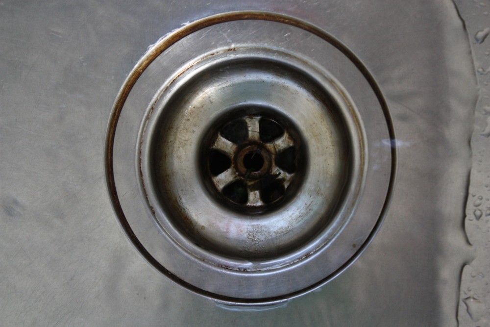
De gootsteen als verspreider van superbacterie
Jaarlijks worden wereldwijd vele mensen tijdens hun opname in het ziekenhuis besmet met een zogenaamde ‘ziekenhuisbacterie’. Dit gaat vaak gepaard met zeer moeilijk te behandelen infecties, die in sommige gevallen fataal kunnen zijn. Maar van waar komen deze ziekteverwekkers? En hoe besmetten ze een patiënt? Apotheker Robin Vanstokstraeten (Vrije Universiteit Brussel) onderzocht tijdens zijn masterproef of de gootstenen in het ziekenhuis hierbij een rol spelen.
Ziekenhuisbacterie
Een ziekenhuisbacterie is een bacterie die patiënten kunnen oplopen tijdens hun hospitalisatie. Vaak gaat dit over zeer resistente bacteriën waartegen weinig soorten antibiotica nog actief zijn. In dat geval wordt er in de volksmond vaak gesproken over een ‘superbacterie’. Dit komt omdat deze beestjes zich aanpassen door manieren te ontwikkelen om het antibioticum steeds te slim af te zijn en te overleven. Deze superbacteriën zijn wereldwijd een groeiend en ernstig probleem, ook in onze Belgische ziekenhuizen.
De gootsteen
Het is belangrijk dit probleem bij de bron aan te pakken en te begrijpen vanwaar deze bacteriën komen. Bacteriën hebben, net zoals alle andere levende beestjes, voedsel en water nodig om te overleven. Deze twee voorwaarden zijn massaal te vinden in de afvoer van een gootsteen: water uit de kraan en voedingsstoffen afkomstig van afval dat in de gootsteen wordt gegoten. Bovendien bezit dit afval in ziekenhuizen vaak grote hoeveelheden antibiotica, waardoor enkel de allersterkste bacteriën in deze gootstenen blijven leven. Daarbovenop produceren deze bacteriën op de wanden van de afvoer een zogenaamde ‘biofilm’. Dit is een uiterst sterke slijmlaag waarin de bacteriën bescherming vinden, hierdoor zijn gecontamineerde gootstenen vaak erg moeilijk bacterievrij te krijgen.
Als deze theorie klopt, en de gootstenen in ziekenhuizen effectief een bron kunnen zijn van deze bacteriën, rijst natuurlijk de vraag hoe patiënten hierdoor besmet worden. Wanneer we de kraan boven een gootstenen laten lopen, wordt er een aerosol gevormd. Dit zijn zeer kleine druppeltjes water die zich via de lucht verspreiden. Deze minuscule druppeltjes kunnen op hun beurt deeltjes, waaronder bacteriën, uit de afvoer vernevelen in de omgeving. Wanneer een gehospitaliseerde patiënt dan zo’n bacterie-bevattende druppel inademt, kan hij geïnfecteerd worden.
Universitair Ziekenhuis Brussel
Helaas kampt ook het UZ Brussel met ziekenhuisbacteriën, in het bijzonder met een superresistente vorm van de soort “Pseudomonas aeruginosa” op de dienst intensieve zorgen. Deze soort is van nature reeds zeer resistent tegen de meest gebruikte antibiotica. Daarbovenop kunnen sommige superresistente varianten het eiwit “VIM” produceren. Dit eiwit zorgt ervoor dat de bacterie nóg resistenter wordt waardoor infecties met deze vorm in sommige gevallen amper nog behandelbaar zijn. Met enige regelmaat worden er patiënten in het UZ Brussel op de dienst intensieve zorgen geïnfecteerd door deze superbacterie. Tot voor kort was de oorzaak van deze infecties nog onbekend.
In het onderzoek van Apr. Vanstokstraeten werden de 36 gootstenen op de dienst intensieve zorgen onderzocht op de aanwezigheid van superresistente varianten van Pseudomonas aeruginosa. Na analyses in het labo werd in 31% van de gootstenen deze superresistente variant gevonden, wat dus bewijst dat deze gevaarlijke beestjes een thuis vinden in de gootstenen van het ziekenhuis.
De gootsteen als verspreider van superbacterie
In een volgend onderdeel van het onderzoek werd het erfelijk materiaal van deze bacteriën uit de gootsteen vergeleken met het erfelijk materiaal van de bacteriën afkomstig van 5 geïnfecteerde patiënten gehospitaliseerd op de intensieve zorgen. Op deze manier kan nagegaan worden of de bacteriën uit de gootsteen dezelfde zijn als deze gevonden bij de patiënten. Na onderzoek in het labo werd duidelijk dat alle 5 de patiënten effectief besmet waren door superresistente kiemen uit de gootsteen. Tijdens het onderzoek bleek ook dat vele andere patiënten besmet waren met andere, minder gevaarlijke bacteriën uit de gootsteen. Vaak staan deze besmette gootstenen net naast het bed van de patiënt, waardoor de kans op infectie aanzienlijk toeneemt. Deze resultaten bewijzen dus dat sommige gootstenen in het ziekenhuis een rol spelen bij de verspreiding van superbacteriën.
Wat is de oplossing?
Vaak kunnen kleine aanpassingen al veel goeds betekenen. Zo speelt het ontwerp van de gootsteen een grote rol: hoe meer het water kan wegspatten en vernevelen, hoe slechter. Uiteraard moet in de toekomst vermeden worden dat er nog afval en antibiotica-bevattend materiaal doorgespoeld wordt in de gootsteen, zodat de groei en selectie van deze superbacteriën vermeden wordt. Verder moeten de zorgverleners op de desbetreffende diensten eens nagaan of het werkelijk nodig is om naast elk bed een gootsteen te zetten. Vaak is één centrale gootsteen per ruimte meer dan voldoende. Ten slotte is een goede hygiëne ook zeer belangrijk: het is van groot belang de gootstenen en zijn afvoer regelmatig en correct te reinigen met de juiste producten. Vaak wordt voor de verwijdering van deze bacteriën en hun biofilm hoge concentraties azijnzuur gebruikt. Sommige ziekenhuizen hebben de gootsteen reeds verbannen uit de patiëntenkamers, met enorme dalingen in het aantal ziekenhuisinfecties als gevolg.
Bibliografie
1. (WHO), World Health Organisation. Health care-associated infections FACT SHEET. [Internet webpage]. 2020.
2. Pseudomonas aeruginosa : new insights into pathogenesis and host defenses. Shaan L. Gellatly, Robert E.W. Hancock. 67, 01 April 2013, Pathogens and Disease, Vol. 3, pp. 159- 173.
3. Todar, Kenneth. Pseudomonas aeruginosa. Todar's Online Textbook of Bacteriology. [Online] [Citaat van: 03 09 2019.] http://textbookofbacteriology.net/pseudomonas.html.
4. Osmosis. Pseudomonas aeruginosa. [Youtube]. 2019.
5. Pseudomonas aeruginosa. Microbiology In Pictures. [Online] 03 09 2019. https://www.microbiologyinpictures.com/pseudomonas-aeruginosa.html.
6. Piérard, Denis. Pathogeen vermogen, virulentie en invasiviteit. Farmaceutische microbiologie. Brussels : VUB, 2019, p. 52.
7. Denis, Piérard. Endotoxines. Farmaceutische microbiologie. Brussels : VUB, 2019, p. 61.
8. Biological Characterization of Endotoxins Released from Antibiotic-Treated Pseudomonas aeruginosa and Escherichia coli. Teruo Kirikae, 1,* Fumiko Kirikae,1 Shinji Saito,1 Kaoru Tominaga, Hirohi Tamura, Yayoi Uemura, Takashi Yokochi, and Masayasu Nakano. 42, May 1998, Antimicrobial Agents and Chemotherapy - American Society for Microbiology, Vol. 5, pp. 1015-1021.
9. The Formation of Biofilms by Pseudomonas aeruginosa: A Review of the Natural and Synthetic Compounds Interfering with Control Mechanisms. Tsiry Rasamiravaka, Quentin Labtani, Pierre Duez, and Mondher El Jaziri. 2015, BioMed Research International, Vol. 2015, p. 17.
10. Educationdimension. Biofilm in the Human Body. Biofilm in Endoscopy. [Online] [Citaat van: 20 9 2019.] https://www.educationaldimensions.com/eLearn/biofilm/body.php.
11. Biofilms. Daniel López, Hera Vlamakis, and Roberto Kolter. July 2010, Cold Spring Harbor Perspectives in Biology.
12. Assembly, structure, function and regulation of type III secretion systems. Wanyin Deng, Natalie C. Marshall, Jennifer L. Rowland, James M. McCoy, Liam J. Worrall, Andrew S. Santos, Natalie C. J. Strynadka & B. Brett Finlay. 15, 12 May 2017, Nature Reviews Microbiology, pp. 323-337.
13. The virulence of Pseudomonas aeruginosa. Pollack, Metthew. 6, 1 September 1984, Reviews of Infectious Diseases.
14. Piérard, Denis. Kapsels van bacteriën. Farmaceutische microbiologie. Brussel : VUB, p. 56.
15. How to manage Pseudomonas aeruginosa infections. Matteo Bassetti, MD, PhD,corresponding author Antonio Vena, MD, Antony Croxatto, PhD, Elda Righi, MD, PhD, and Benoit Guery, MD, PhD. 8, 29 May 2018, Drugs Context.
37
16. Hidron A.I., Edwards J.R., Patel J., Horan T.C., Sievert D.M., Pollock D.A., Fridkin S.K. Nhsn annual update: Antimicrobial resistant pathogens associated with healthcare associated infections: Annual summary of data reported to the national healthcare safety network at the centers for disease control and prevention, 2006–2007. Infect. Control Hosp. Epidemiol. 2008 en 10.1086/591861, 29:996–1011. doi:.
17. Ferroni A., Nguyen L., Pron B., Quesne G., Brusset M.-C., Berche P. Outbreak of nosocomial urinary tract infections due to Pseudomonas aeruginosa in a paediatric surgical unit associated with tap-water contamination. J. Hosp. Infect. 1998 en 10.1016/S0195- 6701(98)90295-X, 39:301–307. doi:.
18. Wingender J. Hygienically relevant microorganisms in biofilms of man-made water systems. In: Flemming H.-C., Wingender J., Szewzyk U., editors. Biofilm Highlights. Volume 5. Springer en Berlin/Heidelberg, Germany: 2011. pp. 189–238.
19. Contamination of the Surfaces of a Health Care Environment by Multidrug-Resistant (MDR) Bacteria. Chaoui L, Mhand R, Mellouki F, Rhallabi N. 29 November 2019, J Microbiol.
20. Outbreak of Multidrug-Resistant Pseudomonas aeruginosa Colonization and Infection Secondary to Imperfect Intensive Care Unit Room Design. Susy Hota, MD, et al. 30, January 2009, Infection control and hospital epidemiology.
21. P. aeruginoas survives in sinks 10 years after hospital outbreak. Bédard, Emilie. 29 July 2015, Infectious Disease News.
22. Generation of Pseudomonas aeruginosa aerosols during handwashing from contaminated sink drains, transmission to hands of hospital personnel, and its prevention by use of a new heating device. Döring G1, Ulrich M, Müller W, Bitzer J, Schmidt-Koenig L, Münst L, Grupp H, Wolz C, Stern M, Botzenhart K. sl : Department of General and Environmental Hygiene, University of Tübingen, Germany., May 1991, pp. 494-505.
23. BiCs symposium 2019. Geyter, Deborah De. 2019. The role of the sink in the transmission of carbapenemase-producing Enterobacteriaceae.
24. Geyter, Deborah De. The sink as a potential source of transmission of carbapenemase- producing Enterobacteriaceae in the intensive care unit of the University Hospital Brussels. Microbiology, VUB. 2015-2016.
25. Post-Outbreak Investigation of Pseudomonas aeruginosa Faucet Contamination by Quantitative Polymerase Chain Reaction and Environmental Factors Affecting Positivity. Emilie Bédard (a1) (a2), Céline Laferrière (a3), Dominique Charron (a1), Cindy Lalancette (a2) ... 36, sl : Cambridge University Press, November 2015, Vol. 11, pp. 1337-1343.
26. Reduced rate of MDROs after introducing ‘water-free patient care’ on a large intensive care unit in the Netherlands. J Hopman1*, R Bos1 , A Voss1 , E Kolwijck2 , P Sturm3 , P Pickkers4 , A Tostmann1 , HVD Hoeven4. June 2015, Antimicrobial resistance and infection control.
27. Influence of the copper-induced viable but non-culturable state on the toxicity of Pseudomonas aeruginosa towards human bronchial epithelial cells in vitro. Dopp E1, Richard J2, Dwidjosiswojo Z3, Simon A4, Wingender J5. sl : ELSEVIER, November 2017, International J Hyg Environ Health, pp. 1363-1369.
28. Whole-genome sequencing identifies highly related Pseudomonas aeruginosa strains in multiple washbasin U-bends at several locations in one hospital: evidence for trafficking
38
of potential pathogens via wastewater pipes. E.M.Moloneya, E.C.Deasya, J.S.Swanb, G.I.Brennanc, M.J.O’Donnella, D.C.Coleman. sl : Elsevier, Received 13 September 2019, Accepted 11 November 2019, Available online 15 November 2019, Journal of Hospital Infection.
29. Piérard, Prof. Dr. Denis. Basisconcepten van ziekte: Microbiologie en infectie. Deel 3: Infectiepreventie. sl : VUB, 2017.
30. The Gale Group, Inc. en 2008., Gale Encyclopedia of Medicine. Copyright.
31. Pseudomonas aeruginosa colonization in the ICU: Prevalence, risk factors and clinical outcomes. Anthony D. Harris, MD MPH,1 Sarah S. Jackson, MPH,1 Gwen Robinson, MPH,1 Lisa Pineles, MA,1 Surbhi Leekha, MD MPH,1 Kerri A Thom, MD MS,1 Yuan Wang, BS,1 Michelle Doll, MD,1 Melinda M. Pettigrew, PhD,2 and J. Kristie Johnson, PhD, D(ABMM)1. May 2016, Infect Control Hosp Epidemiol. , pp. 544-548.
32. Geyter, Prof. Dr. Apr. Klinisch bioloog Deborah De. Mondelinge info dekolonisatie MRSA in het UZ Brussel. Uz Brussel, 26 01 2020.
33. Bundel, Dimitri De. Antimicrobiële middelen. Medicinale chemie. VUB : sn, 2019.
34. Eradicatie P. aeruginosa. Federatie medisch specialisten. [Online] 19 06 2017. https://richtlijnendatabase.nl/richtlijn/bronchiectasieen/p_aeruginosa_… _van_p_aeruginosa.html#tab-content-starting-question.
35. Microbiologie, Belgische Vereniging voor Infectiologie en Klinische. IGGI-Pseudomonas aeruginosa. [Online] [Citaat van: 19 9 2019.] https://www.bvikm.org/.
36. Kahlmeter, Gunnar. EUCAST general and public consultation 9 May – 23 June, 2019 . EUCST. [Online] 10 05 2019. http://www.eucast.org/fileadmin/src/media/PDFs/EUCAST_files/Consultatio… s_and_species_currently_categorised_as_SusceptibleHE_20190509.pdf.
37. Blue laser light inhibits biofilm formation in vitro and in vivo by inducing oxidative stress. Katia Rupel, Luisa Zupin, Giulia Ottaviani, Iris Bertani, Valentina Martinelli, Davide Porrelli, Simone Vodret, Roman Vuerich, Daniel Passos da Silva, Rossana Bussani, Sergio Crovella, Matthew Parsek, Vittorio Venturi, Roberto Di Lenarda, Matteo Biasotto & Serena Zacchigna. 2019, Nature.
38. Fecal Microbiota Transplantation Decreases Intestinal Loads of Multi-Drug Resistant Pseudomonas aeruginosa in Murine Carriers. Mrazek K, Bereswill S, Heimesaat MM. Mar 2019, Eur J Microbiol Immunol., pp. 14-22.
39. Phage therapy against Pseudomonas aeruginosa infections in a cystic fibrosis zebrafish model. Marco Cafora, Gianluca Deflorian, Francesca Forti, Laura Ferrari, Giorgio Binelli, Federica Briani, Daniela Ghisotti & Anna Pistocchi. 06 February 2019, Scientific reports.
40. Honey as an antimicrobial agent against Pseudomonas aeruginosa isolated from infected wounds. Vishnu Prasas Shenoy, Mamatha Balla, PG Shivananda, Indira Bairy. 4, sl : INDUSEM, 2012, Journal of Glabal Infectious Diseases, Vol. 2, pp. 102-105.
41. Multidrug Resistant Bacteria in the Community: Trends and Lessons Learned. Paterson, David van Duin and David. Infect Dis Clin North Am. 2016 Jun; 30(2): 377–390.
39
42. Committee, The Clinical. Antibiotic Resistance Research & Statistics. Clearvue Health. [Online] 18 April 2019. [Citaat van: 19 November 2019.] https://www.clearvuehealth.com/b/antibiotic-resistance-research-statist….
43. Organization, World Health. WHO publishes list of bacteria for which new antibiotics are urgently needed. [Online] 27 Februari 2017. https://www.who.int/news- room/detail/27-02-2017-who-publishes-list-of-bacteria-for-which-new-antibiotics-are- urgently-needed.
44. Antibiotic resistance in Pseudomonas aeruginosa: mechanisms and alternative therapeutic strategies. Zheng Pang, Renee Raudonis, Bernard R.Glick, Tong-JunLin, Zhenyu Cheng. sl : Elsevier, Volume 37, Issue 1, January–February 2019, Pages 177-192 , Biotechnology Advances.
45. Mortality related to Verona Integron-encoded Metallo-β-lactamase-positive Pseudomonas aeruginosa: assessment by a novel clinical tool. Marjolein C. Persoon, Anne F. Voor in ‘t holt, Maurits P. A. van Meer, Karen C. Bokhoven, Diederik Gommers, Margreet C. Vos & Juliëtte A. Severin. sl : Part of Springer Nature, 2019, Antimicrobial Resistance & Infection Control 8.
46. Carbapenemases: the Versatile β-Lactamases. Bush, Anne Marie Queenan* and Karen. July 2007, Clinical Microbiology Reviews, pp. 440-458.
47. Study of the evolution of the susceptibility of Eschericha coli toward beta-lactam inhibitor combinations by MIC and NGS. Belasri, Nassiba. sl : VUB, 2018-2019, Thesis farmacie.
48. Tankeshwar. Oxidase test: Principle, Procedure and oxidase positive organisms. Microbeonline. [Online] 29 December 2012. https://microbeonline.com/oxidase-test- principle-procedure-and-oxidase-positive-organisms/.
49. Analytical profile index. Wikipedia. [Online] 26 9 2018. https://en.wikipedia.org/wiki/Analytical_profile_index.
50. Marie Polet, Nadine Botteldoorn en Katelijne Dierick. MALDI-TOF MS als identificatietool voor voedselpathogenen. NRL Levensmiddelenmicrobiologie, Juliette Wytsmanstraat 14, 1050 Brussel. 2013.
51. Moein et al: "Identification of Appropriate Housekeeping Genes for Gene Expression Analysis in Long-term Hypoxia-treated Kidney Cells." In: Adv Biomed Res., 2017 Feb, 22 en 6:15. [Online]
52. Wikipedia. Minimum spanning tree. Wikipedia. [Online] [Citaat van: 23 1 2020.] https://en.wikipedia.org/wiki/Minimum_spanning_tree.
53. Illumina. Key differences between next-generation sequencing and Sanger sequencing. Illimina NGS. [Online] https://emea.illumina.com/science/technology/next-generation- sequencing/ngs-vs-sanger-sequencing.html.
54. Brussel, UZ. NGS – Sequencing van Massive Parallel Sequencing libraries. Centrum Medische Genetica. Brussel : sn, 2014. p. 12, Analytische procedure.
55. https://pubmlst.org/paeruginosa/. P. aeruginosa PubMLST database. [Online] 2020.
40
56. Hospital Drains as Reservoirs of Pseudomonas aeruginosa: Multiple-Locus Variable- Number of Tandem Repeats Analysis Genotypes Recovered from Faucets, Sink Surfaces and Patients. Cindy Lalancette, Dominique Charron, Céline Laferrière, Patrick Dolcé, Eric Déziel, Michèle Prévost, Emilie Bédard. sl : MDPI, September 2017, Pathogens.
57. Hoxha A, Kärki T, Giambi C, Montano C, Sisto A, Bella A, D'Ancona F: Attributable mortality of carbapenem-resistant Klebsiella pneumoniae infections in a prospective matched cohort study in Italy, 2012-2013.J Hosp Infect. 2016 Jan en 92(1):61-6.
58. Self-disinfecting sink drains reduce the Pseudomonas aeruginosa bioburden in a neonatal intensive care unit. Fusch C1, 2, Pogorzelski D1, Main C2,3, Meyer CL2, El Helou S1,2, Mertz D2,4,5,6. sl : Epub, August 2015, Acta Paediatr., pp. 344-349.
59. Comparison of four methods of hand washing in situations of inadequate water supply. Ogunsola FT1, Adesiji YO. 27 Januari 2008, West Afr J Med.
60. Antibiofilm Properties of Acetic Acid. Thomas Bjarnsholt, Morten Alhede, Peterstrup Jensen, Anne K. NIelsen, Helle Krogh Johansen, Preben Home, Niels Hiby, Michael Givskov, Klaus Kirketerp-Moller. July 2015, Adv Wound Care, pp. 363-372.
61. E. Tacconelli, N. Magrini, Y. Carmeli, S. Harbarth, G. Kahlmeter, J. Kluytmans, M. Mendelson, C. Pulcini, N. Singh, U. Theuretzbacher Global priority list of antibiotic-resistant bacteria to guide research, discovery, and development of new antibiotics World Health Organization (2017), pp. 1-7 (http://www.who.int/medicines/publications/WHO-PPL- Short_Summary_25Feb-ET_NM_WHO.pdf. Last date of access: Nov.18.2018).


