Wat je kunt zien als je niet meer kijkt.
Wat je kunt zien als je niet meer kijkt.
Inleiding
We openen onze ogen en de wereld toont zich aan ons!
Ze toont zich aan ons in kleurrijke beelden… maar hoe ontstaan deze beelden precies? Eigenlijk is er niemand die dat echt weet!
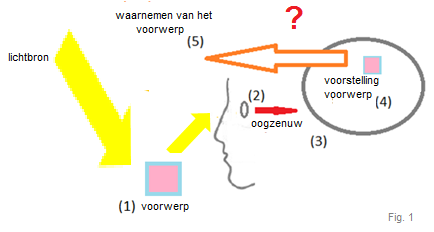
Het enige dat we weten is dat als een lichtstraal op een voorwerp valt, dan reflecteert dat voorwerp die lichtstraal op ons oog. De zenuwen in ons netvlies en in onze hersenen reageren daarop en dat leidt tot een beeld van dat voorwerp in de buitenwereld. Maar hoe de zenuwactiviteit in het oog en in de hersenen wordt omgevormd tot een beeld van de omgeving om ons heen, dat wij dan bovendien nog kunnen waarnemen, is vooralsnog een mysterie. Daarom is de gezichtszin en de werking van het het oog, een favoriet onderzoekdomein van vele wetenschappers (Fig. 1).
Studie
Deze studie onderzocht de gezichtszin bij een patiënt met een bewustzijnsstoornis door hersenschade. Zijn ogen waren intact maar hij was zich niet meer bewust van zijn linker lichaamshelft en hij verwaarloosde de ruimte in het rechter visuele veld. Bij deze patiënt werd onderzocht hoe hij voorwerpen uit de buitenwereld waarnam en of hij nabeelden van die voorwerpen kon zien.
Nabeelden
Een fenomeen dat het zien altijd begeleidt zijn nabeelden (afterimages). Nabeelden kunnen positief of negatief zijn. Een positief nabeeld is een beeld dat verschijnt wanneer men bijvoorbeeld in een lichtbron heeft gekeken. Het nabeeld van de lichtbron zal dan verschijnen als een heldere vlek als men b.v. de ogen sluit. Negatieve of complementaire nabeelden kan men waarnemen als men een korte tijd staart naar een rood vierkant op een witte achtergrond en vervolgens dat vierkant weg neemt en enkel nog naar de witte achtergrond kijkt, dan zal men een groen vierkant waarnemen als het nabeeld van het rode. Een persoon die enkel de witte achtergrond zag zal dat groene vierkant niet waarnemen.
Over deze negatieve nabeelden is nog niet veel geweten. Iedere gezonde persoon kan nabeelden waarnemen, want ze begeleiden àltijd, èlke visuele activiteit, we zijn ons daar alleen niet van bewust tot we er de aandacht op vestigen (Gilroy & Blake, 2005).
Nabeelden worden in de literatuur vaak als een te verwaarlozen zintuigelijk bijverschijnsel beschouwd maar de jongste jaren is aangetoond dat de duur en de intensiteit van negatieve nabeelden samenhangen met vele soorten hogere denkprocessen in de hersenen (Yeonan-Kim, 2019), ze moeten dus een functie hebben!
Ook qua verschijningsvorm zijn nabeelden bijzonder: het zijn geen gewone zintuigelijke waarnemingen maar ook geen louter mentale beelden, want de vorm en de kleur van nabeelden kan rechtstreeks worden beïnvloed door de externe omgeving waarin men een nabeeld waarneemt (Manzotti, 2017). Zo kan een rood voorwerp blauwe, groene of paarse nabeelden opleveren, afhankelijk van de achtergrond waarop het nabeeld wordt waargenomen (Fig.2).
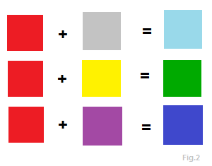
Een louter mentaal beeld zou nooit kunnen worden beïnvloed door de omgeving. Wanneer men zich een beeld voorstelt, louter in gedachten, dan blijft die voorstelling altijd dezelfde ongeacht of men voor een grijze, gele of paarse achtergrond staat, een nabeeld daarentegen verandert daardoor!
Laterale inhibitie
Bij de waarneming van nabeelden zien we dezelfde zenuw-activiteit als bij de waarneming van de voorwerpen die het nabeeld oproepen. Deze zenuwactiviteit wordt laterale inhibitie genoemd en is een soort zenuwactiviteit die we bij de meeste zintuigelijke waarnemingen terugvinden (Yantis, 2014).
Deze studie wou nagaan of er ondanks dezelfde processen in de zenuwen van het oog en in de hersenen, toch een observeerbaar verschil bestaat tussen het zien van zintuigelijk waarneembare objecten rondom ons en het zien van nabeelden.
Experiment
Er werd een experiment opgezet met een patiënt die leed aan een bewustzijnsstoornis door hersenschade in de pariëtale lob. Er werd nagegaan of de hersenschade een invloed had op de waarneming van de objecten om ons heen en/of de nabeelden daarvan.
De gewone objecten waren in dit geval gekleurde vierkanten die in 4 condities werden aangeboden: in het midden (CENTER), het linker (LVF) en het rechter (RVF) gezichtsveld en in beide gezichtsvelden tegelijk (BOTH).
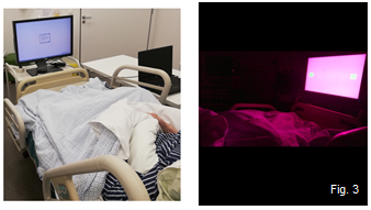
De patiënt moest eerst de blik richten op het vierkant en rapporteren of hij dat kon zien. Het vierkant verdween na 20 sec waarna hij de blik moest richten op een witte achtergrond en vertellen of hij een nabeeld kon waarnemen (Fig. 3).
Bij gezonde personen zal er altijd automatisch een nabeeld zichtbaar worden als men een gekleurd vlak een tijdje laat inwerken op het oog, maar bij deze patiënt verschenen de nabeelden niet automatisch; alle gekleurde vierkanten werden waargenomen, maar hij kon niet alle nabeelden zien, dit laatste duidelijk in relatie tot het hersenletsel, dat hem scheen te verhinderden alle nabeelden waar te nemen.
De patiënt was in staat om in 100% van de gevallen de gewone stimulus te rapporteren, maar 33,3% van de nabeelden werd niet waargenomen.
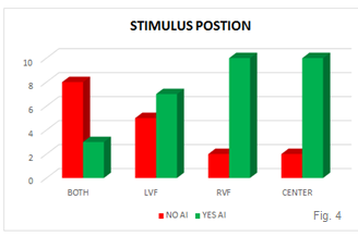
Zoals verwacht bleek de plaats in het gezichtsveld waarin het gekeurde vierkant werd aangeboden ook een rol te spelen: 83% van de nabeelden werd waargenomen wanneer een stimulus werd aangeboden in het midden (CENTER) of aan de rechterkant (RVF) van het scherm. Wanneer een stimulus links (LVF) werd aangeboden, was dit slechts 58% en wanneer twee vierkanten werden aangeboden in beide gezichtsvelden tegelijk (BOTH), liep het aantal gerapporteerde nabeelden terug tot 33%. (Fig.4).
Conclusie
Door deze studie werd voor het eerst de onderscheiding en vergelijking mogelijk van enerzijds waarnemingsprocessen van objecten in de buitenwereld (gekleurde vierkanten) en anderzijds waarnemingsprocessen van de nabeelden.
Er werd aangetoond dat de pariëtale lob een rol speelt bij het waarnemen van nabeelden én er werd geheel nieuw een bewijs geleverd dat twee van elkaar verschillende soorten van bewuste waarneming betrokken zijn bij het zien van de wereld om ons heen: één voor het zien van de voorwerpen zelf en één voor het zien van de nabeelden! Beide waarnemingsprocessen tonen zich mogelijks in dezelfde neurologische activiteit (laterale inhibitie) maar kunnen afzonderlijk van elkaar functioneren of beschadigd zijn, wat werd aangetoond doordat de hersenschade duidelijk een invloed had op die waarnemingsprocessen die bij de nabeelden worden gebruikt, zonder de gewone waarneming van objecten te verstoren.
Bibliografie
Referenties
Allman, J., Miezin, F. & McGuinness, E. (1985). Stimulus specific responses from beyond the classical receptive field: neurophysiological mechanisms for local – global comparisons in visual neurons. Annual Review of Neuroscience, 8, 407–430.
Anstis, S. M. (1975). In Handbook of Psychobiology. Gazzaniga, M. S. & Blakemore, C. (Eds.), (pp. 269–323). New York: Academic
RC Atkinson, R.C., Shiffrin, R.M. (1968). Human memory: A proposed system and its control processes. In The Psychology of Learning and Motivation: advances in research and theory (Vol.2.). K.W. Spence & J.T. Spence (Eds.). New York: Academic Press
Bachmann, T., Mund, C. (2010) Covert spatial attention in search for the location of a color-afterimage patch speeds up its decay from awareness: Introducing a method useful for the study of neural correlates of visual awareness. Vision Research, 50(11), 1048-1053. doi.org/10.1016/j.visres.2010.03.013
Conscious Cogn. 18(4):1039-1048. doi:10.1016/j.concog.2009.09.002
Bakshi, A., Ghosh, K. (2017). A Neural Model of Attention and Feedback for Computing Perceived Brightness in Vision. In Handbook of Neural Computation, (pp. 487–513)
Ball, E. et al. (2017) Does mindfulness meditation improve chronic pain? A systematic review. Current Opinion in Obstetrics and Gynecology, 29 (6), 359-366.
Baltaretu, B.R.(2020). Parietal Cortex Integrates Saccade and object orientation signals to update grasp plans. Journal of Neuroscience DOI: https://doi.org/10.1523/JNEUROSCI.0300-20.2020
Baumgartner, G. (1960). Indirekte Grobenbestimmung der rezeptiven Felder der Retina beim
Menschen mittels der Hermannschen Gittertauschung. Pflugers Archiv für die gesamte PsychoLogie, 272, 21- 22.
Beau Lotto, R., Purves, D. (2002). The empirical basis of color perception. Consciousness and Cognition, 11(4), 609–629. https://doi.org/10.1016/s1053-8100(02)00014-4
Bender, M. B., Kahn, R. L. (1949). After-imagery in defectivefields of vision. Journal of Neurology, Neurosurgery & Psychiatry, 12(3), 196–204.
Bender, M.B., Teuber, H.L., (1946). Phenomena offluctuation, extinction and completion invisual perception. Archives of Neurology And Psychiatry, 55(6), 627. https://doi.org/10.1001/archneurpsyc.1946.02300170075008
Berti, A., Rizzolatti, G. (1992). Visual processing without awareness: Evidence from unilateral neglect
Journal of Cognitive Neuroscience, 4 (4), 345-351.
Betensky, M.G. (1995). What do you see? Phenomenology of therapeutic art expression. London & Bristol: J.Kingsley
Billock, V. A., Gleason, G. A.; Tsou, B.H. (2001). Perception of forbidden colors in retinally stabilized equiluminant images: an indication of softwired cortical color opponency? Journal of the Optical Society of America A, 18(10)
Bisiach E, Luzzatti C., (1978), Unilateral neglect of representational space. Cortex, 14(1),129-33.
Brascamp, J. W., van Boxtel, J. J., Knapen, T., Blake, R. (2010). A dissociation of attention and awareness in phase-sensitive but not phase-insensitive visual channels. Journal of Cognitive Neuroscience, 22, 2326–2344.
Brindley, G.S. (1970). Physiology of the Retina and the Visual Pathway. Baltimore: Williams & Wilkins.
Brindley, G. S. (1962). Two new properties of foveal afterimages and a photochemical hypothesis to explain them. Journal of Physiology, 164, 168–179.
Brown, J. L. (1965). Afterimages. In Vision and visual perception C. H. Graham (Ed.), (pp. 301–302). NY: Wiley.
Brysbaert, M. (2011). Psychologie. London: Academia Press
Burr, D. C., Morrone, M. C., Ross, J. (1994) Selective suppression of the magnocellular visual pathway during saccadic eye movements. Nature, 371, 511–513.
Byrne, A., Hilbert, D. R. (2003). Color realism and color science. Behavioral & Brain Sciences, 26, 3-64.
Cave, R., Batty, M.J. (2006). From searching for features to searching for threat: Drawing the boundary between preattentive and attentive vision. In R. Parasuraman (Ed.),The Attentive Brain (pp. 629-646). Cambridge: MIT Press.
Cheal, J.L., Heisz J.J., Walsh, J.A., Shedden J.M., Rutherford, M.D. (2014), Afterimage induced neural activity during emotional face perception. Brain Res. 2014;1549:11-21. doi:10.1016/j.brainres.2013.12.020
Churchland, P. M. (2005). Chimerical colors: Some phenomenological predictions from cognitive neuroscience. Philosophical Psychology, 18, 527-560.
Cornsweet, T. (1970). Visual perception. New York: Academic Press.
Crane, H. D., Piantanida, T. (1983). On Seeing Reddish Green and Yellowish Blue. Science, 221(4615), 1078–1080.
Daini, R., Angelelli, P., Antonucci, G., Cappa, S. F., Vallar, G. (2002). Exploring the syndrome of spatial unilateral neglect through an illusion of length. Experimental Brain Research, 144(2), 224–237.
Dartnall, H. J. L., Bowmaker, J. K., Mollon, J. D. (1983). Human visual pigments: microspectrophotometric results from the eyes of seven persons. Proceedings of the Royal Society of London. Series B. Biological Sciences, 220(1218), 115–130.
Daw, N. W. (1962). Why After-Images are not Seen in Normal Circumstances. Nature, 196(4860), 1143–1145. https://doi.org/10.1038/1961143a0
Dawson, M. R. W. (1998). Understanding Cognitive Science.Hoboken: John Wiley And Sons Ltd.
Dehaene, S., Changeux, J., Naccache, L., Sackur, J., Sergent, C. (2006). Conscious, preconscious, and subliminal processing: A testable taxonomy. Trends in Cognitive Sciences, 10, 204–211.
De Raedt,R. et al. (2013) Positive emotion broadens attention focus through decreased position-specific spatial encoding in early visual cortex: Evidence from ERPs. Cogn Affect Behav Neurosci 13, 60–79
De Raedt, R. et al (2014) Cognitive control therapy and transcranial direct current stimulation for depression: A randomized, double-blinded, controlled trial. Journal of Affective Disorders, 162 (20), 43-49
Derrington, A. M., Krauskopf, J., & Lennie, P. (1984). Chromatic mechanisms in lateral geniculate nucleus of macaque. Journal of Physiology, 357, 241–265.
De Valois, R. L., Abramov, I. & Jacobs, G. H. (1966). Analysis of response patterns of LGN cells. Journal of the Optical Society of America, 7, 966 – 977.
Driver, J., & Halligan, P. W. (1991). Can visual neglect operate in object-centered co-ordinates? An affirmative single-case study. Cognitive Neuropsychology, 8(6), 475-496.
Driver, J. & Mattingley, J. (1998). Parietal neglect and visual awareness. Nature Neuroscience, 1, 17–22.
J. Driver, J. et. al. (2001) Functional magnetic resonance imaging and evoked potential correlates of conscious and unconscious vision in parietal extinction patients. NeuroImage, 14 (1), 568-575.
Eagleman, D. M., (2001). Visual illusions and neurobiology, Nature Reviews- Neuroscience (2), 920-926.
Egly, R., Driver, J., & Rafal, R. D. (1994). Shifting visual attention between objects and locations: Evidence from normal and parietal lesion subjects. Journal of Experimental Psychology: General, 123(2), 161–177.
Esterman, M., McGlinchey-Berroth, R., Verfaellie, M., Grande, L., Kilduff, P., Milberg, W. (2002) Aware and unaware perception in hemispatial neglect: Evidence from a stem completion priming task. Cortex, 38 (2), 233-246.
Farnè, A., Ponti, F., & Làdavas, E. (1998). In search of biased egocentric reference frames in neglect. Neuropsychologia, 36, 611– 623.
Ferber, S., & Karnath, H. (1999). Parietal and occipital lobe contributions to perception of straight ahead orientation. Journal of Neurology, Neurosurgery & Psychiatry, 67(5), 572–578.
Fermüller, & C. Malm, H. (2004) Uncertainty in visual processes predicts geometrical optical illusions, Vision Research, 44(7), 727-749. https://doi.org/10.1016/j.visres.2003.09.038
Field, G. D., Gauthier, J. L., Sher, A., Greschner, M., Machado, T. A., Jepson, L. H.,Chichilnisky,E. J. (2010). Functional connectivity in the retina at the resolution of photoreceptors. Nature, 467(7316), 673–677
Flavell, J. H. (1979). Metacognition and cognitive monitoring: A new area of cognitive–developmental inquiry. American Psychologist, 34(10), 906–911. https://doi.org/10.1037/0003-066X.34.10.906
Francis, G., Ericson, J. (2004). Using afterimages to test neural mechanisms for perceptual filling-in. Neural Networks, 17(5–6), 737-752.
Ganz, L. (1966a). The mechanism of the figural aftereffect. Psychological Review, 73, 128–150.
Ganz, L. (1966b). Is the figural aftereffect an aftereffect? A review of its intensity, onset, decay, and transfer characteristics. Psychological Bulletin, 66, 151–165.
Gilroy L. A., & Blake R. (2005). The interaction between binocular rivalry and negative afterimages. Current Biology, 15, 1740–1744.
Goethe J.W. (1810) Zur Farbenlehre.Frankfurt/Main: Deutscher Klassikerverlag
Gordon, J. E. (1991). Theories of visual perception. New York: Psychology Press.
Gouras, P. (1968) Identification of cone mechanisms in monkey ganglion cells. Journal of Physiology, 199, 533–547.
Gruber, T.; Müller, M. (2002). Effects of picture repetition on induced gamma band responses, evoked potentials, and phase synchrony in the human EEG. Brain Research. Cognitive Brain Research. 13 (3): 377–392.
Hadjikhani, N., Liu, A. K., Dale, A. M., Cavanagh, P., & Tootell, R. B. (1998). Retinotopy and color sensitivity in human visual cortical area V8. Nature Neuroscience, 1(3), 235–241.
Haeyen, S. (2007). Niet uitleven, maar beleven. Houten: Bohn Stafleu van Loghum
Hart, O., & Nijenhuis, E.R., & Steele, K. (2007). The Haunted Self: Structural Dissociation and the Treatment of Chronic Traumatization. Reehorst: Mens
Hebb, D.O. (1949) The organisation of behaviour. New York: Wiley
Heilman, K. M., Watson, R. T. & Valenstein, E. (1993). Neglect and Related Disorders. New York: Oxford University Press.
Hering, E. (1878). Zur Lehre vom Lichtsinne. Vienna: Carl Gerolds Sohn
Hermann, L. (1870). Eine Erscheinung simultanen Contrastes. Pflüger, Archiv für die Gesammte Physiologie des Menschen und der Tiere, 3(1), 13–15.
Hilton, L., Maher, A. R., Colaiaco, B., Apaydin, E., Sorbero, M. E., Booth, M., Shanman, R. M., & Hempel, S. (2017). Meditation for posttraumatic stress: Systematic review and meta-analysis. Psychological Trauma: Theory, Research, Practice, and Policy, 9(4), 453–460. https://doi.org/10.1037/tra0000180
Hirsch, R. (2004). Exploring Colour Photography: A Complete Guide. London: Laurence King Publishing
Hofer, H. (2005). Organization of the Human Trichromatic Cone Mosaic. Journal of Neuroscience, 25(42), 9669–9679. https://doi.org/10.1523/jneurosci.2414-05.2005
Holmes, G. (1945). Ferrier lecture: the organization of the visual cortex in man.
Proceedings of the Royal Society of London, Series B, 361, 2239–2259.
Hope, A.C.A.(1968). A Simplified Monte Carlo Significance Test Procedure. Journal of the Royal Statistical Society. Series B (Methodological), 30(3), 582-598.
Hsieh, P., & Tse, P. (2006). Illusory color mixing upon perceptual fading and filling-in does not result in ‘forbidden colors’. Vision Research, 46(14), 2251–2258.
Hurvich, L. M. & Jameson, D. (1956) Some quantitative aspects of an opponent-colors theory. IV. A psychological color specification system. Journal of the Optical Society of America, 46, 416–421.
Ito, H. (2012). Cortical shape adaptation transforms a circle into a hexagon: A novel afterimage illusion. Psychological Science, 23(2), 126-132. https://doi.org/10.1177/0956797611422236
Jameson, K., & D'Andrade, R. G. (1997). It's not really red, green, yellow, blue: an inquiry into perceptual color space. Color Categories in Thought and Language, 295–319. https://doi.org/10.1017/cbo9780511519819.014
Jones, P. D., & Holding, D. H. (1975). Extremely long-term persistence of the McCollough effect. Journal of Experimental Psychology: Human Perception and Performance, 1(4), 323–327.
Kabat-Zinn, J. (1990). Full Catastrophe Living: Using the wisdom of your body and mind to face stress, pain, and illness. New York: Dell Publishing
Kalat. W. (2004). Biological psychology. Toronto: Thomso Wadsworth
Kareemulla, S., Khasim, S.M., Samreen, S.S., Siddiqua, H.B., & Khatoon, J.M. (2017) Neuro psychological behavior of hemispatial neglect: a systematic anatomization on its paradigm and rehabilitation approach. Indo American Journal of pharmaceutical sciences, 4(11), 3901-3910.
Karnath, H.O., Schenkel, P., & Fischer, B. (1991). Trunk orientation as the determining factor of the ‘contralateral’deficit in the neglect syndrome and as the physical anchor of the internal representation of body orientation in space. Brain, 114(4), 1997–2014. https://doi.org/10.1093/brain/114.4.1997
Kerkhoff (1999). Multimodal spatial orientation deficits in left-sided visual neglect. Neuropsychologia, 37(12),1387-1405.
Kisler, V.A. (1998). Perceptions and metaperceptions of same-sex social interactions in college women with troubled eating patterns. Dissertation Abstracts International: Section B: The Sciences & Engineering, 58 (9B), 5124.
Kuks, J.B.M.,& Snoek, J.W. (2012) Klinische neurologie. Houten: Springer Media B.V.
E. Làdavas, E., Paladini, R. , Cubelli. R. (1993). Implicit associative priming in a patient with left visual neglect. Neuropsychologia, 31 (12), 1307-1320.
Lazarus,R.S. (1991). Emotion and adaptation. Oxford: Oxford University Press
Lee, B.H., et al. (2004) The Character-line Bisection Task: a new test for hemispatial neglect. Neuropsychologia, 42(12),1715-1724 DOI:10.1016/j.neuropsychologia.2004.02.015
LeDoux, P., Cicchetti, A., Xagoraris, L.M., (1990) The lateral amygdaloid nucleus: sensory interface of the amygdala in fear conditioning. Journal of Neuroscience 10 (4), 1062-1069.
Logan, G. D. (1994) Spatial attention and the apprehension of spatial relations. Journal of Experimental Psychology: Human Perception and Performance, 20, (5), 1015-1036. DOI:10.1037/0096-1523.20.5.1015
Lupyan, G. (2015). Object knowledge changes visual appearance: Semantic effects on color afterimages. Acta Psychologica, 161, 117–130.
Macpherson, F., & Platchias, D. (2013). Hallucination: Philosophy and psychology. Cambridge, MA: MIT Press.
Maffei, L. & Fiorentini, A. (1976). The unresponsive regions of visual cortical receptive fields. Vision Research ,16, 1131–1139.
Manzotti, R. (2017). A Perception-Based Model of Complementary Afterimages. SAGE Open, 7(1). https://doi.org/10.1177/2158244016682478
Marcel, A.J., (1998). Blindsight and shape perception: deficit of visual consciousness or of visual function? Brain, 121(8), 1565–158.
Mattingley, J. B., Davis, G., & Driver, J. (1997). Pre-attentive filling-in of visual surfaces in parietal extinction. Science, 275, 671–674.
Maule, J., Stanworth, K., Pellicano, E. et al. (2018) Color Afterimages in Autistic Adults. Journal of Autism Developmental Disorders 48, 1409–1421 (2018). https://doi.org/10.1007/s10803-016-2786-5
McCollough, C. (1965). Color adaptation of edge-detectors in the human visual system. Science, 149, 1115 – 1116.
McGlinchey-Berroth, R., Milberg, W.P., Verfaellie, M., Alexander, M.P. (1993) Semantic processing in the neglected field: Evidence from a lexical decision task. Cognitive Neuropsychology, 10 (1), 79-108.
McKeefry, D.J. & Zeki, S. (1997) The position and topography of the human colour centre as revealed by functional magnetic resonance imaging. Brain, 120, 2229–2242.
Mepean, J., Phuangsuwan, C., & Ikeda, M. (2017). Chromatic adaptation shown by the adapting and adapted colors in the afterimage. Journal of the color science association of Japan, 41(3), 77-78.
Mesulam, M. (1981). A cortical network for directed attention and unilateral neglect. Annals of Neurology, 10(4), 309–325. https://doi.org/10.1002/ana.410100402
Mikami, K.; Oura, K. (2019) Event Related Hemodynamics and Potentials evoked by Visual Attention Task. Transactions of the Institute of Systems, Control and information engineers 23 (2), p. 63-68
Moscovitch, M., & Behrmann, M. (1994). Coding of Spatial Information in the Somatosensory System: Evidence from Patients with Neglect following Parietal Lobe Damage. Journal of Cognitive Neuroscience, 6(2), 151–155.
Newton, I. (1730). Opticks: Or, a Treatise of the Reflections, Refractions, Inflections, and Colors. London: Printed for William Innys at the West-End of St. Paul's
T. Okada, T., Tanaka, S., Nakai, T., Nishizawa, S., et al. (2000) Naming of animals and tools: A functional magnetic resonance imaging study of categorical differences in the human brain areas commonly used for naming visually presented objects.Neuroscience Letters, 296 (1), 33-36.
Palmer, S. E. (1999). Vision Science: Photons to Phenomenology. Cambridge: Bradford Bokk.
Pöppel, E., Held, R., & Frost, D., (1973). Residual visual function after brain wounds involving the central visual pathways in man. Nature, 243, 295–296.
Posner, M. I., & Petersen, S. E. (1990). The Attention System of the Human Brain. Annual Review of Neuroscience, 13(1), 25–42.
Posner, M. I., Walker, J. A., Friedrich, F. J., & Rafal R. D. (1984) Effects of parietal injury on covert orienting of attention. Journal of Neuroscience, 4, 1863–1874.
Pridmore, R. W. (2011). Complementary colors theory of color vision: Physiology, color mixture, color constancy and color perception. Color Research & Application, 36(6), 394–412.
Pridmore, R. W. (2012). Single cell spectrally opposed responses: opponent colors or complementary colors? Journal of Optics, 42(1), 8–18.
Ptak, R; Valenza,N. (2005). The inferior temporal lobe mediates distracter-resistant visual search of patients with spatial neglect. Journal of Cognitive Neuroscience, 17 (5), 788-799.
Qian, J., Liu, S., & Lei, Q. (2016). Illusory Distance Modulates Perceived Size of Afterimage despite the Disappearance of Depth Cues. PLOS ONE, 11(7).
Rafal, R. (1994). Neglect. Current Opinion in Neurobiology, 4(2), 231–236.
Ratliff, F. (1970). In Ernst Mach Physicist and Philosopher. Cohen, R. S. & Seeger, R. J. (Eds.) (165-184). Dordrecht: Reidel.
Ramel, W., Goldin, P.R., Carmona, P.E. et al. The Effects of Mindfulness Meditation on Cognitive Processes and Affect in Patients with Past Depression. Cognitive Therapy and Research 28, 433–455 (2004). https://doi.org/10.1023/B:COTR.0000045557.15923.96
Revonsuo, A. (2010) Modified from Consciousness: The science of subjectivity. New York: Psychology Press.
Ritschel, T., & Eisemann, E. (2012). A computational model of afterimages. Computer Graphics Forum, 31, 529–534.
Ro, T., Rafal, R.D. (1996). Perception of geometric illusions in hemispatial neglect.Neuropsychologia, 34(10), 973–978.
Robertson, I. H., & Halligan, P. W. (1999). Spatial neglect: A clinical handbook for diagnosis and treatment. Hove: Psychology Press.
Robertson, L. C., & Sagiv, N. (2005). Synesthesia: Perspective from cognitive neuroscience. NY: Oxford University Press.
Röder, B., Rösler, F. & Spence, C. (204).E EE [HTML]arly vision impairs tactile perception in the blind.Current Biology, 20(14), 121-124.
.
Rossi, A. F. & Paradiso, M. A. (1999). Neural correlates of perceived brightness in the retina, lateral geniculate nucleus, and striate cortex. Journal of Neuroscience, 19, 6145–6156.
Rutherford, M.D.; Troubridge, E.K. & Walsh, J. (2012). Visual Afterimages of Emotional Faces in High Functioning Autism. Journal of Autism and Developmental disorders, 42, 221–229 doi.org/10.1007/s10803-011-1233-x
Sabra, A.I., ed. (1989). The Optics of Ibn Al-Haytham. Books I-III. On Direct Vision (London: The Warburg Institute).
Snyder, D.M. (2001). On the Relationship Between Proximal and Distal Stimuli and an Example of Its Significance to Physics. arXiv:physics/9909008 [physics.gen-ph]
Sperandio, I., Lak, A., & Goodale, M. A. (2012). Afterimage size is modulated by size-contrast illusions. Journal of Vision, 12(2), 18.
Sperandio, I., Unwin, K.L., Landry, O. et al. (2017) Size Constancy is Preserved but Afterimages are Prolonged in Typical Individuals with Higher Degrees of Self-Reported Autistic Traits. J Autism Dev Disord 47, 447–459 (2017). https://doi.org/10.1007/s10803-016-2971-6
Spillmann, L. (1994). The Hermann Grid Illusion: A Tool for Studying Human Perceptive Field Organization. Perception, 23(6), 691–708.
Srinivasan, N., & Singh, A. (2017). Concentrative meditation influences visual awareness: a study with color afterimages. Mindfulness, 8(1), 17–26.
Stein, W.J. (1921), Historisch kritische Beitrage zur Entwicklung der neueren Philosophie. Als Inaugural-Dissertation zur Erlangung der Doktorwürde der philosophischen Fakultät der Universität Wien vorgelegt und von dieser approbiert.
Stratton, G. M. (1897). Vision without inversion of the retinal image. Psychological Review, 4, 341–463.
Suarez, J. (2009). Reddish Green: A Challenge for Modal Claims About Phenomenal Structure. Philosophy and Phenomenological Research, 78(2), 346–391.
Sumio, I., Tetsuo, F.†, & Hiroshi T,† (1987) Eye-fixation patterns in homonymous hemianopia and unilateral spatial neglect. Neuropsychologia, 25(4), 675-679.
Sutherland, N. S. (1961). Figural after-effects and apparent size. Quarterly journal of Psychology. 8, 222–228.
Suzuki, S., & Grabowecky, M. (2003). Attention during adaptation weakens negative afterimages. Journal of Experimental Psychology: Human Perception & Performance, 29, 793–807.
Svaetichin, G. (1956). Spectral response curves from single cones. Acta Physiologica – The Scandinavian Physiological Society, 39(134), 17-46.
Thakkar, K. et al. (2020) Visual aftereffects in schizophrenia. DOI10.31234/osf.io/n3j7h
Tant, M. L. M. (2002). Visual performance in homonymous hemianopia: assessment, training and driving (Proefschrift ) Universiteit Groningen, Groningen.
Tham, K. (1996).The baking tray task: a test of spatial neglect. Neuropsychological Rehabilitation, 6(1):19-26. doi: 10.1080/713755496.
Thompson, B. (1802). Conjectures respecting the principles of the harmony of colors. In Philosophical Papers, Vol. 1. London: Cadell and Davies.
Torjussen, T., (1976). Residual function in cortically blind hemifields. Scandinavian Journal of Psychology, 17, 320–322.
Treisman, A. (2006). How the deployment of attention determines what we see. Visual Cognition, 14, (4–8), 411-443.
Tse, P.U., Kohler, P.J., and Reavis, E.A. (2010). Attention modulates perceptual rivalry within after-images. Journal of Vision. 10 (194).
Tsuchiya N., Koch C. (2005). Continuous flash suppression reduces negative afterimages. Nature Neuroscience, 8, 1096–1101.
Valberg, A. (2001). Unique hues: an old problem for a new generation. Vision Research, 41(13), 1645–1657. https://doi.org/10.1016/s0042-6989(01)00041-4
Vallar, G., Daini, R., Antonucci, G. (2000). Processing of illusion of length in spatial hemineglect: a study of line bisection. Neuropsychologia, 38(7),1087-1097.
Van Boxtel, J. J. A., Tsuchiya, N., & Koch, C. (2010). Consciousness and Attention: On Sufficiency and Necessity. Frontiers in Psychology, 1. https://doi.org/10.3389/fpsyg.2010.00217
Van Boxtel, J. J. A., Tsuchiya, N., & Koch, C. (2010b). Opposing effects of attention and consciousness on afterimages. Proceedings of the National Academy of Sciences, 107(19), 8883–8888.
Van Horn, D. R., Francis, G. (2007). Orientation tuning of a two-stimulus afterimage: Implications for theories of filling-in. Advances in Cognitive Psychology. 3(3), 375–387. doi: 10.2478/v10053-008-0002-7
Vizzari, V., Barba, S., Gindri, P., Duca, S., Giobbe, D., Cerrato, P., . . . Torta, D. (2017). Mechanical pinprick pain in patients with unilateral spatial neglect: The influence of space representation on the perception of nociceptive stimuli. European Journal of Pain, 21(4), 738–749.
Viggianoa M.P. et al. (2012) Semantic category effects modulate visual priming in neglect patients. Cortex, 48(9), 1128-1137.
Von Helmholtz, H., & Brüning, J. (2003). Gesammelte Schriften. Hildesheim: Olms-Weidmann.
Von Helmholtz, H. (2000). Treatise Physiological Optics. London: Bloomsbury Academic.
Voss , J.; Paller, K. A. (2009) An electrophysiological signature of unconscious recognition memory. Nature Neuroscience, 12(3): 349 -355.
Vuilleumier, P. & Landis, T. (1998). Illusory contours and spatial neglect. Neuroreport, 9, 2481–2484.
Vuilleumier, P., Sagiv, N., Hazeltine, E., Poldrack, R. A., Swick, D., Rafal, R. D., & Gabrieli, J. D. E. (2001). Neural fate of seen and unseen faces in visuospatial neglect: A combined event-related functional MRI and event-related potential study. Proceedings of the National Academy of Sciences, 98(6), 3495–3500.
Vuilleumier, P., Schwartz, S., Husain, M., Clarke, K. Driver, J. (2001). Implicit processing and learning of visual stimuli in parietal extinction and neglect. Cortex, 37 (5), 741-744.
Wade, N. J., & Verstraten, F. A. J. (1998). In The Motion Aftereffect: a Modern Perspective. Mather, G., Verstraten, F., & Anstis, S. (Eds.), (pp. 1–23). Cambridge, Massachusetts: MIT Press.
Walker, R., Findlay, J.M., Young, A. W., & Welch, J. (1991). Disentangling neglect and hemianopia. Neuropsychologia, 29(10), 1019-1027.
Webster, M. A., Miyahara, E., Malkoc, G., & Raker, V. E. (2000). Variations in normal color vision II Unique hues. Journal of the Optical Society of America, 17(9), 1545.
Weiskrantz, L., (2002). Prime-sight and blindsight. Conscious and cognition, 11, 568–581.
Weiskrantz, L., Cowey, A., & Hodinott-Hill, I., (2002). Prime-sight in a blindsight subject. Nature Neuroscience, 5, 101–102.
Weiss, P.H.; Marshall, J.C. (2000) Neural consequences of acting in near versus far space: a physiological basis for clinical dissociations. Brain, 123(12), 2531–2541. https://doi.org/10.1093/brain/123.12.2531
Werner, J. S., & Bieber, M. L. (1997). Hue opponency: A constraint on color categorization. Behavioral & Brain Sciences, 20, 210-211.
Wheatstone, C. (1838). Contributions to the physiology of vision-Partthe first. On some remarkable, and hitherto unobserved, phenomenaof binocular vision. Philosophical Transactions of the Royal Society 128, 371–394.
Wilson, M. H., & Brocklebank, R., W. (1955). Complementary hues of afterimages. Journal of the Optical Society of America, 45(2), 293-299.
Wilson, B., Cockburn, J., Halligan, P. (1987) Development of a behavioral test of visuospatial neglect. Archives of physical medicine and rehabilitation, 68(2),98-102.
Wilson, H. R. (1997). A neural model of foveal light adaptation and afterimage formation. Visual Neuroscience, 14, 403– 423.
Wuerger, S. M., Atkinson, P., & Cropper, S. (2005). The cone inputs to the unique-hue mechanisms. Vision Research, 45(25-26), 3210–3223. DOI:10.1016/j.visres.2005.06.016
Yantis, S. (2014). Sensation and Perception. New York: Worth Publishers.
Yeonan-Kim, J., Francis, G. (2019). Retinal spatiotemporal dynamics on emergence of visual persistence and afterimages. Psychological Review, 126(3), 374-394. DOI:10.1037/rev0000141
Young, T. (1800). The Bakerian Lecture. On the Theory of Light and Colours. Proceedings of
the Royal Society of London, 1, 63–67. DOI:10.1098/rspl.1800.0044
Zaidi, Q., Ennis, R., Cao, D., & Lee, B. B. (2012). Neural locus of color afterimages. Current Biology, 22, 220–224.
Zeki, S., Cheadle, S., Pepper, J., & Mylonas, D. (2017). The Constancy of Colored After-Images. Frontiers in Human Neuroscience, 11.


