Een dodelijke bacterie als probiotica?
Wist u dat onze lichaamscellen veruit in de minderheid zijn als we dit aantal vergelijken met het aantal microbiële cellen die leven op en in ons lichaam? Meer zelfs, er zijn naar schatting zo een 150 keer meer microbiële genen aanwezig in ons lichaam dan menselijke genen. Dit toont aan hoe noodzakelijk deze micro-organismen zijn voor onze gezondheid. De darmen bevatten de grootste densiteit van bacteriën over heel het lichaam en bevat ook de grootste bacteriële diversiteit. Vele van deze bacteriën zijn goed voor ons, andere zijn gevaarlijk en sommige zijn beiden . Eén van deze controversiële bacteriën is Bacteroides fragilis. In mijn thesis probeerde ik door genoom data te achterhalen waarom deze bacterie zowel een goede als een slechte invloed op ons lichaam kan hebben.
In het verleden was al gevonden dat B. fragilis twee versies van zichzelf heeft. Een versie die het gevaarlijke toxine (Bacteroides fragilis toxin of bft) bevat en een andere versie waarbij deze ontbreekt. De versie die het gen niet heeft, is geassocieerd met het verhelpen van diverse ziektes (zoals multiple sclerose, astma, inflammatoire darmziekten en zelfs autisme) en is daarom ook recent gesuggereerd als een probiotica. Maar er is nog niet veel geweten over de evolutie van de bacterie en voornamelijk hoe het bft gen verspreid is over de wereld.
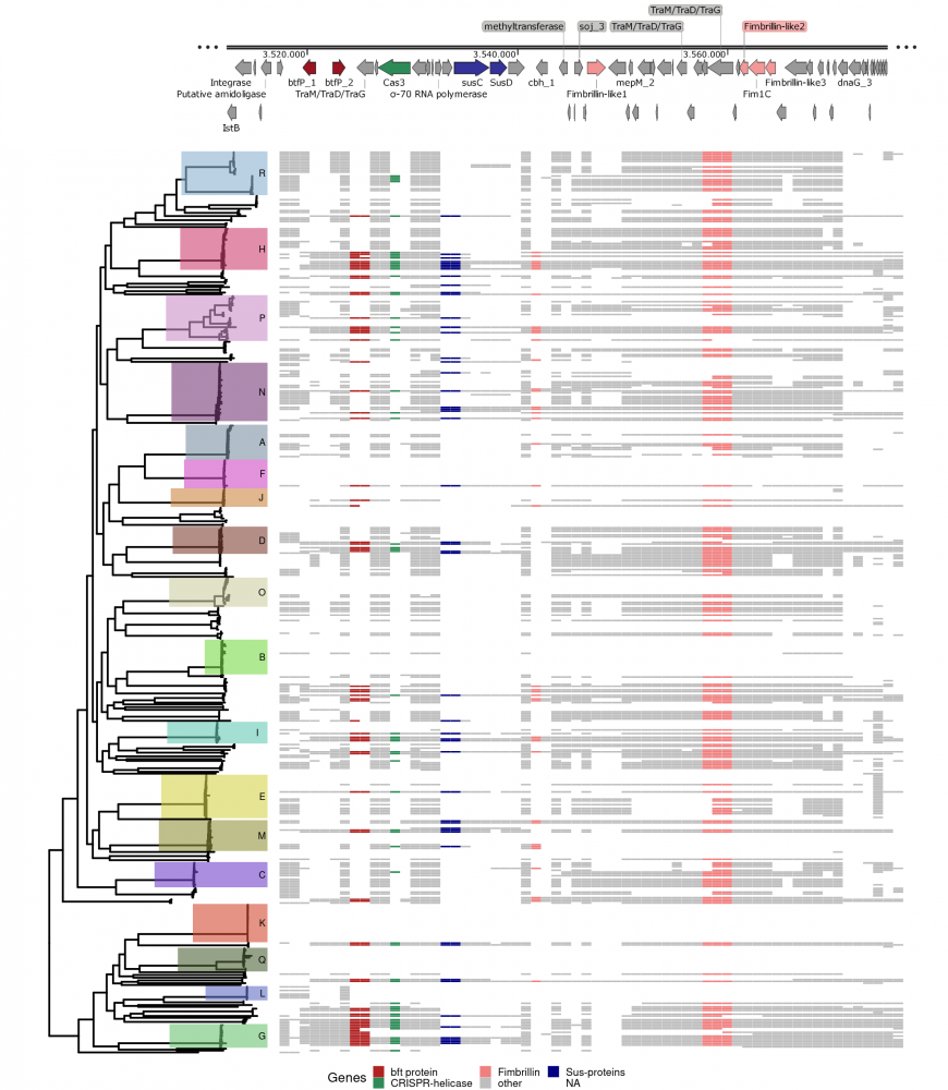
Door gebruik te maken van 500 online beschikbare genomen heb ik een evolutionaire boom kunnen recreëren waarin verschillende afstammingslijnen aanwezig zijn. Elke afstammingslijn bevatte specifieke genen die zowel een ziekte kunnen verhelpen maar ook verergeren. Daarnaast toonde ik ook aan dat de afstammingslijnen bundels van genetische informatie met elkaar uitwisselden (recombinatie) wat van grootte invloed was en is op de evolutie van de bacterie. Deze bundels omvatten antibiotica resistente genen, toxinen zoals het bft gen (afbeelding) maar ook genen die helpen bij de spijsvertering.
In conclusie, de diversiteit van Bacteroides fragilis was in het verleden onderschat. Verder wordt de diversiteit sterk beïnvloed door recombinatie. De bacterie is een potentiële probiotica kandidaat maar de dynamiek rondom B. fragilis voordelige en pathogene eigenschappen moet eerst nog beter worden begrepen voordat er Bacteroides fragilis gebaseerde geneesmiddelen kunnen worden ontwikkeld. Met mijn thesis heb ik hier een bescheiden steentje kunnen bijgedragen.
Bibliografie
1. Quigley, E. M. M. Gut Bacteria in Health and Disease. Gastroenterol. Hepatol. 9, 560–569 (2013).
2. Purcell, R. V. Chapter 4 - Bacteroides fragilis. in Colorectal Neoplasia and the Colorectal Microbiome (ed. Floch, M. H.) 57–77 (Academic Press, 2020). doi:10.1016/B978-0-12-819672-4.00004-0.
3. Sun, F. et al. A potential species of next-generation probiotics? The dark and light sides of Bacteroides fragilis in health. Food Res. Int. 126, 108590 (2019).
4. Zhao, S. et al. Adaptive Evolution within Gut Microbiomes of Healthy People. Cell Host Microbe 25, 656-667.e8 (2019).
5. Pierce, J. V. & Bernstein, H. D. Genomic Diversity of Enterotoxigenic Strains of Bacteroides fragilis. PLOS ONE 11, e0158171 (2016).
6. Magne, F. et al. The Firmicutes/Bacteroidetes Ratio: A Relevant Marker of Gut Dysbiosis in Obese Patients? Nutrients 12, (2020).
7. Wexler, H. M. Bacteroides: the Good, the Bad, and the Nitty-Gritty. Clin. Microbiol. Rev. 20, 593–621 (2007).
8. Elliott, D., Kufera, J. A. & Myers, R. A. The microbiology of necrotizing soft tissue infections. Am. J. Surg. 179, 361–366 (2000).
9. Ghotaslou, R. et al. Mechanisms of Bacteroides fragilis resistance to metronidazole. Infect. Genet. Evol. 64, 156–163 (2018).
10. Ferløv-Schwensen, S. A., Sydenham, T. V., Hansen, K. C. M., Hoegh, S. V. & Justesen, U. S. Prevalence of antimicrobial resistance and the cfiA resistance gene in Danish Bacteroides fragilis group isolates since 1973. Int. J. Antimicrob. Agents 50, 552–556 (2017).
11. Wang, Y. et al. An intestinal commensal symbiosis factor controls neuroinflammation via TLR2-mediated CD39 signalling. Nat. Commun. 5, 4432 (2014).
12. Wang, Y. et al. A commensal bacterial product elicits and modulates migratory capacity of CD39(+) CD4 T regulatory subsets in the suppression of neuroinflammation. Gut Microbes 5, 552–561 (2014).
13. Shen, Y. et al. Outer membrane vesicles of a human commensal mediate immune regulation and disease protection. Cell Host Microbe 12, 509–520 (2012).
14. Chang, Y.-C. et al. TLR2 and interleukin-10 are involved in Bacteroides fragilis-mediated prevention of DSS-induced colitis in gnotobiotic mice. PloS One 12, e0180025 (2017).
15. Round, J. L. & Mazmanian, S. K. Inducible Foxp3+ regulatory T-cell development by a commensal bacterium of the intestinal microbiota. Proc. Natl. Acad. Sci. U. S. A. 107, 12204–12209 (2010).
16. Johnson, J. L., Jones, M. B. & Cobb, B. A. Bacterial capsular polysaccharide prevents the onset of asthma through T-cell activation. Glycobiology 25, 368–375 (2015).
17. Gilbert, J. A., Krajmalnik-Brown, R., Porazinska, D. L., Weiss, S. J. & Knight, R. Towards effective probiotics for autism and other mental disorders? Cell 155, 1446–1448 (2013).
18. Franco, A. A. et al. Molecular evolution of the pathogenicity island of enterotoxigenic Bacteroides fragilis strains. J. Bacteriol. 181, 6623–6633 (1999).
19. Moncrief, J. S. et al. The enterotoxin of Bacteroides fragilis is a metalloprotease. Infect. Immun. 63, 175–181 (1995).
20. Wu, S., Lim, K. C., Huang, J., Saidi, R. F. & Sears, C. L. Bacteroides fragilis enterotoxin cleaves the zonula adherens protein, E-cadherin. Proc. Natl. Acad. Sci. U. S. A. 95, 14979–14984 (1998).
21. Moncrief, J. S., Duncan, A. J., Wright, R. L., Barroso, L. A. & Wilkins, T. D. Molecular characterization of the fragilysin pathogenicity islet of enterotoxigenic Bacteroides fragilis. Infect. Immun. 66, 1735–1739 (1998).
22. Franco, A. A. The Bacteroides fragilis Pathogenicity Island Is Contained in a Putative Novel Conjugative Transposon. J. Bacteriol. 186, 6077–6092 (2004).
23. Van Tassell, R. L., Lyerly, D. M. & Wilkins, T. D. Purification and characterization of an enterotoxin from Bacteroides fragilis. Infect. Immun. 60, 1343–1350 (1992).
24. Franco, A. A. et al. Cloning and characterization of the Bacteroides fragilis metalloprotease toxin gene. Infect. Immun. 65, 1007–1013 (1997).
25. Chung, G.-T. et al. Identification of a Third Metalloprotease Toxin Gene in Extraintestinal Isolates of Bacteroides fragilis. Infect. Immun. 67, 4945–4949 (1999).
26. Zhang, G., Svenungsson, B., Kärnell, A. & Weintraub, A. Prevalence of enterotoxigenic Bacteroides fragilis in adult patients with diarrhea and healthy controls. Clin. Infect. Dis. Off. Publ. Infect. Dis. Soc. Am. 29, 590–594 (1999).
27. Sack, R. B. et al. Enterotoxigenic Bacteroides fragilis: epidemiologic studies of its role as a human diarrhoeal pathogen. J. Diarrhoeal Dis. Res. 10, 4–9 (1992).
28. Ji, D.-D. et al. Prevalence and characterization of enterotoxigenic Bacteroides fragilis and toxigenic Clostridium difficile in a Taipei emergency department. J. Microbiol. Immunol. Infect. 50, 83–89 (2017).
29. Wu, S. et al. The Bacteroides fragilis Toxin Binds to a Specific Intestinal Epithelial Cell Receptor. Infect. Immun. 74, 5382–5390 (2006).
30. Wu, S., Rhee, K.-J., Zhang, M., Franco, A. & Sears, C. L. Bacteroides fragilis toxin stimulates intestinal epithelial cell shedding and γ-secretase-dependent E-cadherin cleavage. J. Cell Sci. 120, 1944–1952 (2007).
31. Rhee, K.-J. et al. Induction of Persistent Colitis by a Human Commensal, Enterotoxigenic Bacteroides fragilis, in Wild-Type C57BL/6 Mice. Infect. Immun. 77, 1708–1718 (2009).
32. Wu, S. et al. A human colonic commensal promotes colon tumorigenesis via activation of T helper type 17 T cell responses. Nat. Med. 15, 1016–1022 (2009).
33. Thiele Orberg, E. et al. The myeloid immune signature of enterotoxigenic Bacteroides fragilis -induced murine colon tumorigenesis. Mucosal Immunol. 10, 421–433 (2017).
34. Kwong, T. N. Y. et al. Association Between Bacteremia From Specific Microbes and Subsequent Diagnosis of Colorectal Cancer. Gastroenterology 155, 383-390.e8 (2018).
35. Zhao, Y. & Lukiw, W. J. Bacteroidetes Neurotoxins and Inflammatory Neurodegeneration. Mol. Neurobiol. 55, 9100–9107 (2018).
36. Mazmanian, S. K., Round, J. L. & Kasper, D. L. A microbial symbiosis factor prevents intestinal inflammatory disease. Nature 453, 620–625 (2008).
37. Sommese, L. et al. Evidence of Bacteroides fragilis Protection from Bartonella henselae-Induced Damage. PLOS ONE 7, e49653 (2012).
38. Sittipo, P. et al. Toll-Like Receptor 2-Mediated Suppression of Colorectal Cancer Pathogenesis by Polysaccharide A From Bacteroides fragilis. Front. Microbiol. 9, (2018).
39. Li, Z. et al. Bioluminescence Imaging to Track Bacteroides fragilis Inhibition of Vibrio parahaemolyticus Infection in Mice. Front. Cell. Infect. Microbiol. 7, (2017).
40. Mazmanian, S. K., Liu, C. H., Tzianabos, A. O. & Kasper, D. L. An Immunomodulatory Molecule of Symbiotic Bacteria Directs Maturation of the Host Immune System. Cell 122, 107–118 (2005).
41. Round, J. L. et al. The Toll-like receptor 2 pathway establishes colonization by a commensal of the human microbiota. Science 332, 974–977 (2011).
42. Chan, J. L. et al. Non-toxigenic Bacteroides fragilis (NTBF) administration reduces bacteria-driven chronic colitis and tumor development independent of polysaccharide A. Mucosal Immunol. 12, 164–177 (2019).
43. Surana, N. K. & Kasper, D. L. The yin yang of bacterial polysaccharides: lessons learned from B. fragilis PSA. Immunol. Rev. 245, 13–26 (2012).
44. Arnolds, K. L., Moreno-Huizar, N., Stanislawski, M. A., Palmer, B. & Lozupone, C. Hemagglutination by B. fragilis is mediated by capsular polysaccharides and is influenced by host ABO blood type. bioRxiv 2020.08.19.258442 (2020) doi:10.1101/2020.08.19.258442.
45. Weng, M. & Walker, W. A. The role of gut microbiota in programming the immune phenotype. J. Dev. Orig. Health Dis. 4, 203–214 (2013).
46. Ewald, D. R. & Sumner, S. C. J. Blood type biochemistry and human disease. WIREs Syst. Biol. Med. 8, 517–535 (2016).
47. Cooling, L. Blood Groups in Infection and Host Susceptibility. Clin. Microbiol. Rev. 28, 801–870 (2015).
48. Rühlemann, M. C. et al. Genome-wide association study in 8,956 German individuals identifies influence of ABO histo-blood groups on gut microbiome. Nat. Genet. 53, 147–155 (2021).
49. Huttenhower, C. et al. Structure, function and diversity of the healthy human microbiome. Nature 486, 207–214 (2012).
50. Eckburg, P. B. et al. Diversity of the Human Intestinal Microbial Flora. Science 308, 1635–1638 (2005).
51. Yatsunenko, T. et al. Human gut microbiome viewed across age and geography. Nature 486, 222–227 (2012).
52. Lay, C. et al. Colonic microbiota signatures across five northern European countries. Appl. Environ. Microbiol. 71, 4153–4155 (2005).
53. Falony, G. et al. Population-level analysis of gut microbiome variation. Science 352, 560–564 (2016).
54. Vandeputte, D. et al. Quantitative microbiome profiling links gut community variation to microbial load. Nature 551, 507–511 (2017).
55. Arumugam, M. et al. Enterotypes of the human gut microbiome. Nature 473, 174–180 (2011).
56. Vieira-Silva, S. et al. Quantitative microbiome profiling disentangles inflammation- and bile duct obstruction-associated microbiota alterations across PSC/IBD diagnoses. Nat. Microbiol. 4, 1826–1831 (2019).
57. Vieira-Silva, S. et al. Statin therapy is associated with lower prevalence of gut microbiota dysbiosis. Nature 581, 310–315 (2020).
58. Valles-Colomer, M. et al. The neuroactive potential of the human gut microbiota in quality of life and depression. Nat. Microbiol. 4, 623–632 (2019).
59. Wybo, I. et al. Differentiation of cfiA-negative and cfiA-positive Bacteroides fragilis isolates by matrix-assisted laser desorption ionization-time of flight mass spectrometry. J. Clin. Microbiol. 49, 1961–1964 (2011).
60. Gutacker, M., Valsangiacomo, C. & Piffaretti, J.-C. Identification of two genetic groups in Bacteroides fragilis by multilocus enzyme electrophoresis: distribution of antibiotic resistance (cfiA, cepA) and enterotoxin (bft) encoding genes. Microbiol. Read. Engl. 146 ( Pt 5), 1241–1254 (2000).
61. Hallatschek, O., Hersen, P., Ramanathan, S. & Nelson, D. R. Genetic drift at expanding frontiers promotes gene segregation. Proc. Natl. Acad. Sci. U. S. A. 104, 19926–19930 (2007).
62. Van Rossum, T., Ferretti, P., Maistrenko, O. M. & Bork, P. Diversity within species: interpreting strains in microbiomes. Nat. Rev. Microbiol. 18, 491–506 (2020).
63. Costea, P. I. et al. Subspecies in the global human gut microbiome. Mol. Syst. Biol. 13, 960 (2017).
64. Retchless, A. C. & Lawrence, J. G. Temporal fragmentation of speciation in bacteria. Science 317, 1093–1096 (2007).
65. Shapiro, B. J. What Microbial Population Genomics Has Taught Us About Speciation. in Population Genomics: Microorganisms (eds. Polz, M. F. & Rajora, O. P.) 31–47 (Springer International Publishing, 2018). doi:10.1007/13836_2018_10.
66. Thomas, C. M. & Nielsen, K. M. Mechanisms of, and barriers to, horizontal gene transfer between bacteria. Nat. Rev. Microbiol. 3, 711–721 (2005).
67. Young, J. P. W. et al. The genome of Rhizobium leguminosarum has recognizable core and accessory components. Genome Biol. 7, R34 (2006).
68. McCoy, R. C. & Akey, J. M. Selection plays the hand it was dealt: evidence that human adaptation commonly targets standing genetic variation. Genome Biol. 18, 139 (2017).
69. Tettelin, H. et al. Genome analysis of multiple pathogenic isolates of Streptococcus agalactiae: implications for the microbial ‘pan-genome’. Proc. Natl. Acad. Sci. U. S. A. 102, 13950–13955 (2005).
70. Golicz, A. A., Bayer, P. E., Bhalla, P. L., Batley, J. & Edwards, D. Pangenomics Comes of Age: From Bacteria to Plant and Animal Applications. Trends Genet. 36, 132–145 (2020).
71. Domingo-Sananes, M. R. & McInerney, J. O. Selection-based model of prokaryote pangenomes. bioRxiv 782573 (2019) doi:10.1101/782573.
72. Colquhoun, R. M. et al. Nucleotide-resolution bacterial pan-genomics with reference graphs. bioRxiv 2020.11.12.380378 (2020) doi:10.1101/2020.11.12.380378.
73. Bolotin, E. & Hershberg, R. Gene Loss Dominates As a Source of Genetic Variation within Clonal Pathogenic Bacterial Species. Genome Biol. Evol. 7, 2173–2187 (2015).
74. McInerney, J. O., McNally, A. & O’Connell, M. J. Why prokaryotes have pangenomes. Nat. Microbiol. 2, 1–5 (2017).
75. Rouli, L., Merhej, V., Fournier, P.-E. & Raoult, D. The bacterial pangenome as a new tool for analysing pathogenic bacteria. New Microbes New Infect. 7, 72–85 (2015).
76. Davies, M. R. et al. Atlas of group A streptococcal vaccine candidates compiled using large-scale comparative genomics. Nat. Genet. 51, 1035–1043 (2019).
77. Poulsen, B. E. et al. Defining the core essential genome of Pseudomonas aeruginosa. Proc. Natl. Acad. Sci. 116, 10072–10080 (2019).
78. Medini, D., Donati, C., Tettelin, H., Masignani, V. & Rappuoli, R. The microbial pan-genome. Curr. Opin. Genet. Dev. 15, 589–594 (2005).
79. Vernikos, G., Medini, D., Riley, D. R. & Tettelin, H. Ten years of pan-genome analyses. Curr. Opin. Microbiol. 23, 148–154 (2015).
80. Vos, M. & Eyre-Walker, A. Are pangenomes adaptive or not? Nat. Microbiol. 2, 1576–1576 (2017).
81. Sela, I., Wolf, Y. I. & Koonin, E. V. Theory of prokaryotic genome evolution. Proc. Natl. Acad. Sci. 113, 11399–11407 (2016).
82. Shapiro, B. J. The population genetics of pangenomes. Nat. Microbiol. 2, 1574–1574 (2017).
83. Bobay, L.-M. & Ochman, H. Factors driving effective population size and pan-genome evolution in bacteria. BMC Evol. Biol. 18, 153 (2018).
84. Ohta, T. Slightly Deleterious Mutant Substitutions in Evolution. Nature 246, 96–98 (1973).
85. Bobay, L.-M. The Prokaryotic Species Concept and Challenges. in The Pangenome: Diversity, Dynamics and Evolution of Genomes (eds. Tettelin, H. & Medini, D.) 21–49 (Springer International Publishing, 2020). doi:10.1007/978-3-030-38281-0_2.
87. Kitts, P. A. et al. Assembly: a resource for assembled genomes at NCBI. Nucleic Acids Res. 44, D73-80 (2016).
88. Youngblut, N. D. et al. Large-Scale Metagenome Assembly Reveals Novel Animal-Associated Microbial Genomes, Biosynthetic Gene Clusters, and Other Genetic Diversity. mSystems 5, (2020).
89. Bolger, A. M., Lohse, M. & Usadel, B. Trimmomatic: a flexible trimmer for Illumina sequence data. Bioinformatics 30, 2114–2120 (2014).
90. Nurk, S. et al. Assembling Genomes and Mini-metagenomes from Highly Chimeric Reads. in Research in Computational Molecular Biology (eds. Deng, M., Jiang, R., Sun, F. & Zhang, X.) 158–170 (Springer, 2013). doi:10.1007/978-3-642-37195-0_13.
91. Gurevich, A., Saveliev, V., Vyahhi, N. & Tesler, G. QUAST: quality assessment tool for genome assemblies. Bioinformatics 29, 1072–1075 (2013).
92. Parks, D. H., Imelfort, M., Skennerton, C. T., Hugenholtz, P. & Tyson, G. W. CheckM: assessing the quality of microbial genomes recovered from isolates, single cells, and metagenomes. Genome Res. 25, 1043 (2015).
93. Jain, C., Rodriguez-R, L. M., Phillippy, A. M., Konstantinidis, K. T. & Aluru, S. High throughput ANI analysis of 90K prokaryotic genomes reveals clear species boundaries. Nat. Commun. 9, 5114 (2018).
94. Carroll, L. M., Wiedmann, M. & Kovac, J. Proposal of a Taxonomic Nomenclature for the Bacillus cereus Group Which Reconciles Genomic Definitions of Bacterial Species with Clinical and Industrial Phenotypes. mBio 11, (2020).
95. Fernández-de-Bobadilla, M. D. et al. PATO: Pangenome Analysis Toolkit. http://biorxiv.org/lookup/doi/10.1101/2021.01.30.428878 (2021) doi:10.1101/2021.01.30.428878.
96. Ondov, B. D. et al. Mash: fast genome and metagenome distance estimation using MinHash. Genome Biol. 17, 132 (2016).
97. Steinegger, M. & Söding, J. MMseqs2 enables sensitive protein sequence searching for the analysis of massive data sets. Nat. Biotechnol. 35, 1026–1028 (2017).
98. Li, H. Minimap2: pairwise alignment for nucleotide sequences. Bioinformatics 34, 3094–3100 (2018).
99. Tonkin-Hill, G. et al. Producing polished prokaryotic pangenomes with the Panaroo pipeline. Genome Biol. 21, 180 (2020).
100. Huerta-Cepas, J. et al. Fast Genome-Wide Functional Annotation through Orthology Assignment by eggNOG-Mapper. Mol. Biol. Evol. 34, 2115–2122 (2017).
101. Huerta-Cepas, J. et al. eggNOG 5.0: a hierarchical, functionally and phylogenetically annotated orthology resource based on 5090 organisms and 2502 viruses. Nucleic Acids Res. 47, D309–D314 (2019).
102. Feldgarden, M. et al. Validating the AMRFinder Tool and Resistance Gene Database by Using Antimicrobial Resistance Genotype-Phenotype Correlations in a Collection of Isolates. Antimicrob. Agents Chemother. 63, (2019).
103. Tonkin-Hill, G., Lees, J. A., Bentley, S. D., Frost, S. D. W. & Corander, J. Fast hierarchical Bayesian analysis of population structure. Nucleic Acids Res. 47, 5539–5549 (2019).
104. Croucher, N. J. et al. Rapid phylogenetic analysis of large samples of recombinant bacterial whole genome sequences using Gubbins. Nucleic Acids Res. 43, e15–e15 (2015).
105. Campello, R. J. G. B., Moulavi, D. & Sander, J. Density-Based Clustering Based on Hierarchical Density Estimates. in Advances in Knowledge Discovery and Data Mining (eds. Pei, J., Tseng, V. S., Cao, L., Motoda, H. & Xu, G.) 160–172 (Springer, 2013). doi:10.1007/978-3-642-37456-2_14.
107. Hadfield, J. et al. Phandango: an interactive viewer for bacterial population genomics. Bioinformatics 34, 292–293 (2018).
108. Quinlan, A. R. & Hall, I. M. BEDTools: a flexible suite of utilities for comparing genomic features. Bioinformatics 26, 841–842 (2010).
109. Brynildsrud, O., Bohlin, J., Scheffer, L. & Eldholm, V. Rapid scoring of genes in microbial pan-genome-wide association studies with Scoary. Genome Biol. 17, 238 (2016).
110. Whelan, F. J., Rusilowicz, M. & McInerney, J. O. Coinfinder: detecting significant associations and dissociations in pangenomes. Microb. Genomics 6, e000338 (2020).
111. Yu, G. Using ggtree to Visualize Data on Tree-Like Structures. Curr. Protoc. Bioinforma. 69, e96 (2020).
112. Bobay, L.-M. & Ochman, H. Biological Species Are Universal across Life’s Domains. Genome Biol. Evol. 9, 491–501 (2017).
113. Lan, R. & Reeves, P. R. When does a clone deserve a name? A perspective on bacterial species based on population genetics. Trends Microbiol. 9, 419–424 (2001).
114. Spratt, B. G. Exploring the Concept of Clonality in Bacteria. in Genomics, Proteomics, and Clinical Bacteriology: Methods and Reviews (eds. Woodford, N. & Johnson, A. P.) 323–352 (Humana Press, 2004). doi:10.1385/1-59259-763-7:323.
115. Truong, D. T., Tett, A., Pasolli, E., Huttenhower, C. & Segata, N. Microbial strain-level population structure and genetic diversity from metagenomes. Genome Res. 27, 626–638 (2017).
116. Kuo, C.-H., Moran, N. A. & Ochman, H. The consequences of genetic drift for bacterial genome complexity. Genome Res. 19, 1450–1454 (2009).
117. Croucher, N. J. et al. Diversification of bacterial genome content through distinct mechanisms over different timescales. Nat. Commun. 5, (2014).
118. Choi, K.-H., Heath, R. J. & Rock, C. O. β-Ketoacyl-Acyl Carrier Protein Synthase III (FabH) Is a Determining Factor in Branched-Chain Fatty Acid Biosynthesis. J. Bacteriol. 182, 365–370 (2000).
119. Lai, C.-Y. & Cronan, J. E. Isolation and Characterization of β-Ketoacyl-Acyl Carrier Protein Reductase (fabG) Mutants of Escherichia coli and Salmonella enterica Serovar Typhimurium. J. Bacteriol. 186, 1869–1878 (2004).
120. Kurokawa, K. et al. Comparative Metagenomics Revealed Commonly Enriched Gene Sets in Human Gut Microbiomes. DNA Res. 14, 169–181 (2007).
121. Merino, S. & Tomás, J. M. Bacterial Capsules and Evasion of Immune Responses. in eLS 1–10 (American Cancer Society, 2015). doi:10.1002/9780470015902.a0000957.pub4.
122. Foley, M. H., Cockburn, D. W. & Koropatkin, N. M. The Sus operon: a model system for starch uptake by the human gut Bacteroidetes. Cell. Mol. Life Sci. CMLS 73, 2603–2617 (2016).
123. Steinert, M., Ramming, I. & Bergmann, S. Impact of Von Willebrand Factor on Bacterial Pathogenesis. Front. Med. 7, (2020).
124. Cha, M.-K., Kim, W.-C., Lim, C.-J., Kim, K. & Kim, I.-H. Escherichia coli Periplasmic Thiol Peroxidase Acts as Lipid Hydroperoxide Peroxidase and the Principal Antioxidative Function during Anaerobic Growth *. J. Biol. Chem. 279, 8769–8778 (2004).
125. Santos-Beneit, F. The Pho regulon: a huge regulatory network in bacteria. Front. Microbiol. 6, (2015).