Chirurg en ingenieur: een ongewone samenwerking?
Een chirurgische ingreep, volledig aangepast aan de unieke anatomie en aan de specifieke noden van iedere patiënt: iets voor de verre toekomst? Gedurende mijn thesisjaar ontwikkelde ik een computermodel van een menselijke schedel om een chirurgische ingreep van het bovenkaakbeen te onderzoeken. In de toekomst kan dit soort onderzoek evolueren tot een semiautomatisch proces waarbij chirurgen, met behulp van CT-scans, de optimale chirurgische procedure kunnen bepalen. Hierdoor kunnen patiënten de beste klinische hulp krijgen.
Een ongewone samenwerking
Alhoewel België tot de absolute top behoort op vlak van gezondheidszorg, is er toch steeds ruimte voor verbetering. Voor bepaalde chirurgische ingrepen is in wetenschappelijke literatuur geen consensus over de optimale methode, en lijkt de behandeling vooral af te hangen van de expertise en de ervaring van de chirurg. Een overduidelijk voorbeeld hiervan is een operatieve ingreep waarbij de bovenkaak wordt verbreed. Tot op heden bestaat er geen conclusie in de klinische literatuur over de optimale methode en welke invloed bepaalde stappen in het proces hebben op het eindresultaat.
Gedurende mijn thesisjaar heb ik, in samenwerking met UZ-Gent, onderzoek uitgevoerd naar een specifieke chirurgische ingreep, namelijk de surgically assisted rapid palatal expansion (SARPE), ook gekend als een bovenkaakverbreding. De ingreep bestaat in eerste instantie uit het losmaken van de bovenkaak van de rest van het aangezicht. Om de bovenkaak effectief te verbreden wordt er gebruikgemaakt van een expansietoestel dat wordt aangebracht op het gehemelte van de patiënt. Dit toestel wordt steeds meer opgespannen, waarbij de twee helften van het bovenkaakbeen langzaam expanderen tot de gewenste breedte.
Er is onder de chirurgen wereldwijd veel discussie over de optimale positie van dit toestel, alsook over welke chirurgische osteotomieën (orthopedische ingrepen waarbij een stuk bot wordt doorgenomen) het belangrijkste zijn om tot een gecontroleerde expansie te komen. Aangezien het belangrijk is om de veiligheid van patiënten niet in gedrang te brengen, kan men moeilijk de invloed van bepaalde stappen uittesten op personen. Om deze reden kan het belangrijk zijn om aan de hand van computermodellen bepaalde nieuwe operaties te simuleren en te kijken wat het effect is van bepaalde handelingen tijdens de operatie.
Computermodel van de menselijke schedel
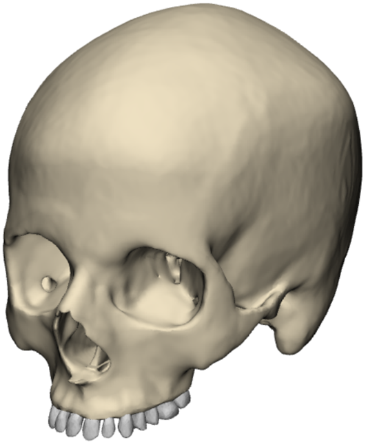
Het eerste doel van dit onderzoek was om na te gaan of er een correct biomechanisch computermodel kan opgesteld worden die de realiteit van de chirurgische ingreep goed kan benaderen. Om dit te bekomen, werd er eerst aan de hand van een CT-scan de unieke anatomie van de patiënt beschouwd, en kon er een digitaal 3D-model gebouwd worden met behulp van specifieke software.
Aangezien een schedel een complexe geometrie heeft werd de chirurgische ingreep gesimuleerd aan de hand van een eindige elementen analyse. Dit houdt in dat het computermodel van de schedel opgedeeld wordt in miljoenen tetraëders, en dat voor elk van deze tetraëders een ingewikkelde vergelijking wordt opgelost die het verband tussen verplaatsing, kracht en spanning weergeeft. Als oplossing van deze complexe berekeningen kan men de verplaatsing van en de spanning in elk van deze tetraëders bekomen.
Het onderzoek
Eens het digitale 3D-model van de schedel opgesteld en getest was, was het belangrijk om tot klinisch relevante data te komen. Aangezien er onzekerheid is bij chirurgen over de ideale positie van het expansietoestel, werd eerst dit onderzocht. Uit de resultaten blijkt dat de positie van dit toestel verregaande gevolgen heeft voor het expansieprofiel van de verbreding van de kaak. Afhankelijk van de initiële misvorming, die uniek is bij iedere patiënt, moet dit toestel anders gepositioneerd zijn op het gehemelte. Deze resultaten tonen direct aan dat een algemene methode onmogelijk voor iedereen tot optimale resultaten zou leiden, en dat er steeds een unieke ingreep nodig is afhankelijk van de noden van de patiënt.
Een tweede parameter die werd onderzocht is de aanwezigheid van bepaalde osteotomieën (orthopedische ingreep waarbij een stuk bot wordt doorgenomen). Wederom werd ondervonden dat afhankelijk van de aanwezigheid van bepaalde osteotomieën een compleet ander expansieprofiel van de bovenkaak bekomen wordt. Afhankelijk van de noden en de specifieke anatomie van de schedel van de patiënt moeten de chirurgen dus een aangepaste ingreep uitvoeren. Een patiëntspecifieke chirurgische ingreep zal steeds de optimale zijn, en aan de hand van computersimulaties kan men nauwkeurig bepalen wat de uitkomst van de ingreep is vooraleer men deze heeft uitgevoerd. Dit zal dan leiden tot verbeterde en preciezere resultaten, zonder dat hierbij moet geëxperimenteerd worden op patiënten. Dit zal zorgen voor een lagere prevalentie van veelvoorkomende complicaties.
En nu?
Dit onderzoek kan gezien worden als startschot voor een verregaande samenwerking tussen chirurgen en biomechanische ingenieurs. Het is duidelijk dat beide disciplines nodig zijn om onze gezondheidszorg te versterken en betere behandelingen te vinden. Aan de ene kant zijn simulaties een sterk middel om te experimenteren met behandelingen zonder hierbij de patiënt potentieel te schaden. Er is echter ook steeds klinische input nodig van chirurgen en artsen, om er zeker van te zijn dat de juiste klinische vraagstukken gesimuleerd en opgelost worden, en dat deze ook op een juiste manier geïnterpreteerd worden.
De eerste resultaten van deze samenwerking tonen onmiddellijk aan dat er op dit moment nog heel wat verbeteringen mogelijk zijn op vlak van patiëntspecifieke behandelingen. Verdere research kan de invloed van nog andere parameters onderzoeken en meerdere schedels kunnen onderzocht worden. Op deze manier zou er een handleiding opgesteld kunnen worden om de chirurgen te assisteren in het kiezen van een bepaalde behandeling.
In de verdere toekomst zouden veel van deze stappen ook geautomatiseerd kunnen worden, en de werkwijze zou kunnen uitgebreid worden naar andere toepassingen binnen de gezondheidszorg (ontwerp van implantaten en andere chirurgische ingrepen). Na het nemen van de CT-scans zouden de simulaties automatisch kunnen verlopen, waarbij de chirurg enkel nog de resultaten van de simulaties moet interpreteren en op basis hiervan de optimale ingreep selecteert. Op deze manier kan elk individueel geval op een systematische manier geëvalueerd worden en kunnen de beste resultaten bereikt worden. Zo kan de samenwerking tussen disciplines leiden tot een revolutie in de gezondheidszorg, waarbij patiënt specifieke behandelingen de nieuwe norm worden.
Bibliografie
- [1] R. Nowak, A. Strza lkowska, and E. Zawi´slak, “Treatment options and limitations in transverse maxillary deficiency,” Dental and Medical Problems, vol. 52, no. 4, p. 389–400, 2015.
- [2] Innovative pespectives in oral and maxillofacial surgery. Springer Nature, 2021.
- [3] M. C. D. Andrucioli and M. A. N. Matsumoto, “Transverse maxillary deficiency: treatment alternatives in face of early skeletal maturation,” Dental Press Journal of Orthodontics, vol. 25, no. 1, p. 70–79, 2020.
- [4] K. Jha and M. Adhikari, “Surgically assisted rapid palatal expansion for transverse maxillary discrepancy in adults - case report,” International Journal of Surgery Case Reports, vol. 90, p. 106687, 2022.
- [5] K. Manzella, L. Franchi, and T. Al-Jewair, “Correction of maxillary transverse deficiency in growing patients with permanent dentitions,” Journal of clinical orthodontics : JCO, vol. 52, pp. 148–156, 03 2018.
- [6] Philippe, “Palatal expansion (rapid maxillary expansion): B¨ucco,” Sep 2019. [Online]. Available: https://www.orthodontisteenligne.com/en/ palatal-expansion-rapid-maxillary-expansion/
- [7] “Medical definition of suture,” Mar 2021. [Online]. Available: https: //www.medicinenet.com/suture/definition.htm
- [8] “Bones of the skull.” [Online]. Available: https://teachmeanatomy.info/head/ osteology/skull/
- [9] “What is rapid maxillary expansion treatment?” Aug 2021. [Online]. Available: https://orthodonticsaustralia.org.au/rapid-maxillary-expansion-treatmen…
- [10] A. Agarwal, “Maxillary expansion,” International Journal of Clinical Pediatric Dentistry, vol. 3, no. 3, p. 139–146, 2010. 101 BIBLIOGRAPHY
- [11] V. Shetty, J. Caridad, A. A. Caputo, and S. J. Chaconas, “Biomechanical rationale for surgical-orthodontic expansion of the adult maxilla,” Journal of Oral and Maxillofacial Surgery, vol. 52, no. 7, p. 742–749, 1994.
- [12] M. Koudstaal, L. Poort, K. V. D. Wal, E. Wolvius, B. Prahl-Andersen, and A. Schulten, “Surgically assisted rapid maxillary expansion (sarme): a review of the literature,” International Journal of Oral and Maxillofacial Surgery, vol. 34, no. 7, p. 709–714, 2005.
- [13] S. Bartlett, M. Ehrenfeld, G. Mast, and A. Sugar, “Surgically assisted rapid palatal expansion.” [Online]. Available: https://surgeryreference.aofoundation. org/cmf/orthognathic/maxilla/maxilla-transverse-hypoplasia-of-maxilla/ surgically-assisted-rapid-palatal-expansion#osteotomy
- [14] S. Shyam Sundar, B. Nandlal, D. Saikrishna, and G. Mallesh, “Finite element analysis: A maxillofacial surgeon’s perspective,” Journal of Maxillofacial and Oral Surgery, vol. 11, no. 2, p. 206–211, 2011.
- [15] D. Bignotti, A. Gracco, G. Bruno, and A. D. Stefani, “Fem in orthodontics: a review of the palatal expander clinical investigation,” ORAL amp; Implantology, p. 161–167, Feb 2019.
- [16] “1.4d: Body planes and sections,” Aug 2020. [Online]. Available: https: //med.libretexts.org/
- [17] R. Bailey, “Anatomical directional terms and body planes,” Aug 2020. [Online]. Available: https://www.thoughtco.com/ anatomical-directional-terms-and-body-planes-373204
- [18] H. Gray, Gray’s anatomy, 41st ed. Medina University Press International, 2020.
- [19] N. S. Norton and F. H. Netter, Netters head and neck anatomy for dentistry. Elsevier., 2017.
- [20] D. A. Samra and R. Hadad, “Midpalatal suture: evaluation of the morphological maturation stages via bone density,” Progress in Orthodontics, vol. 19, no. 1, 2018.
- [21] B. S. Knapp, By:, and S. Knapp, “Sphenoid bone - the definitive guide,” Aug 2020. [Online]. Available: https://biologydictionary.net/sphenoid-bone/ #body-of-the-sphenoid-bone
- [22] “The sphenoid bone.” [Online]. Available: https://teachmeanatomy.info/head/ osteology/sphenoid-bone/
- [23] Atlas of operative oral and maxillofacial surgery. WILEY-BLACKWELL, 2015. 102 BIBLIOGRAPHY
- [24] K. F. Linnau, R. B. Stanley, D. K. Hallam, J. A. Gross, and F. Mann, “Imaging of high-energy midfacial trauma: What the surgeon needs to know,” European Journal of Radiology, vol. 48, no. 1, p. 17–32, 2003.
- [25] S. Celin, Fractures of the upper facial and midfacial skeleton, ser. Myers EN ed., Operative Otoolaryngology Head and Neck Surgery. WB Saunders Company, 1997.
- [26] “Tooth.” [Online]. Available: https://www.britannica.com/science/ tooth-anatomy
- [27] T. W. Oates and D. L. Cochran, “Dental applications of bone biology,” Topics in Bone Biology, p. 129–140.
- [28] O. Team, “Bone matrix.” [Online]. Available: https://www.orthobullets.com/ basic-science/9003/bone-matrix
- [29] W. Murphy, J. Black, and G. W. Hastings, Handbook of Biomaterial Properties. Springer, 2016.
- [30] “Bone morphology.” [Online]. Available: https://www.britannica.com/science/ bone-anatomy/Bone-morphology
- [31] G. Osterhoff, E. F. Morgan, S. J. Shefelbine, L. Karim, L. M. McNamara, and P. Augat, “Bone mechanical properties and changes with osteoporosis,” Injury, vol. 47, 2016.
- [32] J. Peterson, Q. Wang, and P. C. Dechow, “Material properties of the dentate maxilla,” The Anatomical Record Part A: Discoveries in Molecular, Cellular, and Evolutionary Biology, vol. 288A, no. 9, p. 962–972, 2006.
- [33] B. Melsen, “Palatal growth studied on human autopsy material,” American Journal of Orthodontics, vol. 68, no. 1, p. 42–54, 1975.
- [34] F. Angelieri, L. H. Cevidanes, L. Franchi, J. R. Gon¸calves, E. Benavides, and J. A. M. Jr, “Midpalatal suture maturation: Classification method for individual assessment before rapid maxillary expansion,” American Journal of Orthodontics and Dentofacial Orthopedics, vol. 144, no. 5, p. 759–769, 2013.
- [35] F. Angelieri, L. Franchi, L. Cevidanes, J. Gon¸calves, M. Nieri, L. Wolford, and J. Mcnamara, “Cone beam computed tomography evaluation of midpalatal suture maturation in adults,” International Journal of Oral and Maxillofacial Surgery, vol. 46, no. 12, p. 1557–1561, 2017. 103 BIBLIOGRAPHY
- [36] L. Suri and P. Taneja, “Surgically assisted rapid palatal expansion: A literature review,” American Journal of Orthodontics and Dentofacial Orthopedics, vol. 133, no. 2, p. 290–302, 2008.
- [37] H. Korbmacher, A. Schilling, K. P¨uschel, M. Amling, and B. Kahl-Nieke, “Agedependent three-dimensional microcomputed tomography analysis of the human midpalatal suture*,” Journal of Orofacial Orthopedics / Fortschritte der Kieferorthop¨adie, vol. 68, no. 5, p. 364–376, 2007.
- [38] M. C. Andrucioli and M. A. Matsumoto, “Transverse maxillary deficiency: Treatment alternatives in face of early skeletal maturation,” Dental Press Journal of Orthodontics, vol. 25, no. 1, p. 70–79, 2020.
- [39] D. S. F. R. D. Assis, T. A. Xavier, P. Y. Noritomi, and E. S. Gon¸cales, “Finite element analysis of bone stress after sarpe,” Journal of Oral and Maxillofacial Surgery, vol. 72, no. 1, 2014.
- [40] S. Menon, R. Manerikar, and R. Sinha, “Surgical management of transverse maxillary deficiency in adults,” Journal of Maxillofacial and Oral Surgery, vol. 9, no. 3, p. 241–246, 2010.
- [41] A. Kapetanovi´c, C. I. Theodorou, S. J. Berg´e, J. G. Schols, and T. Xi, “Efficacy of miniscrew-assisted rapid palatal expansion (marpe) in late adolescents and adults: A systematic review and meta-analysis,” European Journal of Orthodontics, vol. 43, no. 3, p. 313–323, 2021.
- [42] J. Y. Jeon, S.-H. Choi, C. J. Chung, and K.-J. Lee, “The success and effectiveness of miniscrew-assisted rapid palatal expansion are age- and sex-dependent,” Clinical Oral Investigations, 2021.
- [43] C. Marchetti, M. Pironi, A. Bianchi, and A. Musci, “Surgically assisted rapid palatal expansion vs. segmental le fort i osteotomy: Transverse stability over a 2-year period,” Journal of Cranio-Maxillofacial Surgery, vol. 37, no. 2, p. 74–78, 2009.
- [44] A. Y. Saga, O. M. Antelo, C. M. de Araujo, I. T. Maruo, and O. M. Tanaka, “Sarpe treatment of a class iii malocclusion with maxillary transverse deficiency, short roots, gingival recession, and alveolar bone loss: Long-term stability,” AJODO Clinical Companion, 2022.
- [45] S. Brody-Camp and R. Winters, Craniofacial Distraction Osteogenesis. StatPearls Publishing, 2022. 104 BIBLIOGRAPHY
- [46] T. Starch-Jensen and T. L. Blæhr, “Transverse expansion and stability after segmental le fort i osteotomy versus surgically assisted rapid maxillary expansion: A systematic review,” Journal of Oral and Maxillofacial Research, vol. 7, no. 4, 2016.
- [47] S. Bartlett, M. Ehrenfeld, G. Mast, and A. Sugar, “Planning of orthognatic surgery.” [Online]. Available: https://surgeryreference.aofoundation.org/cmf/orthognathic/ further-reading/planning-of-orthognathic-surgery#3d-virtual-planning
- [48] “Chapter 9 - orthognathic surgery,” in Clinical Review of Oral and Maxillofacial Surgery (Second Edition), second edition ed., S. C. Bagheri, Ed. St. Louis (MO): Mosby, 2014, pp. 293–332. [Online]. Available: https://www.sciencedirect.com/science/article/pii/B9780323171267000091
- [49] A. Sygouros, M. Motro, F. Ugurlu, and A. Acar, “Surgically assisted rapid maxillary expansion: Cone-beam computed tomography evaluation of different surgical techniques and their effects on the maxillary dentoskeletal complex,” American Journal of Orthodontics and Dentofacial Orthopedics, vol. 146, no. 6, p. 748–757, 2014.
- [50] S. Chamberland and W. R. Proffit, “Closer look at the stability of surgically assisted rapid palatal expansion,” Journal of Oral and Maxillofacial Surgery, vol. 66, no. 9, p. 1895–1900, 2008.
- [51] M. Mu˜noz-Pereira, O. Haas-Junior, L. Da Silva Meirelles, A. Machado-Fern´andez, R. Guijarro-Mart´ınez, F. Hern´andez-Alfaro, R. de Oliveira, and R. Pagnoncelli, “Stability and surgical complications of tooth-borne and bone-borne appliances in surgical assisted rapid maxillary expansion: A systematic review,” British Journal of Oral and Maxillofacial Surgery, vol. 59, no. 2, 2021.
- [52] S. Chamberland and W. R. Proffit, “Short-term and long-term stability of surgically assisted rapid palatal expansion revisited,” American Journal of Orthodontics and Dentofacial Orthopedics, vol. 139, no. 6, 2011. [53] “transpalatal distractor. a bone-borne modular distraction device for surgically assisted, rapid, palatal expansion.”
- [54] “Hyrax,” Mar 2018. [Online]. Available: https://www.johnsdental.com/ lateral-expansion/hyrax/
- [55] M. Koudstaal, E. Wolvius, A. Schulten, W. Hop, and K. van der Wal, “Stability, tipping and relapse of bone-borne versus tooth-borne surgically assisted rapid maxillary expansion; a prospective randomized patient trial,” International Journal of Oral and Maxillofacial Surgery, vol. 38, no. 4, p. 308–315, 2009. 105 BIBLIOGRAPHY
- [56] M. Kr¨usi, T. Eliades, and S. N. Papageorgiou, “Are there benefits from using bone-borne maxillary expansion instead of tooth-borne maxillary expansion? a systematic review with meta-analysis,” Progress in Orthodontics, vol. 20, no. 1, 2019.
- [57] M. Mommaerts, “Transpalatal distraction as a method of maxillary expansion,” British Journal of Oral and Maxillofacial Surgery, vol. 37, no. 4, p. 268–272, 1999.
- [58] V. P. Curvˆello, F. P. Valarelli, L. N. Pegoraro, R. H. Can¸cado, and T. M. Oliveira, “The use of bone-borne distractor for correcting the maxillary transverse discrepancy in an adult patient,” Iranian Journal of Orthodontics, vol. In Press, no. In Press, 2016.
- [59] F. Kunz, C. Linz, G. Baunach, H. B¨ohm, and P. Meyer-Marcotty, “Expansion patterns in surgically assisted rapid maxillary expansion,” Journal of Orofacial Orthopedics / Fortschritte der Kieferorthop¨adie, vol. 77, no. 5, p. 357–365, 2016.
- [60] M. Smeets, O. Da Costa Senior, S. Eman, and C. Politis, “A retrospective analysis of the complication rate after sarpe in 111 cases, and its relationship to patient age at surgery,” Journal of Cranio-Maxillofacial Surgery, vol. 48, no. 5, p. 467–471, 2020.
- [61] G. Dergin, S. Aktop, A. Varol, F. Ugurlu, and H. Garip, “Complications related to surgically assisted rapid palatal expansion,” Oral Surgery, Oral Medicine, Oral Pathology and Oral Radiology, vol. 119, no. 6, p. 601–607, 2015.
- [62] S. Braun, J. Bottrel, K.-G. Lee, J. J. Lunazzi, and H. L. Legan, “The biomechanics of rapid maxillary sutural expansion,” American Journal of Orthodontics and Dentofacial Orthopedics, vol. 118, no. 3, p. 257–261, 2000.
- [63] F. Jiang, K. Kula, and J. Chen, “Estimating the location of the center of resistance of canines,” The Angle Orthodontist, vol. 86, no. 3, p. 365–371, 2015.
- [64] C. Matteini and M. Y. Mommaerts, “Posterior transpalatal distraction with pterygoid disjunction: A short-term model study,” American Journal of Orthodontics and Dentofacial Orthopedics, vol. 120, no. 5, p. 498–502, 2001.
- [65] C.-H. Chung, “Dental tipping and rotation immediately after surgically assisted rapid palatal expansion,” The European Journal of Orthodontics, vol. 25, no. 4, p. 353–358, 2003.
- [66] F. K. Byloff, “Skeletal and dental changes following surgically assisted rapid palatal expansion,” The European Journal of Orthodontics, vol. 26, no. 4, p. 403–409, 2004. 106 BIBLIOGRAPHY
- [67] A. Anttila, “Feasibility and long-term stability of surgically assisted rapid maxillary expansion with lateral osteotomy,” The European Journal of Orthodontics, vol. 26, no. 4, p. 391–395, 2004.
- [68] J. L. Berger, V. Pangrazio-Kulbersh, T. Borgula, and R. Kaczynski, “Stability of orthopedic and surgically assisted rapid palatal expansion over time,” American Journal of Orthodontics and Dentofacial Orthopedics, vol. 114, no. 6, p. 638–645, 1998.
- [69] R. A. Bays and J. M. Greco, “Surgically assisted rapid palatal expansion: An outpatient technique with long-term stability,” Journal of Oral and Maxillofacial Surgery, vol. 50, no. 2, p. 110–113, 1992.
- [70] C.-H. Chung, “Dental tipping and rotation immediately after surgically assisted rapid palatal expansion,” The European Journal of Orthodontics, vol. 25, no. 4, p. 353–358, 2003.
- [71] N. Sharma, A. Ray, K. Shukla, S. Sharma, S. Pradhan, A. Srivastva, and L. Aggarwal, “Automated medical image segmentation techniques,” Journal of Medical Physics, vol. 35, no. 1, p. 3, 2010.
- [72] Unknown, “Cone-beam ct.” [Online]. Available: https://www.azsintjan.be/nl/ diensten/radiologie/campus-sint-jan/onderzoeken/cone-beam-ct
- [73] J. Li, M. Erdt, F. Janoos, T.-c. Chang, and J. Egger, “Medical image segmentation in oral-maxillofacial surgery,” Computer-Aided Oral and Maxillofacial Surgery, p. 1–27, 2021.
- [74] “What are hounsfied units (hu)?” [Online]. Available: https://www.materialise. com/
- [75] G. Swennen, 3D virtual treatment planning of orthognathic surgery a step-by-step approach for orthodontists and surgeons. Springer Berlin, 2018.
- [76] J. J.-C. Kuo, “Validating the 3d and 2d mandibular plane to the frankfort plane for craniofacial measurement,” Taiwanese Journal of Orthodontics, vol. 31, no. 3, p. 142–152, 2019.
- [77] “A three-dimensional cephalometric analysis.”
- [78] E. Zawi´slak, A. Olejnik, R. Fratczak, and R. Nowak, “Impact of osteotomy in surgically assisted rapid maxillary expansion using tooth-borne appliance on the formation of stresses and displacement patterns in the facial skeleton—a study using finite element analysis (fea),” Applied Sciences, vol. 10, no. 22, p. 8261, 2020. 107 BIBLIOGRAPHY
- [79] R. Nowak, A. Olejnik, H. Gerber, R. Fratczak, and E. Zawi´slak, “Comparison of tooth- and bone-borne appliances on the stress distributions and displacement patterns in the facial skeleton in surgically assisted rapid maxillary expansion—a finite element analysis (fea) study,” Materials, vol. 14, no. 5, p. 1152, 2021.
- [80] S. C. Lee, J. H. Park, M. Bayome, K. B. Kim, E. A. Araujo, and Y.-A. Kook, “Effect of bone-borne rapid maxillary expanders with and without surgical assistance on the craniofacial structures using finite element analysis,” American Journal of Orthodontics and Dentofacial Orthopedics, vol. 145, no. 5, p. 638–648, 2014.
- [81] Y.-R. Zhang, W. Du, X.-D. Zhou, and H.-Y. Yu, “Review of research on the mechanical properties of the human tooth,” International Journal of Oral Science, vol. 6, no. 2, p. 61–69, 2014.
- [82] P. Gautam, A. Valiathan, and R. Adhikari, “Stress and displacement patterns in the craniofacial skeleton with rapid maxillary expansion: A finite element method study,” American Journal of Orthodontics and Dentofacial Orthopedics, vol. 132, no. 1, 2007.
- [83] K. Srinivas, “Abaqus - tips and tricks vol 1 - finite element analysis fea consulting services: Hyperelastic thermoplastics rubber composite material fatigue testing laboratory,” Dec 2017. [Online]. Available: https://advanses.com/ abaqus-tips-and-tricks-vol-1/
- [84] S. M¨ohlhenrich, A. Modabber, K. Kniha, F. Peters, T. Steiner, F. H¨olzle, U. Fritz, and S. Raith, “Simulation of three surgical techniques combined with two different bone-borne forces for surgically assisted rapid palatal expansion of the maxillofacial complex: A finite element analysis,” International Journal of Oral and Maxillofacial Surgery, vol. 46, no. 10, p. 1306–1314, 2017.
- [85] “What is von mises stress in fea?: Simwiki,” Sep 2021. [Online]. Available: https://www.simscale.com/docs/simwiki/fea-finite-element-analysis/ what-is-von-mises-stress/
- [86] W. Biederman, “Rapid correction of class iii malocclusion by midpalatal expansion,” American Journal of Orthodontics, vol. 63, no. 1, p. 47–55, 1973.
- [87] C. A. Landes, K. Laudemann, F. Sch¨ubel, O. Petruchin, M. Mack, S. Kopp, and R. A. Sader, “Comparison of tooth- and bone-borne devices in surgically assisted rapid maxillary expansion by three-dimensional computed tomography monitoring,” Journal of Craniofacial Surgery, vol. 20, no. 4, p. 1132–1141, 2009.
- [88] R. A. Wertz, “Skeletal and dental changes accompanying rapid midpalatal suture opening,” American Journal of Orthodontics, vol. 58, no. 1, p. 41–66, 1970. 108 BIBLIOGRAPHY
- [89] M. Zandi, A. Miresmaeili, and A. Heidari, “Short-term skeletal and dental changes following bone-borne versus tooth-borne surgically assisted rapid maxillary expansion: A randomized clinical trial study,” Journal of Cranio-Maxillofacial Surgery, vol. 42, no. 7, p. 1190–1195, 2014.
- [90] P. X. Pinto, M. Y. Mommaerts, G. Wreakes, and W. V. Jacobs, “Immediate postexpansion changes following the use of the transpalatal distractor,” Journal of Oral and Maxillofacial Surgery, vol. 59, no. 9, p. 994–1000, 2001.
- [91] M. J. Mirzaali, J. J. Schwiedrzik, S. Thaiwichai, J. P. Best, J. Michler, P. K. Zysset, and U. Wolfram, “Mechanical properties of cortical bone and their relationships with age, gender, composition and microindentation properties in the elderly,” Bone, vol. 93, p. 196–211, 2016.
- [92] D. Roylance, Engineering Viscoelasticity. MIT, 2001.
- [93] D. Deligianni, A. Maris, and Y. Missirlis, “Stress relaxation behaviour of trabecular bone specimens,” Journal of Biomechanics, vol. 27, no. 12, p. 1469–1476, 1994.
- [94] R. D. Edgar I., A. H. Jos´e J., R. C. Osvaldo, J. A. Victor H., and O. P. Armando, “Viscoelastic characterization of bovine trabecular bone samples,” International Journal of Mechanical and Mechatronics Engineering, vol. 9, no. 5, 2015.
- [95] U. Wolfram and J. Schwiedrzik, “Post-yield and failure properties of cortical bone,” BoneKEy Reports, vol. 5, 2016.
- [96] J. Schwiedrzik, R. Raghavan, A. B¨urki, V. LeNader, U. Wolfram, J. Michler, and P. Zysset, “In situ micropillar compression reveals superior strength and ductility but an absence ofnbsp;damage innbsp;lamellar bone,” Nature Materials, vol. 13, no. 7, p. 740–747, 2014.
- [97] N. Rodriguez-Florez, M. L. Oyen, and S. J. Shefelbine, “Insight into differences in nanoindentation properties of bone,” Journal of the Mechanical Behavior of Biomedical Materials, vol. 18, p. 90–99, 2013.
- [98] Rodr´ıguez-Navarro and F. Gonzalez-Valverde, “Unilateral blindness after orthognathic surgery: Hypotensive anaesthesia is not the primary cause,” International Journal of Oral and Maxillofacial Surgery, vol. 47, no. 1, p. 79–82, 2018.
- [99] U. A. Han, Y. Kim, and J. U. Park, “Three-dimensional finite element analysis of stress distribution and displacement of the maxilla following surgically assisted rapid maxillary expansion,” Journal of Cranio-Maxillofacial Surgery, vol. 37, no. 3, p. 145–154, 2009. 109 BIBLIOGRAPHY
- [100] G. Ramieri, M. Spada, M. Austa, S. Bianchi, and S. Berrone, “Transverse maxillary distraction with a bone-anchored appliance: Dento-periodontal effects and clinical and radiological results,” International Journal of Oral and Maxillofacial Surgery, vol. 34, no. 4, p. 357–363, 2005.
- [101] R. M. Nada, P. S. Fudalej, T. J. Maal, S. J. Berg´e, Y. A. Mostafa, and A. M. Kuijpers-Jagtman, “Three-dimensional prospective evaluation of tooth-borne and bone-borne surgically assisted rapid maxillary expansion,” Journal of CranioMaxillofacial Surgery, vol. 40, no. 8, p. 757–762, 2012.
- [102] M. P. Huizinga, J. W. Meulstee, P. U. Dijkstra, R. H. Schepers, and J. Jansma, “Bone-borne surgically assisted rapid maxillary expansion: A retrospective threedimensional evaluation of the asymmetry in expansion,” Journal of CranioMaxillofacial Surgery, vol. 46, no. 8, p. 1329–1335, 2018.
- [103] B. C. do Egito Vasconcelos, A. F. Caubi, E. Dias, C. A. Lago, and G. G. Porto, “Surgically assisted rapid maxillary expasion: A preliminar study,” Brazilian Journal of Otorhinolaryngology, vol. 72, no. 4, p. 457–461, 2006.
- [104] J. Verstraaten, A. M. Kuijpers-Jagtman, M. Y. Mommaerts, S. J. Berg´e, R. M. Nada, and J. G. Schols, “A systematic review of the effects of bone-borne surgical assisted rapid maxillary expansion,” Journal of Cranio-Maxillofacial Surgery, vol. 38, no. 3, p. 166–174, 2010.
- [105] S. M¨ohlhenrich, K. Ernst, F. Peters, K. Kniha, S. Chhatwani, A. Prescher, G. Danesh, F. H¨olzle, and A. Modabber, “Immediate dental and skeletal influence of distractor position on surgically assisted rapid palatal expansion with or without pterygomaxillary disjunction,” International Journal of Oral and Maxillofacial Surgery, vol. 50, no. 5, p. 649–656, 2021.
- [106] K. Laudemann, O. Petruchin, M. G. Mack, S. Kopp, R. Sader, and C. A. Landes, “Evaluation of surgically assisted rapid maxillary expansion with or without pterygomaxillary disjunction based upon preoperative and post-expansion 3d computed tomography data,” Oral and Maxillofacial Surgery, vol. 13, no. 3, p. 159–169, 2009.
- [107] C. Holberg, S. Steinh¨auser, and I. Rudzki, “Surgically assisted rapid maxillary expansion: Midfacial and cranial stress distribution,” American Journal of Orthodontics and Dentofacial Orthopedics, vol. 132, no. 6, p. 776–782, 2007.
- [108] M. Dalband, J. Kashani, and H. Hashemzehi, “Three-dimensional finite element analysis of stress distribution and displacement of the maxilla following surgically assisted rapid maxillary expansion with tooth- and bone-borne devices,” Journal of Dentistry, Tehran University of Medical Sciences, vol. 12, no. 4, Apr 2015. 110 BIBLIOGRAPHY
- [109] I. Elkenawy, L. Fijany, O. Colak, N. A. Paredes, A. Gargoum, S. Abedini, D. Cantarella, R. Dominguez-Mompell, L. Sfogliano, W. Moon, and et al., “An assessment of the magnitude, parallelism, and asymmetry of micro-implant-assisted rapid maxillary expansion in non-growing patients,” Progress in Orthodontics, vol. 21, no. 1, 2020.
- [110] N.-R. Choi, S.-H. Shin, S.-S. Kim, G. Sandor, and Y.-D. Kim, “Healing pattern of intentional pterygoid plate fracture after posterior movement of maxilla through le fort i osteotomy,” Journal of Cranio-Maxillofacial Surgery, vol. 46, no. 10, p. 1828–1833, 2018.


