Cholera: een ver-van-mijn-bed show?
21 000 tot 143 000, dat is het aantal cholera slachtoffers dat de WHO elk jaar registreert. Waarschijnlijk is dit nog maar het topje van de ijsberg, want een groot deel van de sterfgevallen wordt niet gerapporteerd. Er is dus dringend een nieuwe behandeling nodig om te voorkomen dat cholera nog meer doden maakt.
Cholera, oorlog en wapens
Cholera is een ziekte die voorkomt in regio’s met slechte hygiënische omstandigheden en zonder toegang tot proper water en voedsel. Deze beschrijving correleren we spontaan met ontwikkelingslanden. Een algemene perceptie is dat cholera uitbraken in Europa behoren tot onze geschiedenisboeken. Maar de ziekte is dichterbij dan we denken. Ook landen in oorlog en gebieden waar een natuurramp heeft plaatsgevonden zijn gevoelig voor cholera uitbraken. Een voorbeeld hiervan is de oorlog in Oekraïne. In bezette gebieden zoals Marioepol zijn al enkele cholera gevallen bekend. Wegens de beperkte toegang tot water en medicijnen zou de situatie snel kunnen escaleren tot een grote cholera uitbraak.
Cholera wordt veroorzaakt door de Vibrio cholerae (V. cholerae) bacterie, die van nature in water verblijft. Wanneer iemand besmet water of voedsel consumeert, komt de bacterie in de darmen terecht. Om hier te overleven en de ziekte te kunnen veroorzaken, moet V. cholerae opboksen tegen een leger van goede bacteriën die de darmen beschermen tegen indringers. Als wapen gebruikt V. cholerae een soort harpoen die opgebouwd is uit verschillende onderdelen.
Twee belangrijke onderdelen van het afschietmechanisme van deze harpoen zijn VipA en VipB. Als VipA en VipB niet met elkaar in contact staan, werkt de harpoen niet, en kan V. cholerae het leger goede bacteriën in de darmen niet meer verslaan. De cholera bacterie verlaat de darmen dan weer, zonder dat je ziek wordt. Het blokkeren van het VipA-VipB contact zou dus kunnen dienen als nieuwe behandeling van cholera.
Bacteriën: de beste chemici ter wereld
In mijn masterthesis ben ik een zoektocht gestart naar een component om dit VipA-VipB contact te voorkomen. De medicijnen die vandaag de dag op de markt bestaan, zijn hier te klein voor. Maar de natuur heeft een alternatief voor deze te kleine medicijnen die het contact wel kan verhinderen, genaamd cyclische peptiden. Dit zijn ronde ketens van aminozuren, de bouwstenen van elk levend wezen. Er is alleen één groot probleem. De structuur van deze cyclische peptiden is vaak zo complex dat ze niet nagemaakt kan worden door een chemicus in een labo. Dit verhindert hun gebruik als medicijnen. In mijn thesis ontwikkelde ik een oplossing voor dit probleem door gebruik te maken van de beste chemici in de wereld, namelijk bacteriën.
Bacteriën kunnen complexe structuren gemakkelijk bouwen, zonder dat wij moeten ingrijpen tijdens het proces. We moeten alleen de nodige voedingsstoffen voorzien en zorgen dat de juiste genetische code aanwezig is om het cyclisch peptide te genereren. Ik heb mij gefocust op één specifiek cyclisch peptide genaamd subtilosine A. Normaal wordt dit peptide aangemaakt door de Bacillus subtilis bacterie. Het enige probleem is dat deze bacterie subtilosine A produceert in te lage hoeveelheden, en dat deze bacterie moeilijk aangepast kan worden. Daarom heb ik in mijn thesis de subtilosine A genetische code geïntroduceerd in een andere bacterie, genaamd Escherichia coli (E. coli).
E. coli is een modelorganisme waarover al veel geweten is en waarvan we de eigenschappen gemakkelijk kunnen veranderen. Je kan E. coli vergelijken met een fabriek waarin je de assemblagelijnen kan optimaliseren om de productie van subtilosine A te verbeteren. Tijdens mijn masterthesis heb ik de basis voor deze assemblagelijn gelegd. Ik ben er als eerste in geslaagd om subtilosine A in zijn actieve vorm te produceren in E. coli. In de toekomst zal ik deze bacteriële fabriek nog verder aanpassen om een optimaal productiesysteem te bekomen. Aangezien E. coli van nature voorkomt in de darmen, kunnen we deze bacterie in het finale systeem gebruiken als postbode om subtilosine A rechtstreeks op deze locatie af te leveren.
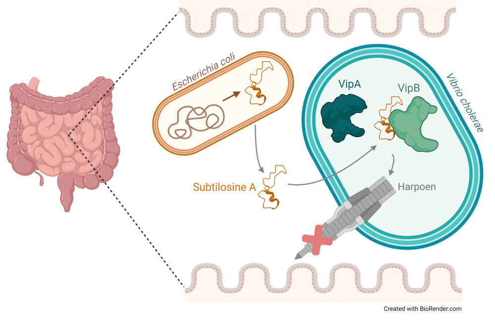
Finaal systeem – E. coli levert subtilosine A af in de darmen zodat deze voorkomt dat V. cholerae zijn harpoen kan gebruiken.
Meer varianten resulteert in een hogere kans op slagen
Vandaag worden nieuwe medicijnen ontwikkeld door varianten van bestaande medicijnen te creëren. Ik wil van deze werkwijze gebruik maken om een subtilosine A variant te vinden die het VipA-VipB contact kan blokkeren, en zo als behandeling tegen de cholera ziekte kan dienen. De natuurlijke activiteit van subtilosine A is het vernietigen van bepaalde bacteriën, maar de component kan ook nieuwe activiteiten verkrijgen wanneer deze aangepast wordt. In de toekomst wil ik dus een systeem creëren om op korte tijd heel veel varianten van subtilosine A te creëren, deze in E. coli te produceren en ze te testen voor hun potentieel om het VipA-VipB contact te verhinderen.
Hoe vinden we de juiste variant?
Om de enorme hoeveelheid gecreëerde varianten op een snelle manier te kunnen testen, is een systeem nodig dat dit toelaat. De opbouw van dit systeem ben ik tijdens mijn masterthesis gestart. Ik heb hiervoor opnieuw de E. coli fabriek ingeschakeld, waarin ik zowel de subtilosine A varianten, als VipA en VipB zal produceren. Om dit systeem te kunnen gebruiken, heb ik tijdens mijn thesis eerst getest of het VipA-VipB contact nog steeds opgaat, wanneer deze onderdelen niet meer in V. cholerae maar in E. coli worden aangemaakt. Daarna ben ik gestart met de opbouw van het uiteindelijke systeem. Hierin zal de E. coli bacterie sterven wanneer de subtilosine A variant het VipA-VipB contact niet kan blokkeren. Maar wanneer een subtilosine A variant het VipA-VipB contact kan voorkomen, zal E. coli overleven en groeien. Op deze manier kan ik gemakkelijk de subtilosine A variant vinden die ik nodig heb.
Ik hoop dat mijn masterthesis uiteindelijk zal bijdragen tot een nieuw medicijn voor de behandeling van de cholera ziekte. Ik hoop dat zo de sterftecijfers zullen dalen en wie weet wordt cholera dan een begrip dat enkel in geschiedenisboeken voorkomt. Op deze manier zal cholera niet alleen voor jou, maar ook voor alle andere mensen op de wereld een ver-van-het-bed show worden.
Bibliografie
1. Clemens JD, Nair GB, Ahmed T, Qadri F, Holmgren J. Cholera. The Lancet. 2017;390:1539–49.
2. Chaudhuri K, Chatterjee SN. Cholera Toxins. Berlin, Germany: Springer; 2009. 321p.
3. Cho JY, Liu R, Macbeth JC, Hsiao A. The Interface of Vibrio cholerae and the Gut Microbiome. Gut Microbes. 2021;13(1):1937015.
4. Bröms JE, Ishikawa T, Wai SN, Sjöstedt A. A functional VipA-VipB interaction is required for the type VI secretion system activity of Vibrio cholerae O1 strain A1552. BMC Microbiology. 2013;13(1):96.
5. Yang X, Lennard KR, He C, Walker MC, Ball AT, Doigneaux C, et al. A lanthipeptide library used to identify a protein-protein interaction inhibitor. Nature chemical biology. 2018;14(4):375–80.
6. Arnison PG, Bibb MJ, Bierbaum G, Bowers AA, Bugni TS, Bulaj G, et al. Ribosomally synthesized and post-translationally modified peptide natural products: overview and recommendations for a universal nomenclature. Natural Product Reports. 2013;30(1):108–60.
7. Jermyn WS, O’Shea YA, Quirke AM, Boyd EF. Genomics and the Evolution of Pathogenic Vibrio cholerae. In: Chan VL, Sherman PM, Bourke B, editors. Bacterial Genomes and Infectious Diseases. Totowa, NJ, US: Humana Press; 2006. p. 227– 54.
8. Vanden Broeck D, Horvath C, De Wolf MJS. Vibrio cholerae: Cholera toxin. The International Journal of Biochemistry & Cell Biology. 2007;39(10):1771–5.
9. Faruque SM, Albert MJ, Mekalanos JJ. Epidemiology, Genetics, and Ecology of Toxigenic Vibrio cholerae. Microbiology and Molecular Biology Reviews. 1998;62(4):1301–14.
10. Borgeaud S, Metzger LC, Scrignari T, Blokesch M. The type VI secretion system of Vibrio cholerae fosters horizontal gene transfer. Science. 2015;347(6217):63–7.
11. Alavi S, Mitchell JD, Cho JY, Liu R, Macbeth JC, Hsiao A. Interpersonal Gut Microbiome Variation Drives Susceptibility and Resistance to Cholera Infection. Cell. 2020;181(7):1533–46.
12. Khuntia HK, Ramamurthy T, Bal M, Pati S, Ranjit M. Decades of cholera in Odisha, India (1993–2015): lessons learned and the ways forward. Epidemiology & Infection. 2021;149:e148, 1–8.
13. Marin MA, Thompson CC, Freitas FS, Fonseca EL, Aboderin AO, Zailani SB, et al. Cholera Outbreaks in Nigeria Are Associated with Multidrug Resistant Atypical El Tor and Non-O1/Non-O139 Vibrio cholerae. PLOS Neglected Tropical Diseases. 2013;7(2):e2049.
14. Ma AT, McAuley S, Pukatzki S, Mekalanos JJ. Translocation of a Vibrio cholerae Type VI Secretion Effector Requires Bacterial Endocytosis by Host Cells. Cell Host & Microbe. 2009;5(3):234–43.
15. Unterweger D, Miyata ST, Bachmann V, Brooks TM, Mullins T, Kostiuk B, et al. The Vibrio cholerae type VI secretion system employs diverse effector modules for intraspecific competition. Nature communications. 2014;5:3549.
16. Safa A, Nair GB, Kong RYC. Evolution of new variants of Vibrio cholerae O1. Trends in Microbiology. 2009;18(1):46–54.
17. Ishikawa T, Sabharwal D, Bröms J, Milton DL, Sjöstedt A, Uhlin BE, et al. Pathoadaptive Conditional Regulation of the Type VI Secretion System in Vibrio cholerae O1 Strains. Infection and Immunity. 2012;80(2):575–84.
18. Sánchez J, Holmgren J. Cholera toxin - a foe & a friend. The Indian journal of Medical Research. 2011;133:153–63.
19. Nair GB, Faruque SM, Bhuiyan NA, Kamruzzaman M, Siddique AK, Sack DA. New Variants of Vibrio cholerae O1 Biotype El Tor with Attributes of the Classical Biotype from Hospitalized Patients with Acute Diarrhea in Bangladesh. Journal of Clinical Microbiology. 2002;40(9):3296–9.
20. Pukatzki S, Ma AT, Sturtevant D, Krastins B, Sarracino D, Nelson WC, et al. Identification of a conserved bacterial protein secretion system in Vibrio cholerae using the Dictyostelium host model system. PNAS. 2006;103(5):1528–33.
21. Breen P, Winters AD, Theis KR, Withey JH. The Vibrio cholerae Type Six Secretion System Is Dispensable for Colonization but Affects Pathogenesis and the Structure of Zebrafish Intestinal Microbiome. Infection and Immunity. 2021;89(9):e0015121.
22. Sun K, Bröms J, Lavander M, Gurram BK, Enquist PA, Andersson CD, et al. Screening for Inhibition of Vibrio cholerae VipA-VipB Interaction Identifies Small- Molecule Compounds Active against Type VI Secretion. Antimicrobial Agents and Chemotherapy. 2014;58(7):4123–30.
23. Ali M, Nelson AR, Lopez AL, Sack DA. Updated Global Burden of Cholera in Endemic Countries. PLOS Neglected Tropical Diseases. 2015;9(6):e0003832.
24. Zhang XY, Brunet YR, Logger L, Douzi B, Cambillau C, Journet L, et al. Dissection of the TssB-TssC Interface during Type VI Secretion Sheath Complex Formation. PLOS ONE. 2013;8(11):e81074.
25. Bernal P, Furniss RCD, Fecht S, Leung RCY, Spiga L, Mavridou DAI, et al. A novel stabilization mechanism for the type VI secretion system sheath. PNAS. 2021;118(7):e2008500118.
26. Basler M, Pilhofer M, Henderson GP, Jensen GJ, Mekalanos JJ. Type VI secretion requires a dynamic contractile phage tail-like structure. Nature. 2012;483(7388):182–6.
27. Salmond RJ, Luross JA, Williams NA. Immune modulation by the cholera-like enterotoxins. Expert reviews in molecular medicine. 2002;4(21):1–16.
28. Bingle LEH, Bailey CM, Pallen MJ. Type VI secretion: a beginner’s guide. Current Opinion in Microbiology. 2008;11(1):3–8.
29. Bönemann G, Pietrosiuk A, Mogk A. Tubules and donuts: a type VI secretion story. Molecular Microbiology. 2010;76(4):815–21.
30. Boyer F, Fichant G, Berthod J, Vandenbrouck Y, Attree I. Dissecting the bacterial type VI secretion system by a genome wide in silico analysis: what can be learned from available microbial genomic resources? BMC Genomics. 2009;10(1):104.
31. Coulthurst S. The Type VI secretion system: a versatile bacterial weapon. Microbiology (Society for General Microbiology). 2019;165(5):503–15.
32. Gallegos-Monterrosa R, Coulthurst SJ. The ecological impact of a bacterial weapon: microbial interactions and the Type VI secretion system. FEMS Microbiology Reviews. 2021;45(6):1.
33. Jani AJ, Cotter PA. Type VI Secretion: Not Just for Pathogenesis Anymore. Cell Host & Microbe. 2010;8(1):2–6.
34. Leiman PG, Basler M, Ramagopal UA, Bonanno JB, Sauder JM, Pukatzki S, et al. Type VI secretion apparatus and phage tail-associated protein complexes share a common evolutionary origin. PNAS. 2009;106(11):4154–9.
35. Pukatzki S, Ma AT, Revel AT, Sturtevant D, Mekalanos JJ. Type VI secretion system translocates a phage tail spike-like protein into target cells where it cross-links actin. PNAS. 2007;104(39):15508–13.
36. Pukatzki S, McAuley SB, Miyata ST. The type VI secretion system: translocation of effectors and effector-domains. Current Opinion in Microbiology. 2009;12(1):11–7.
37. Schwarz S, Hood RD, Mougous JD. What is type VI secretion doing in all those bugs? Trends in Microbiology. 2010;18(12):531–7.
38. Zoued A, Brunet YR, Durand E, Aschtgen MS, Logger L, Douzi B, et al. Architecture and assembly of the Type VI secretion system. Biochimica et Biophysica Acta (BBA) - Molecular Cell Research. 2014;1843(8):1664–73.
39. Cianfanelli FR, Monlezun L, Coulthurst SJ. Aim, Load, Fire: The Type VI Secretion System, a Bacterial Nanoweapon. Trends in Microbiology. 2016;24(1):51–62.
40. Ho BT, Dong TG, Mekalanos JJ. A View to a Kill: The Bacterial Type VI Secretion System. Cell Host & Microbe. 2014;15(1):9–21.
41. Cherrak Y, Flaugnatti N, Durand E, Journet L, Cascales E. Structure and Activity of the Type VI Secretion System. Microbiology Spectrum. 2019;7(4):7.4.11.
42. Jurėnas D, Journet L. Activity, delivery, and diversity of Type VI secretion effectors. Molecular Microbiology. 2021;115(3):383–94.
43. Wang J, Brodmann M, Basler M. Assembly and Subcellular Localization of Bacterial Type VI Secretion Systems. Annual review of microbiology. 2019;73(1):621–38.
44. Cherrak Y, Rapisarda C, Pellarin R, Bouvier G, Bardiaux B, Allain F, et al. Biogenesis and structure of a type VI secretion baseplate. Nature Microbiology. 2018;3(12):1404–16.
45. Ishikawa T, Rompikuntal PK, Lindmark B, Milton DL, Wai SN. Quorum Sensing Regulation of the Two hcp Alleles in Vibrio cholerae O1 Strains. PLOS ONE. 2009;4(8):e6734.
46. Shneider MM, Buth SA, Ho BT, Basler M, Mekalanos JJ, Leiman PG. PAAR-repeat proteins sharpen and diversify the type VI secretion system spike. Nature. 2013;500(7462):350–3.
47. Kudryashev M, Wang RYR, Brackmann M, Scherer S, Maier T, Baker D, et al. Structure of the Type VI Secretion System Contractile Sheath. Cell. 2015;160(5):952–62.
48. Primrose SB, Twyman RM. Principles of gene manipulation and genomics. 7th ed. Oxford, UK: Blackwell Publishing; 2006. 644 p.
49. Lu H, Zhou Q, He J, Jiang Z, Peng C, Tong R, et al. Recent advances in the development of protein–protein interactions modulators: mechanisms and clinical trials. Signal Transduction and Targeted Therapy. 2020;5(1):213.
50. Milroy LG, Grossmann TN, Hennig S, Brunsveld L, Ottmann C. Modulators of Protein–Protein Interactions. Chemical Reviews. 2014;114(9):4695–748.
51. Wendt MD. Protein-Protein Interactions as Drug Targets. In: Wendt MD, editor. Protein-Protein Interactions. Berlin, Heidelberg, Germany: Springer; 2012. p. 1–55. (Topics in Medicinal Chemistry).
52. Fry DC. Targeting Protein-Protein Interactions for Drug Discovery. In: Meyerkord CL, Fu H, editors. Protein-Protein Interactions: Methods and Applications. New York, NY, US: Springer; 2015. p. 93–106. (Methods in Molecular Biology).
53. Rosell M, Fernández-Recio J. Hot-spot analysis for drug discovery targeting protein-protein interactions. Expert Opinion on Drug Discovery. 2018;13(4):327–38.
54. Stynen B, Tournu H, Tavernier J, Van Dijck P. Diversity in Genetic In Vivo Methods for Protein-Protein Interaction Studies: from the Yeast Two-Hybrid System to the Mammalian Split-Luciferase System. Microbiology and Molecular Biology Reviews. 2012;76(2):331–82.
55. Phizicky EM, Fields S. Protein-protein interactions: methods for detection and analysis. Microbiological Reviews. 1995;59(1):94–123.
56. Gao M, Cheng K, Yin H. Targeting protein−protein interfaces using macrocyclic peptides. Peptide Science. 2015;104(4):310–6.
57. Loregian A, Palù G. Disruption of protein–protein interactions: Towards new targets for chemotherapy. Journal of Cellular Physiology. 2005;204(3):750–62.
58. Cardote TAF, Ciulli A. Cyclic and Macrocyclic Peptides as Chemical Tools To Recognise Protein Surfaces and Probe Protein–Protein Interactions. ChemMedChem. 2016;11(8):787–94.
59. Smith MC, Gestwicki JE. Features of protein-protein interactions that translate into potent inhibitors: topology, surface area and affinity. Expert reviews in molecular medicine. 2012;14:e16.
60. Arkin MR, Tang Y, Wells JA. Small-Molecule Inhibitors of Protein-Protein Interactions: Progressing toward the Reality. Chemistry & Biology. 2014;21(9):1102–14.
61. Jiang Y, Long H, Zhu Y, Zeng Y. Macrocyclic peptides as regulators of protein-protein interactions. Chinese Chemical Letters. 2018;29(7):1067–73.
62. Bruzzoni-Giovanelli H, Alezra V, Wolff N, Dong CZ, Tuffery P, Rebollo A. Interfering peptides targeting protein–protein interactions: the next generation of drugs? Drug Discovery Today. 2018;23(2):272–85.
63. Giordanetto F, Kihlberg J. Macrocyclic Drugs and Clinical Candidates: What Can Medicinal Chemists Learn from Their Properties? Journal of Medicinal Chemistry. 2014;57(2):278–95.
64. Nevola L, Giralt E. Modulating protein–protein interactions: the potential of peptides. Chemical Communications. 2015;51(16):3302–15.
65. Karimova G, Pidoux J, Ullmann A, Ladant D. A bacterial two-hybrid system based on a reconstituted signal transduction pathway. PNAS. 1998;95(10):5752–6.
66. Di Lallo G, Castagnoli L, Ghelardini P, Paolozzi L. A two-hybrid system based on chimeric operator recognition for studying protein homo/heterodimerization in Escherichia coli. Microbiology. 2001;147(6):1651–6.
67. Battesti A, Bouveret E. The bacterial two-hybrid system based on adenylate cyclase reconstitution in Escherichia coli. Methods. 2012;58(4):325–34.
68. Karimova G, Ullmann A, Ladant D. [5] A bacterial two-hybrid system that exploits a cAMP signaling cascade in Escherichia coli. In: Thorner J, Emr SD, Abelson JN, editors. Methods in Enzymology. Academic Press; 2000. p. 59–73. (Applications of Chimeric Genes and Hybrid Proteins - Part C: Protein-Protein Interactions and Genomics; vol. 328).
69. Meng X, Smith RM, Giesecke AV, Keith Joung J, Wolfe SA. Counter-selectable marker for bacterial-based interaction trap systems. BioTechniques. 2006;40(2):179–84.
70. Karimova G, Gauliard E, Davi M, Ouellette SP, Ladant D. Protein–Protein Interaction: Bacterial Two-Hybrid. In: Journet L, Cascales E, editors. Bacterial Protein Secretion Systems: Methods and Protocols. New York, NY, US: Springer; 2017. p. 159–76. (Methods in Molecular Biology).
71. Olson MG, Goldammer M, Gauliard E, Ladant D, Ouellette SP. A Bacterial Adenylate Cyclase-Based Two-Hybrid System Compatible with Gateway® Cloning. In: Oñate- Sánchez L, editor. Two-Hybrid Systems: Methods and Protocols. New York, NY, US: Springer; 2018. p. 75–96. (Methods in Molecular Biology).
72. Euromedex. BACTH System Kit: Bacterial Adenylate Cyclase Two-Hybrid System Kit. Euromedex, Souffelweyersheim, France; n.d.
73. Kjelstrup S, Hansen PMP, Thomsen LE, Hansen PR, Løbner-Olesen A. Cyclic Peptide Inhibitors of the β-Sliding Clamp in Staphylococcus aureus. PLOS ONE. 2013;8(9):e72273.
74. Horswill AR, Savinov SN, Benkovic SJ. A systematic method for identifying small-molecule modulators of protein–protein interactions. PNAS. 2004;101(44):15591–6.
75. Kurniyati K, Li C. pyrF as a Counterselectable Marker for Unmarked Genetic Manipulations in Treponema denticola. Applied and Environmental Microbiology. 2016;82(4):1346–52.
76. Tavassoli A, Lu Q, Gam J, Pan H, Benkovic SJ, Cohen SN. Inhibition of HIV Budding by a Genetically Selected Cyclic Peptide Targeting the Gag−TSG101 Interaction. ACS Chemical Biology. 2008;3(12):757–64.
77. Sohrabi C, Foster A, Tavassoli A. Methods for generating and screening libraries of genetically encoded cyclic peptides in drug discovery. Nature reviews Chemistry. 2020;4(2):90–101.
78. Lennard KR, Tavassoli A. Peptides Come Round: Using SICLOPPS Libraries for Early Stage Drug Discovery. Chemistry: A European Journal. 2014;20(34):10608–14.
79. Castillo F, Tavassoli A. Genetic Selections with SICLOPPS Libraries: Toward the Identification of Novel Protein–Protein Interaction Inhibitors and Chemical Tools. In: Goetz G, editor. Cyclic Peptide Design. New York, NY, US: Springer; 2019. p. 317-28. (Methods in Molecular Biology).
80. Tavassoli A, Benkovic SJ. Genetically Selected Cyclic-Peptide Inhibitors of AICAR Transformylase Homodimerization. Angewandte Chemie. 2005;117(18):2820–3.
81. Gang D, Kim DW, Park HS. Cyclic Peptides: Promising Scaffolds for Biopharmaceuticals. Genes. 2018;9(11):557.
82. Renukuntla J, Vadlapudi AD, Patel A, Boddu SHS, Mitra AK. Approaches for enhancing oral bioavailability of peptides and proteins. International Journal of Pharmaceutics. 2013;447(1):75–93.
83. Scapin G, Yang X, Prosise WW, McCoy M, Reichert P, Johnston JM, et al. Structure of full-length human anti-PD1 therapeutic IgG4 antibody pembrolizumab. Nature Structural & Molecular Biology. 2015;22(12):953–8.
84. Passioura T, Katoh T, Goto Y, Suga H. Selection-Based Discovery of Druglike Macrocyclic Peptides. Annual Review of Biochemistry. 2014;83(1):727–52.
85. Ortega MA, van der Donk WA. New Insights into the Biosynthetic Logic of Ribosomally Synthesized and Post-translationally Modified Peptide Natural Products. Cell Chemical Biology. 2016;23(1):31–44.
86. Sikandar A, Koehnke J. The role of protein–protein interactions in the biosynthesis of ribosomally synthesized and post-translationally modified peptides. Natural Product Reports. 2019;36(11):1576–88.
87. Yang X, van der Donk WA. Ribosomally Synthesized and Post-Translationally Modified Peptide Natural Products: New Insights into the Role of Leader and Core Peptides during Biosynthesis. Chemistry. 2013;19(24):7662–77.
88. Benjdia A, Berteau O. Radical SAM Enzymes and Ribosomally‐Synthesized and Post‐translationally Modified Peptides: A Growing Importance in the Microbiomes. Frontiers in Chemistry. 2021;9:678068.
89. Cao L, Do T, Link AJ. Mechanisms of action of ribosomally synthesized and posttranslationally modified peptides (RiPPs). Journal of industrial microbiology & biotechnology. 2021;48(3–4):kuab005.
90. Hudson GA, Mitchell DA. RiPP antibiotics: biosynthesis and engineering potential. Current Opinion in Microbiology. 2018;45:61–9.
91. Duperthuy M. Antimicrobial Peptides: Virulence and Resistance Modulation in Gram-Negative Bacteria. Microorganisms. 2020;8(2):280.
92. Huang F, Teng K, Liu Y, Cao Y, Wang T, Ma C, et al. Bacteriocins: Potential for Human Health. Oxidative Medicine and Cellular Longevity. 2021;2021:5518825.
93. Meade E, Slattery MA, Garvey M. Bacteriocins, Potent Antimicrobial Peptides and the Fight against Multi Drug Resistant Species: Resistance Is Futile? Antibiotics. 2020;9(1):32.
94. Li Y, Rebuffat S. The manifold roles of microbial ribosomal peptide–based natural products in physiology and ecology. Journal of Biological Chemistry. 2020;295(1):34–54.
95. Montalbán-López M, Scott TA, Ramesh S, Rahman IR, van Heel AJ, Viel JH, et al. New developments in RiPP discovery, enzymology and engineering. Natural Product Reports. 2021;38(1):130–239.
96. Zhang Y, Chen M, Bruner SD, Ding Y. Heterologous Production of Microbial Ribosomally Synthesized and Post-translationally Modified Peptides. Frontiers in Microbiology. 2018;9:1801.
97. Acedo JZ, Chiorean S, Vederas JC, van Belkum MJ. The expanding structural variety among bacteriocins from Gram-positive bacteria. FEMS microbiology reviews. 2018;42(6):805–28.
98. Chen Y, Wang J, Li G, Yang Y, Ding W. Current Advancements in Sactipeptide Natural Products. Frontiers in Chemistry. 2021;9:595991.
99. Mahanta N, Hudson GA, Mitchell DA. Radical S-Adenosylmethionine Enzymes Involved in RiPP Biosynthesis. Biochemistry. 2017;56(40):5229–44.
100. Rebuffat S. Ribosomally synthesized peptides, foreground players in microbial interactions: recent developments and unanswered questions. Natural Product Reports. 2022;39(2):273–310.
101. Fu Y, Jaarsma AH, Kuipers OP. Antiviral activities and applications of ribosomally synthesized and post-translationally modified peptides (RiPPs). Cellular and Molecular Life Sciences. 2021;78(8):3921–40.
102. Thennarasu S, Lee DK, Poon A, Kawulka KE, Vederas JC, Ramamoorthy A. Membrane permeabilization, orientation, and antimicrobial mechanism of subtilosin A. Chemistry and Physics of Lipids. 2005;137(1–2):38–51.
103. Kawulka KE, Sprules T, Diaper CM, Whittal RM, McKay RT, Mercier P, et al. Structure of Subtilosin A, a Cyclic Antimicrobial Peptide from Bacillus subtilis with Unusual Sulfur to α-Carbon Cross-Links: Formation and Reduction of α-Thio-α-Amino Acid Derivatives. Biochemistry. 2004;43(12):3385–95.
104. Huang T, Geng H, Miyyapuram VR, Sit CS, Vederas JC, Nakano MM. Isolation of a Variant of Subtilosin A with Hemolytic Activity. Journal of Bacteriology. 2009;191(18):5690–6.
105. Negash AW, Tsehai BA. Current Applications of Bacteriocin. International Journal of Microbiology. 2020;2020:4374891.
106. Zheng G, Yan LZ, Vederas JC, Zuber P. Genes of the sbo-alb Locus of Bacillus subtilis Are Required for Production of the Antilisterial Bacteriocin Subtilosin. Journal of Bacteriology. 1999;181(23):7346–55.
107. Zheng G, Hehn R, Zuber P. Mutational Analysis of the sbo-alb Locus of Bacillus subtilis: Identification of Genes Required for Subtilosin Production and Immunity. Journal of Bacteriology. 2000;182(11):3266–73.
108. Sutyak KE, Anderson RA, Dover SE, Feathergill KA, Aroutcheva AA, Faro S, et al. Spermicidal Activity of the Safe Natural Antimicrobial Peptide Subtilosin. Infectious Diseases in Obstetrics and Gynecology. 2008;2008:540758.
109. Shelburne CE, An FY, Dholpe V, Ramamoorthy A, Lopatin DE, Lantz MS. The spectrum of antimicrobial activity of the bacteriocin subtilosin A. Journal of Antimicrobial Chemotherapy. 2007;59(2):297–300.
110. Funk MA, van der Donk WA. Ribosomal Natural Products, Tailored To Fit. Accounts of Chemical Research. 2017;50(7):1577–86.
111. Oman TJ, van der Donk WA. Follow the leader: the use of leader peptides to guide natural product biosynthesis. Nature chemical biology. 2010;6(1):9–18.
112. Budisa N. Expanded genetic code for the engineering of ribosomally synthetized and post-translationally modified peptide natural products (RiPPs). Current Opinion in Biotechnology. 2013;24(4):591–8.
113. Himes PM, Allen SE, Hwang S, Bowers AA. Production of Sactipeptides in Escherichia coli: Probing the Substrate Promiscuity of Subtilosin A Biosynthesis. ACS Chemical Biology. 2016;11(6):1737–44.
114. Grove TL, Himes PM, Hwang S, Yumerefendi H, Bonanno JB, Kuhlman B, et al. Structural Insights into Thioether Bond Formation in the Biosynthesis of Sactipeptides. Journal of the American Chemical Society. 2017;139(34):11734–44.
115. Benjdia A, Guillot A, Lefranc B, Vaudry H, Leprince J, Berteau O. Thioether bond formation by SPASM domain radical SAM enzymes: Cα H-atom abstraction in subtilosin A biosynthesis. Chemical Communications. 2016;52(37):6249–52.
116. Flühe L, Knappe TA, Gattner MJ, Schäfer A, Burghaus O, Linne U, et al. The radical SAM enzyme AlbA catalyzes thioether bond formation in subtilosin A. Nature Chemical Biology. 2012;8(4):350–7.
117. Stein T, Düsterhus S, Stroh A, Entian KD. Subtilosin Production by Two Bacillus subtilis Subspecies and Variance of the sbo-alb Cluster. Applied and Environmental Microbiology. 2004;7(4):2349–53.
118. Torres NI, Sutyak Noll K, Xu S, Li J, Huang Q, Sinko PJ, et al. Safety, Formulation and In Vitro Antiviral Activity of the Antimicrobial Peptide Subtilosin Against Herpes Simplex Virus Type 1. Probiotics and Antimicrobial Proteins. 2013;5(1):26–35.
119. Algburi A, Zehm S, Netrebov V, Bren AB, Chistyakov V, Chikindas ML. Subtilosin Prevents Biofilm Formation by Inhibiting Bacterial Quorum Sensing. Probiotics and Antimicrobial Proteins. 2017;9(1):81–90.
120. Silkin L, Hamza S, Kaufman S, Cobb SL, Vederas JC. Spermicidal bacteriocins: Lacticin 3147 and subtilosin A. Bioorganic & Medicinal Chemistry Letters. 2008;18(10):3103–6.
121. Corless EI, Mettert EL, Kiley PJ, Antony E. Elevated Expression of a Functional Suf Pathway in Escherichia coli BL21(DE3) Enhances Recombinant Production of an Iron-Sulfur Cluster-Containing Protein. Journal of Bacteriology. 2020;202(3):e00496-19.
122. Mamantopoulos M, Frising UC, Asaoka T, van Loo G, Lamkanfi M, Wullaert A. El Tor Biotype Vibrio cholerae Activates the Caspase-11-Independent Canonical Nlrp3 and Pyrin Inflammasomes. Frontiers in Immunology. 2019;10:2463.
123. Mülhardt C, Beese EW. Molecular Biology and Genomics. Burlington, MA, US: Academic Press; 2007. 257 p.
124. Camacho EM, Mesa-Pereira B, Medina C, Flores A, Santero E. Engineering Salmonella as intracellular factory for effective killing of tumour cells. Scientific Reports. 2016;6(1):30591.
125. Park D, Swayambhu G, Pfeifer BA. Heterologous biosynthesis as a platform for producing new generation natural products. Current Opinion in Biotechnology. 2020;66:123–30.
126. Kaur J, Kumar A, Kaur J. Strategies for optimization of heterologous protein expression in E. coli: Roadblocks and reinforcements. International Journal of Biological Macromolecules. 2018;106:803–22.
127. Huo L, Hug JJ, Fu C, Bian X, Zhang Y, Müller R. Heterologous expression of bacterial natural product biosynthetic pathways. Natural Product Reports. 2019;36(10):1412–36.
128. Cui Y, Luo L, Wang X, Lu Y, Yi Y, Shan Y, et al. Mining, heterologous expression, purification, antibactericidal mechanism, and application of bacteriocins: A review. Comprehensive Reviews in Food Science and Food Safety. 2021;20(1):863–99.
129. antiSMASH. antiSMASH bacterial version [Internet]. n.d. [cited 2022 May 1]. Available from: https://antismash.secondarymetabolites.org/#!/start
130. Cheung-Lee WL, Parry ME, Cartagena AJ, Darst SA, Link AJ. Discovery and structure of the antimicrobial lasso peptide citrocin. Journal of Biological Chemistry. 2019;294(17):6822–30.
131. Cheung-Lee WL, Parry ME, Zong C, Cartagena AJ, Darst SA, Connell ND, et al. Discovery of Ubonodin, an Antimicrobial Lasso Peptide Active against Members of the Burkholderia cepacia Complex. ChemBioChem. 2020;21(9):1335–40.
132. Cao L, Beiser M, Koos JD, Orlova M, Elashal HE, Schröder HV, et al. Cellulonodin-2 and Lihuanodin: Lasso Peptides with an Aspartimide Post-Translational Modification. Journal of the American Chemical Society. 2021;143(30):11690–702.
133. Sutyak KE, Wirawan RE, Aroutcheva AA, Chikindas ML. Isolation of the Bacillus subtilis antimicrobial peptide subtilosin from the dairy product-derived Bacillus amyloliquefaciens. Journal of Applied Microbiology. 2007;104(4):1067–74.
134. van Kuijk S, Noll KS, Chikindas ML. The species-specific mode of action of the antimicrobial peptide subtilosin against Listeria monocytogenes Scott A. Letters in Applied Microbiology. 2011;54(1):52–8.
135. Benjdia A, Balty C, Berteau O. Radical SAM Enzymes in the Biosynthesis of Ribosomally Synthesized and Post-translationally Modified Peptides (RiPPs). Frontiers in Chemistry. 2017;5:87.
136. Outten FW, Djaman O, Storz G. A suf operon requirement for Fe–S cluster assembly during iron starvation in Escherichia coli. Molecular Microbiology. 2004;52(3):861–72.
137. National Library of Medicine. National Center for Biotechnology Information [Internet]. n.d. [cited 2022 Apr 23]. Available from: https://www.ncbi.nlm.nih.gov/
138. Pagala VR, High AA, Wang X, Tan H, Kodali K, Mishra A, et al. Quantitative Protein Analysis by Mass Spectrometry. In: Meyerkord CL, Fu H, editors. Protein-Protein Interactions: Methods and Applications. New York, NY, US: Springer; 2015. p. 281–305. (Methods in Molecular Biology).
139. Mesa-Pereira B, Rea MC, Cotter PD, Hill C, Ross RP. Heterologous Expression of Biopreservative Bacteriocins With a View to Low Cost Production. Frontiers in Microbiology. 2018;9:1654.
140. Peti W, Page R. Strategies to maximize heterologous protein expression in Escherichia coli with minimal cost. Protein Expression and Purification. 2007;51(1):1–10.
141. Cárcel-Márquez J, Flores A, Martín-Cabello G, Santero E, Camacho EM. Development of an inducible lytic system for functional metagenomic screening. Scientific Reports. 2019;9(1):3887.
142. Swiss Institute of Bioinformatics. Holin/endolysin/spanin cell lysis [Internet]. ViralZone. n.d. [cited 2022 May 9]. Available from: https://viralzone.expasy.org/4056
143. Khochamit N, Siripornadulsil S, Sukon P, Siripornadulsil W. Antibacterial activity and genotypic-phenotypic characteristics of bacteriocin-producing Bacillus subtilis KKU213: potential as a probiotic strain. Microbiological Research. 2015;170:36–50.
144. Turovskiy Y, Cheryian T, Algburi A, Wirawan RE, Takhistov P, Sinko PJ, et al. Susceptibility of Gardnerella vaginalis biofilms to natural antimicrobials subtilosin, ε-poly-L-lysine, and lauramide arginine ethyl ester. Infectious diseases in obstetrics and gynecology. 2012;2012:284762.
145. Rani RP, Anandharaj M, Hema S, Deepika R, Ravindran AD. Purification of Antilisterial Peptide (Subtilosin A) from Novel Bacillus tequilensis FR9 and Demonstrate Their Pathogen Invasion Protection Ability Using Human Carcinoma Cell Line. Frontiers in Microbiology. 2016;7:1910.
146. García-Fruitós E, Sabate R, de Groot NS, Villaverde A, Ventura S. Biological role of bacterial inclusion bodies: a model for amyloid aggregation. The FEBS Journal. 2011;278(14):2419–27.
147. Puckett MC. Hexahistidine (6xHis) Fusion-Based Assays for Protein-Protein Interactions. In: Meyerkord CL, Fu H, editors. Protein-Protein Interactions: Methods and Applications. New York, NY, US: Springer; 2015. p. 365–70. (Methods in Molecular Biology).
148. Butt TR, Edavettal SC, Hall JP, Mattern MR. SUMO fusion technology for difficult-to-express proteins. Protein Expression and Purification. 2005;43(1):1–9.
149. Borkowski O, Ceroni F, Stan GB, Ellis T. Overloaded and stressed: whole-cell considerations for bacterial synthetic biology. Current Opinion in Microbiology. 2016;33:123–30.
150. Shi Y, Yang X, Garg N, van der Donk WA. Production of Lantipeptides in Escherichia coli. Journal of the American Chemical Society. 2011;133(8):2338–41.
151. STRING Consortium. STRING: functional protein association networks [Internet]. 2022 [cited 2022 Apr 23]. Available from: https://string-db.org/
152. Szklarczyk D, Jensen LJ. Protein-Protein Interaction Databases. In: Meyerkord CL, Fu H, editors. Protein-Protein Interactions: Methods and Applications. New York, NY, US: Springer; 2015. p. 39–56. (Methods in Molecular Biology).
153. Clark DP. The fermentation pathways of Escherichia coli. FEMS Microbiology Reviews. 1989;63(3):223–34.
154. Hengen PN. Purification of His-Tag fusion proteins from Escherichia coli. Trends in Biochemical Sciences. 1995;20(7):285–6.
155. Bönemann G, Pietrosiuk A, Diemand A, Zentgraf H, Mogk A. Remodelling of VipA/VipB tubules by ClpV-mediated threading is crucial for type VI protein secretion. The EMBO journal. 2009;28(4):315–25.
156. Chung HS, Raetz CRH. Interchangeable Domains in the Kdo Transferases of Escherichia coli and Haemophilus influenzae. Biochemistry. 2010;49(19):4126–37.
157. Kube S, Kapitein N, Zimniak T, Herzog F, Mogk A, Wendler P. Structure of the VipA/B Type VI Secretion Complex Suggests a Contraction-State-Specific Recycling Mechanism. Cell Reports. 2014;8(1):20–30.
158. National Library of Medicine. BLAST: Basic Local Alignment Search Tool [Internet]. n.d. [cited 2022 May 1]. Available from: https://blast.ncbi.nlm.nih.gov/Blast.cgi
159. Zhang X, Bremer H. Control of the Escherichia coli rrnB P1 Promoter Strength by ppGpp. Journal of Biological Chemistry. 1995;270(19):11181–9.
160. Rowe SM, Spring DR. The role of chemical synthesis in developing RiPP antibiotics. Chemical Society Reviews. 2021;50(7):4245–58.
161. Burkhart BJ, Kakkar N, Hudson GA, van der Donk WA, Mitchell DA. Chimeric Leader Peptides for the Generation of Non-Natural Hybrid RiPP Products. ACS Central Science. 2017;3(6):629–38.
162. Bouet JY, Nordström K, Lane D. Plasmid partition and incompatibility – the focus shifts. Molecular Microbiology. 2007;65(6):1405–14.


