
De veranderingen in het celmembraan van Schwann cellen in de ziekte van Charcot-Marie-Tooth type 1A: een patiënt-in-een-schaal onderzoeksmethode
- TimVangansewinkel
- EstherWolfs
- JeroenBogie
- HanneJeurissen
- KarenLibberecht
- SteffieHasevoets
- SamVanherle
- MelanieLoix
Ongeveer 3 miljoen mensen wereldwijd lijdt aan de ziekte van Charcot-Marie-Tooth (CMT), een genetische zenuwziekte die we reeds bijna 150 jaar al kennen. Ondanks deze confronterende cijfers, is er tot op de dag van vandaag nog steeds geen enkele therapie die werkt. 60 tot 70% van de CMT-patiënten heeft de meest voorkomende variant, CMT type 1A (CMT1A). Ondanks vele pogingen om een eerste medicament aan deze grootste groep patiënten te kunnen voorzien, is er tot op heden nog geen enkele goedgekeurd of werkend in de mens. Het verstaan van de ziekte CMT1A, en vooral de rol van de extra kopie van het PMP22-gen hierin, is een cruciaal knelpunt op dit moment. De gevolgen van de PMP22-genduplicatie verstaan in menselijke CMT1A celmodellen is de topic van mijn thesis.
Ziekte van Charcot-Marie-Watte?
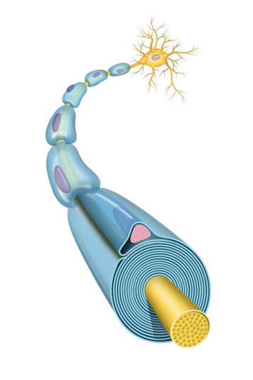
CMT1A komt ongeveer bij 1 op 4000 mensen voor. CMT1A patiënten hebben vooral bewegings- en gevoelsproblemen in de armen en benen, vaak beginnende al in de eerste tien levensjaren. De ziekte blijft aanwezig gedurende heel hun leven en kan in sommige gevallen leiden tot leven in een rolstoel. CMT1A wordt veroorzaakt door een kapotte insulerende laag rond de zenuw, vergelijkbaar met een stroomkabel. Deze laag heet myeline en wordt gemaakt door gespecialiseerde cellen, Schwann cellen genaamd. Schade in de myeline zorgt ervoor dat de zenuwen minder goed werken en leidt tot de symptomen. Hoe de Schwann cellen exact verstoord worden door de PMP22-genduplicatie, en door welke tot dusver onbekende mechanismen dit gebeurt, is nog steeds een belangrijke vraag zonder antwoord.
Schwann cellen en bindweefsel
In mijn thesis hebben we vooral gekeken naar het celmembraan van de Schwann cel, aangezien deze uiteindelijk ook de myelinelaag vormt. In onze onderzoeksgroep gebruiken wij voornamelijk menselijke stamcellen om een representatief ziektemodel te vormen als een soort patiënt-in-een-schaal. Stamcellen zijn cellen die nog in een heleboel andere cellen omgezet kunnen worden, een beetje zoals een cadeaukaart die je nog kan inruilen voor een cadeau naar keuze. Tot dusver is een belangrijke reden waardoor er geen medicament voor CMT1A is, de tekortkoming van de diermodellen geweest. Om erachter te komen wat er fout gaat in het celmembraan van Schwann cellen van CMT1A-patiënten, hebben we stamcellen van de huid van CMT1A patiënten gebruikt die wij in het labo dan omvormen tot Schwann cellen. Daarnaast hebben wij in het laboratorium ook tandstamcellen uit menselijke wijsheidstanden waar we het genetisch defect van CMT1A in aangebracht hebben. Om te bepalen hoezeer deze veranderd zijn, vergelijken we de zieke cellen met gezonde cellen van dezelfde patiënt. In mijn experimenten zagen we dat in de CMT1A Schwann cellen er grote veranderingen zijn in het celmembraan. Zo zien we dat integrines, een groepje moleculen die op het celmembraan zitten die zorgen voor de interactie met bindweefsel in de zenuw, sterk verschillen in hoeveelheid in CMT1A Schwann cellen ten opzichte van de gezonde cellen. Dit hebben we bepaald aan de hand van technieken zoals immunocytochemie, een microscopie techniek om de molecule te bekijken en de hoeveelheid eiwit te bepalen, en qPCR, waarmee je de hoeveelheid RNA kan bepalen die uiteindelijk omgezet wordt in de integrine eiwitten. Ook zien we dat celmigratie minder vlot verloopt in CMT1A Schwann cellen, wat ook aangeeft dat er een verminderde interactie is met het bindweefsel. Dit deden we door de cellen met een microscoop op te volgen voor meerdere dagen met foto’s om de zoveel minuten. Omdat de interactie met bindweefsel essentieel is voor het vormen van het myelinelaagje, vermoeden wij dus met deze experimenten dat de verstoring van deze integrines bijdraagt tot de kapotte myelinelaag die we zien in CMT1A-patiënten.
Vetten
Naast de integrines, moeten Schwann cellen ook genoeg en vooral ook de juiste vetten hebben om hun myelinelaag rond de zenuwen te maken. Omdat myeline voor het overgrote deel uit vetten bestaat, zorgen veranderingen hierin dus voor een onstabiele myeline laag. In een experiment waarbij we specifiek naar de vetten in het celmembraan kijken, zien we dat er een verandering is in celmembraanpolariteit. Dit geeft ons al een idee over de samenstelling aan vetten van het celmembraan en wijst op fouten in cholesterol, veranderingen in verzadigde en onverzadigde vetten, etc. Normaal gezien zijn er ook een soort vet-eilandjes aanwezig op het celmembraan, met daarin een heleboel eiwitten die hier samenwerken om efficiënt te zorgen voor de interactie van de cel met hun bindweefsel-omgeving. Wanneer we met super-resolutie microscopie kijken naar het celmembraan, zien we dat deze vet-eilandjes minder bewegen in CMT1A. Dit kan bijdragen aan een minder flexibele interactie met de bindweefselomgeving en kan zo zorgen voor minder goede vorming van myeline. Tot slot zien we bij het bekijken van vetdruppels — dienend als vetopslag voor later gebruik — in de Schwann cellen dat in CMT1A er veel minder opslag is van vetten, waardoor de cellen dus minder kunnen terugvallen op hun reserves. Dit wijst er allemaal op dat de myeline laag in CMT1A dus waarschijnlijk ook niet goed gevormd wordt door een sterk veranderd profiel aan vetsoorten en vetopslag.
Wat is de volgende stap?
Alle bevindingen in mijn thesis wijzen erop dat in CMT1A Schwann cellen een verstoorde interactie plaats vindt met het bindweefsel van de zenuw, en dat het celmembraan al verstoring heeft in zijn vetprofiel en membraanvetten zelfs voor myeline vorming. Een belangrijke volgende stap is het begrijpen hoe de aanmaak van vetten exact verstoord is, welke vetten vooral aangetast zijn en welke vetten gunstig zijn om te herstellen voor functioneel zenuwherstel. Daarnaast is het verstaan van de verandering in het integrine profiel belangrijk in verschillende stadia van de zenuwontwikkeling, zodat we deze kunnen verbeteren en zo de vorming van myeline kunnen stimuleren. De puzzel van CMT1A is dus nog niet opgelost, maar hiermee zijn er een aantal belangrijke puzzelstukken gelegd die weg maken voor vervolgonderzoek en mogelijks een toekomstige therapie voor CMT1A patiënten.
Bibliografie
1. Kazamel M, Boes CJ. Charcot Marie Tooth disease (CMT): historical perspectives and evolution. J Neurol. 2015;262(4):801-5.
2. van Paassen BW, van der Kooi AJ, van Spaendonck-Zwarts KY, Verhamme C, Baas F, de Visser M. PMP22 related neuropathies: Charcot-Marie-Tooth disease type 1A and Hereditary Neuropathy with liability to Pressure Palsies. Orphanet J Rare Dis. 2014;9:38.
3. Vallat JM, Mathis S, Funalot B. The various Charcot-Marie-Tooth diseases. Curr Opin Neurol. 2013;26(5):473-80.
4. Pareyson D, Marchesi C. Diagnosis, natural history, and management of Charcot-Marie-Tooth disease. Lancet Neurol. 2009;8(7):654-67.
5. Boutary S, Echaniz-Laguna A, Adams D, Loisel-Duwattez J, Schumacher M, Massaad C, et al. Treating PMP22 gene duplication-related Charcot-Marie-Tooth disease: the past, the present and the future. Transl Res. 2021;227:100-11.
6. Pantera H, Shy ME, Svaren J. Regulating PMP22 expression as a dosage sensitive neuropathy gene. Brain Res. 2020;1726:146491.
7. Gautier B, Hajjar H, Soares S, Berthelot J, Deck M, Abbou S, et al. AAV2/9-mediated silencing of PMP22 prevents the development of pathological features in a rat model of Charcot-Marie-Tooth disease 1 A. Nat Commun. 2021;12(1):2356.
8. Hartmannsberger B, Doppler K, Stauber J, Schlotter-Weigel B, Young P, Sereda MW, et al. Intraepidermal nerve fibre density as biomarker in Charcot-Marie-Tooth disease type 1A. Brain Commun. 2020;2(1):fcaa012.
9. Berciano J, Gallardo E, Garcia A, Pelayo-Negro AL, Infante J, Combarros O. New insights into the pathophysiology of pes cavus in Charcot-Marie-Tooth disease type 1A duplication. J Neurol. 2011;258(9):1594-602.
10. Azevedo H, Pupe C, Pereira R, Nascimento OJM. Pain in Charcot-Marie-Tooth disease: an update. Arq Neuropsiquiatr. 2018;76(4):273-6.
11. Burns J, Ryan MM, Ouvrier RA. Quality of life in children with Charcot-Marie-Tooth disease. J Child Neurol. 2010;25(3):343-7.
12. Cordeiro JL, Marques W, Hallak JE, Osorio FL. Charcot-Marie-Tooth disease, psychiatric indicators and quality of life: a systematic review. ASN Neuro. 2014;6(3):185-92.
13. Taniguchi JB, Elui VM, Osorio FL, Hallak JE, Crippa JA, Machado-de-Sousa JP, et al. Quality of life in patients with Charcot-Marie-Tooth disease type 1A. Arq Neuropsiquiatr. 2013;71(6):392-6.
14. Amici SA, Dunn WA, Jr., Murphy AJ, Adams NC, Gale NW, Valenzuela DM, et al. Peripheral myelin protein 22 is in complex with alpha6beta4 integrin, and its absence alters the Schwann cell basal lamina. J Neurosci. 2006;26(4):1179-89.
15. Maier M, Berger P, Suter U. Understanding Schwann cell-neurone interactions: the key to Charcot-Marie-Tooth disease? J Anat. 2002;200(4):357-66.
16. Rosso G, Young P, Shahin V. Implications of Schwann Cells Biomechanics and Mechanosensitivity for Peripheral Nervous System Physiology and Pathophysiology. Front Mol Neurosci. 2017;10:345.
17. Taveggia C. Schwann cells-axon interaction in myelination. Curr Opin Neurobiol. 2016;39:24-9.
18. Mittendorf KF, Marinko JT, Hampton CM, Ke Z, Hadziselimovic A, Schlebach JP, et al. Peripheral myelin protein 22 alters membrane architecture. Sci Adv. 2017;3(7):e1700220.
19. Marinko JT, Kenworthy AK, Sanders CR. Peripheral myelin protein 22 preferentially partitions into ordered phase membrane domains. Proc Natl Acad Sci U S A. 2020;117(25):14168-77.
20. Lee S, Amici S, Tavori H, Zeng WM, Freeland S, Fazio S, et al. PMP22 is critical for actin-mediated cellular functions and for establishing lipid rafts. J Neurosci. 2014;34(48):16140-52.
21. Zhou Y, Miles JR, Tavori H, Lin M, Khoshbouei H, Borchelt DR, et al. PMP22 Regulates Cholesterol Trafficking and ABCA1-Mediated Cholesterol Efflux. J Neurosci. 2019;39(27):5404-18.
22. Di Tomaso MV, Vazquez Alberdi L, Olsson D, Cancela S, Fernandez A, Rosillo JC, et al. Colocalization Analysis of Peripheral Myelin Protein-22 and Lamin-B1 in the Schwann Cell Nuclei of Wt and TrJ Mice. Biomolecules. 2022;12(3).
23. Fortun J, Go JC, Li J, Amici SA, Dunn WA, Jr., Notterpek L. Alterations in degradative pathways and protein aggregation in a neuropathy model based on PMP22 overexpression. Neurobiol Dis. 2006;22(1):153-64.
24. Johnson JS, Roux KJ, Fletcher BS, Fortun J, Notterpek L. Molecular alterations resulting from frameshift mutations in peripheral myelin protein 22: implications for neuropathy severity. J Neurosci Res. 2005;82(6):743-52.
25. Tobler AR, Notterpek L, Naef R, Taylor V, Suter U, Shooter EM. Transport of Trembler-J mutant peripheral myelin protein 22 is blocked in the intermediate compartment and affects the transport of the wild-type protein by direct interaction. J Neurosci. 1999;19(6):2027-36.
26. Chernousov MA, Yu WM, Chen ZL, Carey DJ, Strickland S. Regulation of Schwann cell function by the extracellular matrix. Glia. 2008;56(14):1498-507.
27. Muppirala AN, Limbach LE, Bradford EF, Petersen SC. Schwann cell development: From neural crest to myelin sheath. Wiley Interdiscip Rev Dev Biol. 2021;10(5):e398.
28. Vega-Lopez GA, Cerrizuela S, Tribulo C, Aybar MJ. Neurocristopathies: New insights 150 years after the neural crest discovery. Dev Biol. 2018;444 Suppl 1:S110-S43.
29. Radomska KJ, Topilko P. Boundary cap cells in development and disease. Curr Opin Neurobiol. 2017;47:209-15.
30. Jessen KR, Mirsky R. Schwann Cell Precursors; Multipotent Glial Cells in Embryonic Nerves. Front Mol Neurosci. 2019;12:69.
31. Juliano RL, Haskill S. Signal transduction from the extracellular matrix. J Cell Biol. 1993;120(3):577-85.
32. Feltri ML, Scherer SS, Nemni R, Kamholz J, Vogelbacker H, Scott MO, et al. Beta 4 integrin expression in myelinating Schwann cells is polarized, developmentally regulated and axonally dependent. Development. 1994;120(5):1287-301.
33. Pellegatta M, De Arcangelis A, D'Urso A, Nodari A, Zambroni D, Ghidinelli M, et al. alpha6beta1 and alpha7beta1 integrins are required in Schwann cells to sort axons. J Neurosci. 2013;33(46):17995-8007.
34. Lefcort F, Venstrom K, McDonald JA, Reichardt LF. Regulation of expression of fibronectin and its receptor, alpha 5 beta 1, during development and regeneration of peripheral nerve. Development. 1992;116(3):767-82.
35. Pereira JA, Lebrun-Julien F, Suter U. Molecular mechanisms regulating myelination in the peripheral nervous system. Trends Neurosci. 2012;35(2):123-34.
36. Nodari A, Zambroni D, Quattrini A, Court FA, D'Urso A, Recchia A, et al. Beta1 integrin activates Rac1 in Schwann cells to generate radial lamellae during axonal sorting and myelination. J Cell Biol. 2007;177(6):1063-75.
37. Shen Y, Cheng Z, Chen S, Zhang Y, Chen Q, Yi S. Dysregulated miR-29a-3p/PMP22 Modulates Schwann Cell Proliferation and Migration During Peripheral Nerve Regeneration. Mol Neurobiol. 2022;59(2):1058-72.
38. Poitelon Y, Kopec AM, Belin S. Myelin Fat Facts: An Overview of Lipids and Fatty Acid Metabolism. Cells. 2020;9(4).
39. Fledrich R, Abdelaal T, Rasch L, Bansal V, Schutza V, Brugger B, et al. Targeting myelin lipid metabolism as a potential therapeutic strategy in a model of CMT1A neuropathy. Nat Commun. 2018;9(1):3025.
40. Visigalli D, Capodivento G, Basit A, Fernandez R, Hamid Z, Pencova B, et al. Exploiting Sphingo- and Glycerophospholipid Impairment to Select Effective Drugs and Biomarkers for CMT1A. Front Neurol. 2020;11:903.
41. Levental I, Levental KR, Heberle FA. Lipid Rafts: Controversies Resolved, Mysteries Remain. Trends Cell Biol. 2020;30(5):341-53.
42. Zhou Y, Borchelt D, Bauson JC, Fazio S, Miles JR, Tavori H, et al. Subcellular diversion of cholesterol by gain- and loss-of-function mutations in PMP22. Glia. 2020;68(11):2300-15.
43. Fledrich R, Stassart RM, Sereda MW. Murine therapeutic models for Charcot-Marie-Tooth (CMT) disease. Br Med Bull. 2012;102:89-113.
44. Juneja M, Burns J, Saporta MA, Timmerman V. Challenges in modelling the Charcot-Marie-Tooth neuropathies for therapy development. J Neurol Neurosurg Psychiatry. 2019;90(1):58-67.
45. Huang Z, Powell R, Phillips JB, Haastert-Talini K. Perspective on Schwann Cells Derived from Induced Pluripotent Stem Cells in Peripheral Nerve Tissue Engineering. Cells. 2020;9(11).
46. Martens W, Sanen K, Georgiou M, Struys T, Bronckaers A, Ameloot M, et al. Human dental pulp stem cells can differentiate into Schwann cells and promote and guide neurite outgrowth in an aligned tissue-engineered collagen construct in vitro. FASEB J. 2014;28(4):1634-43.
47. Sanen K, Paesen R, Luyck S, Phillips J, Lambrichts I, Martens W, et al. Label-free mapping of microstructural organisation in self-aligning cellular collagen hydrogels using image correlation spectroscopy. Acta Biomater. 2016;30:258-64.
48. Duval K, Grover H, Han LH, Mou Y, Pegoraro AF, Fredberg J, et al. Modeling Physiological Events in 2D vs. 3D Cell Culture. Physiology (Bethesda). 2017;32(4):266-77.
49. Lam D, Enright HA, Peters SKG, Moya ML, Soscia DA, Cadena J, et al. Optimizing cell encapsulation condition in ECM-Collagen I hydrogels to support 3D neuronal cultures. J Neurosci Methods. 2020;329:108460.
50. Sezgin E, Kaiser HJ, Baumgart T, Schwille P, Simons K, Levental I. Elucidating membrane structure and protein behavior using giant plasma membrane vesicles. Nat Protoc. 2012;7(6):1042-51.
51. Sezgin E, Waithe D, Bernardino de la Serna J, Eggeling C. Spectral imaging to measure heterogeneity in membrane lipid packing. Chemphyschem. 2015;16(7):1387-94.
52. Amici SA, Dunn WA, Jr., Notterpek L. Developmental abnormalities in the nerves of peripheral myelin protein 22-deficient mice. J Neurosci Res. 2007;85(2):238-49.
53. Poitelon Y, Matafora V, Silvestri N, Zambroni D, McGarry C, Serghany N, et al. A dual role for Integrin alpha6beta4 in modulating hereditary neuropathy with liability to pressure palsies. J Neurochem. 2018;145(3):245-57.
54. Previtali SC, Dina G, Nodari A, Fasolini M, Wrabetz L, Mayer U, et al. Schwann cells synthesize alpha7beta1 integrin which is dispensable for peripheral nerve development and myelination. Mol Cell Neurosci. 2003;23(2):210-8.
55. Heermann S, Schwab MH. Molecular control of Schwann cell migration along peripheral axons: keep moving! Cell Adh Migr. 2013;7(1):18-22.
56. Solovieva T, Bronner M. Reprint of: Schwann cell precursors: Where they come from and where they go. Cells Dev. 2021;168:203729.
57. Kaukua N, Shahidi MK, Konstantinidou C, Dyachuk V, Kaucka M, Furlan A, et al. Glial origin of mesenchymal stem cells in a tooth model system. Nature. 2014;513(7519):551-4.
58. Gaudreault M, Vigneault F, Gingras ME, Leclerc S, Carrier P, Germain L, et al. Transcriptional regulation of the human alpha6 integrin gene by the transcription factor NFI during corneal wound healing. Invest Ophthalmol Vis Sci. 2008;49(9):3758-67.
59. Linde A. The extracellular matrix of the dental pulp and dentin. J Dent Res. 1985;64 Spec No:523-9.
60. Furlan A, Adameyko I. Schwann cell precursor: a neural crest cell in disguise? Dev Biol. 2018;444 Suppl 1:S25-S35.
61. Hagedorn L, Suter U, Sommer L. P0 and PMP22 mark a multipotent neural crest-derived cell type that displays community effects in response to TGF-beta family factors. Development. 1999;126(17):3781-94.
62. Wulf P, Bernhardt RR, Suter U. Characterization of peripheral myelin protein 22 in zebrafish (zPMP22) suggests an early role in the development of the peripheral nervous system. J Neurosci Res. 1999;57(4):467-78.
63. Amaro M, Reina F, Hof M, Eggeling C, Sezgin E. Laurdan and Di-4-ANEPPDHQ probe different properties of the membrane. J Phys D Appl Phys. 2017;50(13):134004.
64. Yu W, So PT, French T, Gratton E. Fluorescence generalized polarization of cell membranes: a two-photon scanning microscopy approach. Biophys J. 1996;70(2):626-36.
65. Bogie JFJ, Grajchen E, Wouters E, Corrales AG, Dierckx T, Vanherle S, et al. Stearoyl-CoA desaturase-1 impairs the reparative properties of macrophages and microglia in the brain. J Exp Med. 2020;217(5).
66. Fagone P, Jackowski S. Phosphatidylcholine and the CDP-choline cycle. Biochim Biophys Acta. 2013;1831(3):523-32.
67. Levental I, Veatch S. The Continuing Mystery of Lipid Rafts. J Mol Biol. 2016;428(24 Pt A):4749-64.
68. Santos G, Diaz M, Torres NV. Lipid Raft Size and Lipid Mobility in Non-raft Domains Increase during Aging and Are Exacerbated in APP/PS1 Mice Model of Alzheimer's Disease. Predictions from an Agent-Based Mathematical Model. Front Physiol. 2016;7:90.
69. Wang N, Westerterp M. ABC Transporters, Cholesterol Efflux, and Implications for Cardiovascular Diseases. Adv Exp Med Biol. 2020;1276:67-83.
70. Dawaliby R, Trubbia C, Delporte C, Noyon C, Ruysschaert JM, Van Antwerpen P, et al. Phosphatidylethanolamine Is a Key Regulator of Membrane Fluidity in Eukaryotic Cells. J Biol Chem. 2016;291(7):3658-67.
71. Olzmann JA, Carvalho P. Dynamics and functions of lipid droplets. Nat Rev Mol Cell Biol. 2019;20(3):137-55.
72. Penno A, Hackenbroich G, Thiele C. Phospholipids and lipid droplets. Biochim Biophys Acta. 2013;1831(3):589-94.
73. Smolic T, Zorec R, Vardjan N. Pathophysiology of Lipid Droplets in Neuroglia. Antioxidants (Basel). 2021;11(1).
74. de Macedo CS, Lara FA, Pinheiro RO, Schmitz V, de Berredo-Pinho M, Pereira GM, et al. New insights into the pathogenesis of leprosy: contribution of subversion of host cell metabolism to bacterial persistence, disease progression, and transmission. F1000Res. 2020;9.
75. Lewandowski CT, Laham MS, Thatcher GRJ. Remembering your A, B, C's: Alzheimer's disease and ABCA1. Acta Pharm Sin B. 2022;12(3):995-1018.
76. Maqdasy S, Trousson A, Tauveron I, Volle DH, Baron S, Lobaccaro JM. Once and for all, LXRalpha and LXRbeta are gatekeepers of the endocrine system. Mol Aspects Med. 2016;49:31-46.
77. Janani C, Ranjitha Kumari BD. PPAR gamma gene--a review. Diabetes Metab Syndr. 2015;9(1):46-50.
78. Kidani Y, Bensinger SJ. Liver X receptor and peroxisome proliferator-activated receptor as integrators of lipid homeostasis and immunity. Immunol Rev. 2012;249(1):72-83.
79. Rao MJ, Goodman JM. Seipin: harvesting fat and keeping adipocytes healthy. Trends Cell Biol. 2021;31(11):912-23.
80. Ito D, Suzuki N. Seipinopathy: a novel endoplasmic reticulum stress-associated disease. Brain. 2009;132(Pt 1):8-15.
81. Castelnovo LF, Bonalume V, Melfi S, Ballabio M, Colleoni D, Magnaghi V. Schwann cell development, maturation and regeneration: a focus on classic and emerging intracellular signaling pathways. Neural Regen Res. 2017;12(7):1013-23.
82. Hilkens P, Gervois P, Fanton Y, Vanormelingen J, Martens W, Struys T, et al. Effect of isolation methodology on stem cell properties and multilineage differentiation potential of human dental pulp stem cells. Cell Tissue Res. 2013;353(1):65-78.
83. Burg T, Rossaert E, Moisse M, Van Damme P, Van Den Bosch L. Histone Deacetylase Inhibition Regulates Lipid Homeostasis in a Mouse Model of Amyotrophic Lateral Sclerosis. Int J Mol Sci. 2021;22(20).
84. Baron DM, Fenton AR, Saez-Atienzar S, Giampetruzzi A, Sreeram A, Shankaracharya, et al. ALS-associated KIF5A mutations abolish autoinhibition resulting in a toxic gain of function. Cell Rep. 2022;39(1):110598.


