Spreeuwen: Meesters in transformatie
Spreeuwen, de zangvogels die alom bekend staan om hun imitatietalent en melodieuze deuntjes, blijken niet alleen te schitteren met hun kleurrijke verenkleed, maar ook met veranderingen in hun hersenstructuur en -activiteit. Recente bevindingen tonen aan dat deze vogels niet alleen hun verenkleed transformeren van somber winterzwart naar sprankelend zomergroen, maar dat ook hun hersenen veranderen in lijn met de seizoenen.
Tijd voor verandering
De fysieke transformatie van de spreeuwen begint in het broedseizoen, wanneer ze hun veren tooien met een diepzwarte tint doorspekt met schakeringen van paarsgroen om een gepaste partner aan te trekken. Wanneer de winter op zijn einde komt, ondergaan hun veren subtiele wijzigingen. De witte, winter spikkels verdwijnen als sneeuw voor de zon. Ook de kleur van de snavel verandert van inktachtig zwart naar een opvallende gele kleur naarmate het broedseizoen nadert. Tijdens het broedeizoen is het mogelijk om het geslacht van de vogel te achterhalen door middel van de snavelkleur. De basis van de snavel kleurt dan blauw bij mannetjes en roze bij vrouwtjes. Zijn spreeuwen de basis van onze stereotype genderkleuren?
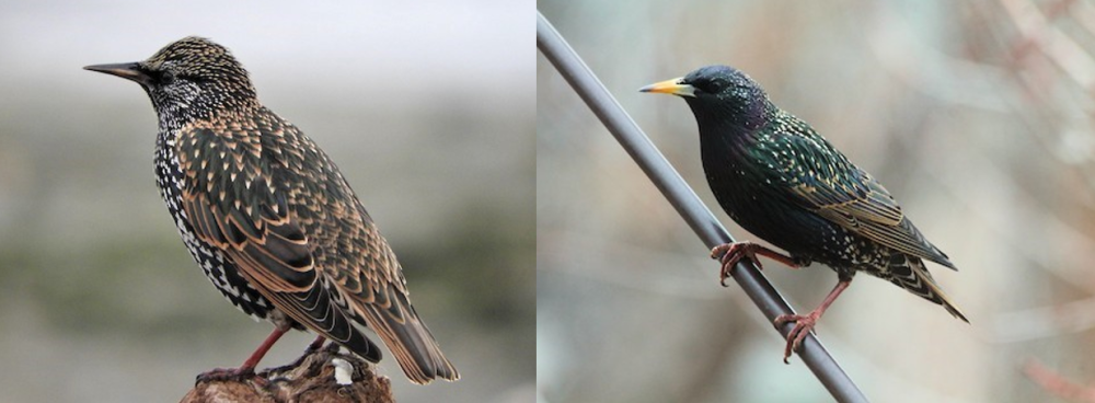
Spreeuw buiten het broedseizoen © Gerald McKeating (links) Mannelijke spreeuw in het broedseizoen © Lorraine Lenning (rechts)
Naast de fysieke transformatie ontdekten onderzoekers nog een ander interessant aspect van deze vogels: hun zanggedrag. Spreeuwen staan bekend om hun vermogen geluiden na te bootsen en hun melodieën te veranderen. De onderzoekers merkten op dat de frequentie en complexiteit van hun liedjes variëren afhankelijk van het geslacht en het seizoen. In het broedseizoen zingen mannelijke spreeuwen intensiever, dit brengt veranderingen in hun hersenstructuur met zich mee.
De reorganisatie van de hersenstructuur wordt neuroplasticiteit genoemd. Dit fenomeen komt voor bij opgroeiende kinderen, alsook na ongevallen of als deel van het normale leerproces bij volwassenen. Bij spreeuwen wordt de neuroplasticiteit gestimuleerd door de hoeveelheid daglicht. Wanneer er meer dan 12 uur licht per dag is, breekt het broedseizoen aan. Met als gevolg de uiterlijke veranderingen, alsook een vergroting van het zangcontrole systeem in de hersenen.
Hersenen in beeld
Onderzoekers hebben inzicht gekregen in de correlatie tussen de seizoensgebonden veranderingen in het zanggedrag van spreeuwen en de structuur van hun hersenen. Via geavanceerde beeldvorming technieken zoals Magnetic Resonance Imaging (MRI) hebben ze de mogelijkheid om niet alleen de hersenstructuur en -volume te observeren, maar ook om te onderzoeken welke delen van de hersenen actief zijn tijdens verschillende taken.
Dit gebeurt door middel van magnetische signalen. Het type weefsel beïnvloedt de magnetische signalen waardoor deze van elkaar kunnen worden onderscheiden. De activiteit kan gemeten worden aan de hand van de doorbloeding van de verschillende delen van de hersenen. Wanneer cellen actiever zijn, hebben ze meer zuurstof nodig, bijgevolg zal er meer bloed naar die regio gestuurd worden. De hoeveelheid zuurstof in de regio bepaalt de sterkte van het signaal.
Resting-state functionele MRI (rs-fMRI) is een methode waarbij hersenactiviteit wordt gemeten terwijl individuen in rust zijn. Dit heeft al geleid tot opmerkelijke inzichten bij zowel mensen als knaagdieren. Met deze techniek kunnen verschillende neurale netwerken aangetoond worden wanneer men in rust is. Deze netwerken kunnen variëren tussen gezonde en zieke individuen, waardoor deze techniek in de toekomst als diagnose tool gebruikt zou kunnen worden voor bepaalde ziekten zoals de ziekte van Alzheimer. Dit rs-fMRI onderzoek is het eerste dat deze techniek toepast in spreeuwen. Hierdoor kunnen de netwerken en hun seizoenale veranderingen in het spreeuwenbrein waargenomen worden.
De veranderingen in zanggedrag en hersenstructuur doen vermoeden dat de seizoenen impact hebben op de functionele aspecten van hun hersenen. Wanneer de structuur van de hersenen verandert heeft dit vaak effect op de functie het brein zoals bijvoorbeeld bij hersentrauma. Dit heeft geleid tot een uitgebreid onderzoeksproject waarbij gekeken wordt of de seizoensgebonden verschillen in zanggedrag kunnen worden vastgesteld aan de hand van rs-fMRI.
Uit de resultaten van het rs-fMRI onderzoek blijkt dat, vooral in mannelijke spreeuwen, het zangcontrole systeem actiever is in het broedseizoen dan daarbuiten. Dit bevestigt de voorgenoemde hypothese dat de functionele aspecten van de hersenen zich ook aanpassen aan de seizoen veranderingen. Naast het zangcontrole systeem, zijn er nog andere, minder onderzochte netwerken die dezelfde trend vertonen, waaruit blijkt dat het spreeuwenbrein over het algemeen actiever is in het broedseizoen.
Dit onderzoek toont aan dat spreeuwen een terugkerende neuroplasticiteit ervaren waardoor de dieren een goed model kunnen zijn voor het ontrafelen van de onderliggende mechanismen van neuroplasticiteit. Dit zou er in de toekomst voor kunnen zorgen dat neuroplasticiteit gemanipuleerd kan worden en een oplossing kunnen bieden voor kinderen met ontwikkelingsstoornissen en blinde/dove personen.
Al met al onthult dit boeiende onderzoek niet alleen de betoverende transformatie van spreeuwen in de loop van de seizoenen, maar werpt het ook nieuw licht op de complexe relatie tussen gedrag, fysiologie en hersenfunctie in het vogelrijk. De veelzijdige spreeuw blijft ons verbazen, zowel met zijn kleurrijk uiterlijk als met de intrigerende geheimen die in hun zingende hersenen verborgen liggen.
Bibliografie
Theofanopoulou, C., Boeckx, C. & Jarvis, E. D. A hypothesis on a role of oxytocin in the social mechanisms of speech and vocal learning. Proc Biol Sci 284, doi:10.1098/rspb.2017.0988 (2017). 2 De Groof, G. & Van der Linden, A. Love songs, bird brains and diffusion tensor imaging. NMR Biomed 23, 873-883, doi:10.1002/nbm.1551 (2010). 3 Orije, J. & Van der Linden, A. A brain for all seasons: An in vivo MRI perspective on songbirds. J Exp Zool A Ecol Integr Physiol 337, 967-984, doi:10.1002/jez.2650 (2022). 4 L, L. L. Birdsong and Human Speech and Language: What the Zebra Finch Uses, Loses, and Regains. (2012). 5 Panaitof, S. C. A songbird animal model for dissecting the genetic bases of autism spectrum disorder. Dis Markers 33, 241-249, doi:10.3233/DMA-2012-0918 (2012). 6 Layden, E. A., Schertz, K. E., London, S. E. & Berman, M. G. Interhemispheric functional connectivity in the zebra finch brain, absent the corpus callosum in normal ontogeny. Neuroimage 195, 113-127, doi:10.1016/j.neuroimage.2019.03.064 (2019). 7 Kubikova, L. & Kostal, L. Dopaminergic system in birdsong learning and maintenance. J Chem Neuroanat 39, 112-123, doi:10.1016/j.jchemneu.2009.10.004 (2010). 8 Moorman, S. et al. Human-like brain hemispheric dominance in birdsong learning. Proc Natl Acad Sci U S A 109, 12782-12787, doi:10.1073/pnas.1207207109 (2012). 9 Stinnett, T. J., Reddy, V. & Zabel, M. K. in StatPearls (2022). 10 Binder, J. R. Current Controversies on Wernicke's Area and its Role in Language. Curr Neurol Neurosci Rep 17, 58, doi:10.1007/s11910-017-0764-8 (2017). 11 Sherman, S. M. Thalamic relay functions. Prog Brain Res 134, 51-69, doi:10.1016/s0079- 6123(01)34005-0 (2001). 12 Jarvis, E. D. Learned birdsong and the neurobiology of human language. Ann N Y Acad Sci 1016, 749- 777, doi:10.1196/annals.1298.038 (2004). 13 Pinaud, R. & Terleph, T. A. A songbird forebrain area potentially involved in auditory discrimination and memory formation. J Biosci 33, 145-155, doi:10.1007/s12038-008-0030-y (2008). 14 Szaflarski, J. P. et al. Language lateralization in left-handed and ambidextrous people: fMRI data. Neurology 59, 238-244, doi:10.1212/wnl.59.2.238 (2002). 15 Hamaide, J. et al. In vivo assessment of the neural substrate linked with vocal imitation accuracy. Elife 9, doi:10.7554/eLife.49941 (2020). 16 Jonckers, E., Gunturkun, O., De Groof, G., Van der Linden, A. & Bingman, V. P. Network structure of functional hippocampal lateralization in birds. Hippocampus 25, 1418-1428, doi:10.1002/hipo.22462 (2015). 17 Riters, L. V., Eens, M., Pinxten, R. & Ball, G. F. Seasonal changes in the densities of alpha(2) noradrenergic receptors are inversely related to changes in testosterone and the volumes of song control nuclei in male European starlings. J Comp Neurol 444, 63-74, doi:10.1002/cne.10131 (2002). 18 Pavlova, D. Z., Pinxten, R. & Eens, M. Seasonal singing patterns and individual consistency in song activity in female European starlings (Sturnus vulgaris). Behaviour 144, 663-680, doi:Doi 10.1163/156853907781347835 (2007). 19 Pavlova, D., Pinxten, R. & Eens, M. Female song in European Starlings: Sex differences, complexity, and composition. Condor 107, 559-569, doi:Doi 10.1650/0010-5422(2005)107[0559:Fsiess]2.0.Co;2 (2005). 20 Chaiken, M. L., Böhner Jörg. SONG LEARNING AFTER ISOLATION IN THE OPENENDED LEARNER THE EUROPEAN STARLING: DISSOCIATION OF IMITATION AND SYNTACTIC DEVELOPMENT. (2007). 21 Lehongre, K., P. Lenouvel, T. Draganoiu, C. Del Negro. Long-term effect of isolation rearing conditions on songs of an ‘open-ended’ song learner species, the Canary. (2006). 22 Williams, H. Birdsong and singing behavior. Ann N Y Acad Sci 1016, 1-30, doi:10.1196/annals.1298.029 (2004). 23 Williams, H. Birdsong and singing behavior. Ann Ny Acad Sci 1016, 1-30, doi:10.1196/annals.1298.029 (2004). Master thesis | Silke Lemmens | 39 24 Nottebohm, F., Nottebohm, M. E. & Crane, L. Developmental and seasonal changes in canary song and their relation to changes in the anatomy of song-control nuclei. Behav Neural Biol 46, 445-471, doi:10.1016/s0163-1047(86)90485-1 (1986). 25 Eens, M., Pinxten, R. & Verheyen, R. F. Song Learning in Captive European Starlings, Sturnus-Vulgaris. Animal Behaviour 44, 1131-1143, doi:Doi 10.1016/S0003-3472(05)80325-2 (1992). 26 Hindmarsh, A. M. Vocal Mimicry in Starlings. Behaviour 90, 302-324, doi:Doi 10.1163/156853984x00182 (1984). 27 George, I. & Cousillas, H. How social experience shapes song representation in the brain of starlings. J Physiol Paris 107, 170-177, doi:10.1016/j.jphysparis.2012.12.002 (2013). 28 Poirier, C. et al. Direct social contacts override auditory information in the song-learning process in starlings (Sturnus vulgaris). J Comp Psychol 118, 179-193, doi:10.1037/0735-7036.118.2.179 (2004). 29 Nicholls, T. J., Goldsmith, A. R. & Dawson, A. Photorefractoriness in birds and comparison with mammals. Physiol Rev 68, 133-176, doi:10.1152/physrev.1988.68.1.133 (1988). 30 Gwinner, E. Circannual rhythms in birds. Curr Opin Neurobiol 13, 770-778, doi:10.1016/j.conb.2003.10.010 (2003). 31 Magee, J. C. & Grienberger, C. Synaptic Plasticity Forms and Functions. Annu Rev Neurosci 43, 95-117, doi:10.1146/annurev-neuro-090919-022842 (2020). 32 Patton, M. H., Blundon, J. A. & Zakharenko, S. S. Rejuvenation of plasticity in the brain: opening the critical period. Curr Opin Neurobiol 54, 83-89, doi:10.1016/j.conb.2018.09.003 (2019). 33 Cisneros-Franco, J. M., Voss, P., Thomas, M. E. & de Villers-Sidani, E. Critical periods of brain development. Handb Clin Neurol 173, 75-88, doi:10.1016/B978-0-444-64150-2.00009-5 (2020). 34 Knudsen, E. I. Sensitive periods in the development of the brain and behavior. J Cogn Neurosci 16, 1412-1425, doi:10.1162/0898929042304796 (2004). 35 Zhou, Q., Tao, H. W. & Poo, M. M. Reversal and stabilization of synaptic modifications in a developing visual system. Science 300, 1953-1957, doi:10.1126/science.1082212 (2003). 36 Friedmann, N. & Rusou, D. Critical period for first language: the crucial role of language input during the first year of life. Curr Opin Neurobiol 35, 27-34, doi:10.1016/j.conb.2015.06.003 (2015). 37 Fitzpatrick, E. Neurocognitive development in congenitally deaf children. Handb Clin Neurol 129, 335- 356, doi:10.1016/B978-0-444-62630-1.00019-6 (2015). 38 Kral, A. & Sharma, A. Developmental neuroplasticity after cochlear implantation. Trends in Neurosciences 35, 111-122, doi:10.1016/j.tins.2011.09.004 (2012). 39 Holmes, J. M. & Levi, D. M. Treatment of amblyopia as a function of age. Vis Neurosci 35, E015, doi:10.1017/S0952523817000220 (2018). 40 Ismail, F. Y., Fatemi, A. & Johnston, M. V. Cerebral plasticity: Windows of opportunity in the developing brain. Eur J Paediatr Neurol 21, 23-48, doi:10.1016/j.ejpn.2016.07.007 (2017). 41 Roder, B. & Kekunnaya, R. Visual experience dependent plasticity in humans. Curr Opin Neurobiol 67, 155-162, doi:10.1016/j.conb.2020.11.011 (2021). 42 Hamaide, J., De Groof, G. & Van der Linden, A. Neuroplasticity and MRI: A perfect match. Neuroimage 131, 13-28, doi:10.1016/j.neuroimage.2015.08.005 (2016). 43 De Groof, G. et al. Structural changes between seasons in the songbird auditory forebrain. J Neurosci 29, 13557-13565, doi:10.1523/JNEUROSCI.1788-09.2009 (2009). 44 Villemagne, V. L. et al. Molecular Imaging Approaches in Dementia. Radiology 298, 517-530, doi:10.1148/radiol.2020200028 (2021). 45 Dorbala, S. et al. Single Photon Emission Computed Tomography (SPECT) Myocardial Perfusion Imaging Guidelines: Instrumentation, Acquisition, Processing, and Interpretation. J Nucl Cardiol 25, 1784-1846, doi:10.1007/s12350-018-1283-y (2018). 46 Flohr, T. et al. Photon-counting CT review. Phys Med 79, 126-136, doi:10.1016/j.ejmp.2020.10.030 (2020). 47 Fonti, R., Conson, M. & Del Vecchio, S. PET/CT in radiation oncology. Semin Oncol 46, 202-209, doi:10.1053/j.seminoncol.2019.07.001 (2019). 48 Zeng, D. et al. Basis and current state of computed tomography perfusion imaging: a review. Phys Med Biol 67, doi:10.1088/1361-6560/ac8717 (2022). 49 Yousaf, T., Dervenoulas, G. & Politis, M. Advances in MRI Methodology. Int Rev Neurobiol 141, 31-76, doi:10.1016/bs.irn.2018.08.008 (2018). 50 Grover, V. P. et al. Magnetic Resonance Imaging: Principles and Techniques: Lessons for Clinicians. J Clin Exp Hepatol 5, 246-255, doi:10.1016/j.jceh.2015.08.001 (2015). Master thesis | Silke Lemmens | 40 51 Weiger, M. & Pruessmann, K. P. Short-T(2) MRI: Principles and recent advances. Prog Nucl Magn Reson Spectrosc 114-115, 237-270, doi:10.1016/j.pnmrs.2019.07.001 (2019). 52 Gauthier, C. J. & Fan, A. P. BOLD signal physiology: Models and applications. Neuroimage 187, 116- 127, doi:10.1016/j.neuroimage.2018.03.018 (2019). 53 Logothetis, N. K. What we can do and what we cannot do with fMRI. Nature 453, 869-878, doi:10.1038/nature06976 (2008). 54 Rangaprakash, D., Tadayonnejad, R., Deshpande, G., O'Neill, J. & Feusner, J. D. FMRI hemodynamic response function (HRF) as a novel marker of brain function: applications for understanding obsessivecompulsive disorder pathology and treatment response. Brain Imaging Behav 15, 1622-1640, doi:10.1007/s11682-020-00358-8 (2021). 55 Rangaprakash, D., Wu, G. R., Marinazzo, D., Hu, X. & Deshpande, G. Hemodynamic response function (HRF) variability confounds rng-state fMRI functional connectivity. Magn Reson Med 80, 1697-1713, doi:10.1002/mrm.27146 (2018). 56 Jackson, G. D., Badawy, R. & Gotman, J. Functional magnetic resonance imaging: focus localization. Handb Clin Neurol 107, 369-385, doi:10.1016/B978-0-444-52898-8.00023-9 (2012). 57 Dupuy, S. L. et al. MRI detection of hypointense brain lesions in patients with multiple sclerosis: T1 spin-echo vs. gradient-echo. Eur J Radiol 84, 1564-1568, doi:10.1016/j.ejrad.2015.05.004 (2015). 58 Poustchi-Amin, M., Mirowitz, S. A., Brown, J. J., McKinstry, R. C. & Li, T. Principles and applications of echo-planar imaging: a review for the general radiologist. Radiographics 21, 767-779, doi:10.1148/radiographics.21.3.g01ma23767 (2001). 59 Behroozi, M. et al. In vivo measurement of T(1) and T(2) relaxation times in awake pigeon and rat brains at 7T. Magn Reson Med 79, 1090-1100, doi:10.1002/mrm.26722 (2018). 60 De Groof, G. et al. Functional MRI and functional connectivity of the visual system of awake pigeons. Behav Brain Res 239, 43-50, doi:10.1016/j.bbr.2012.10.044 (2013). 61 Van Ruijssevelt, L., Hamaide, J., Van Gurp, M. T., Verhoye, M. & Van der Linden, A. Auditory evoked BOLD responses in awake compared to lightly anaesthetized zebra finches. Sci Rep 7, 13563, doi:10.1038/s41598-017-13014-x (2017). 62 Steiner, A. R., Rousseau-Blass, F., Schroeter, A., Hartnack, S. & Bettschart-Wolfensberger, R. Systematic Review: Anesthetic Protocols and Management as Confounders in Rodent Blood Oxygen Level Dependent Functional Magnetic Resonance Imaging (BOLD fMRI)-Part B: Effects of Anesthetic Agents, Doses and Timing. Animals (Basel) 11, doi:10.3390/ani11010199 (2021). 63 Kelly, C. & Castellanos, F. X. Strengthening connections: functional connectivity and brain plasticity. Neuropsychol Rev 24, 63-76, doi:10.1007/s11065-014-9252-y (2014). 64 Fox, M. D. & Greicius, M. Clinical applications of resting state functional connectivity. Front Syst Neurosci 4, 19, doi:10.3389/fnsys.2010.00019 (2010). 65 Raichle, M. E. & Mintun, M. A. Brain work and brain imaging. Annu Rev Neurosci 29, 449-476, doi:10.1146/annurev.neuro.29.051605.112819 (2006). 66 Raichle, M. E. Neuroscience. The brain's dark energy. Science 314, 1249-1250 (2006). 67 Jahanian, H. et al. Advantages of short repetition time resting-state functional MRI enabled by simultaneous multi-slice imaging. J Neurosci Methods 311, 122-132, doi:10.1016/j.jneumeth.2018.09.033 (2019). 68 Grandjean, J. et al. Psilocybin exerts distinct effects on resting state networks associated with serotonin and dopamine in mice. Neuroimage 225, doi:ARTN 117456 10.1016/j.neuroimage.2020.117456 (2021). 69 Vasilkovska, T. et al. Resting-state fMRI reveals longitudinal alterations in brain network connectivity in the zQ175DN mouse model of Huntington's disease. Neurobiol Dis 181, 106095, doi:10.1016/j.nbd.2023.106095 (2023). 70 Liang, Z., Liu, X. & Zhang, N. Dynamic resting state functional connectivity in awake and anesthetized rodents. Neuroimage 104, 89-99, doi:10.1016/j.neuroimage.2014.10.013 (2015). 71 Behroozi, M., Strockens, F., Helluy, X., Stacho, M. & Gunturkun, O. Functional Connectivity Pattern of the Internal Hippocampal Network in Awake Pigeons: A Resting-State fMRI Study. Brain Behav Evol 90, 62-72, doi:10.1159/000475591 (2017). 72 Baggio, H. C., Segura, B. & Junque, C. Resting-state functional brain networks in Parkinson's disease. CNS Neurosci Ther 21, 793-801, doi:10.1111/cns.12417 (2015). 73 Xing, C. et al. Abnormal Static and Dynamic Functional Network Connectivity in Patients With Presbycusis. Front Aging Neurosci 13, 774901, doi:10.3389/fnagi.2021.774901 (2021). Master thesis | Silke Lemmens | 41 74 Stacho, M. et al. A cortex-like canonical circuit in the avian forebrain. Science 369, doi:10.1126/science.abc5534 (2020). 75 Marzluff, J. M., Miyaoka, R., Minoshima, S. & Cross, D. J. Brain imaging reveals neuronal circuitry underlying the crow's perception of human faces. Proc Natl Acad Sci U S A 109, 15912-15917, doi:10.1073/pnas.1206109109 (2012). 76 Gunturkun, O. & Bugnyar, T. Cognition without Cortex. Trends Cogn Sci 20, 291-303, doi:10.1016/j.tics.2016.02.001 (2016). 77 Layden, E. A., Li, H. B., Schertz, K. E., Berman, M. G. & London, S. E. Experience selectively alters functional connectivity within a neural network to predict learned behavior in juvenile songbirds. Neuroimage 222, doi:ARTN 117218 10.1016/j.neuroimage.2020.117218 (2020). 78 De Groof, G. et al. Functional MRI and functional connectivity of the visual system of awake pigeons. Behavioural Brain Research 239, 43-50, doi:10.1016/j.bbr.2012.10.044 (2013). 79 Orije, J. et al. In vivo online monitoring of testosterone-induced neuroplasticity in a female songbird. Horm Behav 118, 104639, doi:10.1016/j.yhbeh.2019.104639 (2020). 80 Tong, Y., Hocke, L. M. & Frederick, B. B. Low Frequency Systemic Hemodynamic "Noise" in Resting State BOLD fMRI: Characteristics, Causes, Implications, Mitigation Strategies, and Applications. Front Neurosci 13, 787, doi:10.3389/fnins.2019.00787 (2019). 81 Wu, Z., Huang, N. E., Long, S. R. & Peng, C. K. On the trend, detrending, and variability of nonlinear and nonstationary time series. Proc Natl Acad Sci U S A 104, 14889-14894, doi:10.1073/pnas.0701020104 (2007). 82 Lee, D., Jang, C. & Park, H. J. Multivariate detrending of fMRI signal drifts for real-time multiclass pattern classification. Neuroimage 108, 203-213, doi:10.1016/j.neuroimage.2014.12.062 (2015). 83 Li, J. et al. Global signal regression strengthens association between resting-state functional connectivity and behavior. Neuroimage 196, 126-141, doi:10.1016/j.neuroimage.2019.04.016 (2019). 84 Chang, C. et al. Tracking brain arousal fluctuations with fMRI. Proc Natl Acad Sci U S A 113, 4518-4523, doi:10.1073/pnas.1520613113 (2016). 85 Calhoun, V. D., Adali, T., Pearlson, G. D. & Pekar, J. J. A method for making group inferences from functional MRI data using independent component analysis. Hum Brain Mapp 14, 140-151, doi:10.1002/hbm.1048 (2001). 86 De Groof, G. et al. A three-dimensional digital atlas of the starling brain. Brain Struct Funct 221, 1899- 1909, doi:10.1007/s00429-015-1011-1 (2016). 87 Zhao, W. et al. Consistency of independent component analysis for FMRI. J Neurosci Methods 351, 109013, doi:10.1016/j.jneumeth.2020.109013 (2021). 88 Jarvis, E. D. et al. Avian brains and a new understanding of vertebrate brain evolution. Nature Reviews Neuroscience 6, 151-159, doi:10.1038/nrn1606 (2005). 89 Watanabe, S. Effects of ectostriatal lesions on discriminations of conspecific, species and familiar objects in pigeons. Behav Brain Res 81, 183-188, doi:10.1016/s0166-4328(96)89079-6 (1996). 90 Watanabe, S. Effect of lesions in the ectostriatum and Wulst on species and individual discrimination in pigeons. Behav Brain Res 49, 197-203, doi:10.1016/s0166-4328(05)80165-2 (1992). 91 Bennett, A. T., Cuthill, I. C., Partridge, J. C. & Lunau, K. Ultraviolet plumage colors predict mate preferences in starlings. Proc Natl Acad Sci U S A 94, 8618-8621, doi:10.1073/pnas.94.16.8618 (1997). 92 Oberlander, J. G., Schlinger, B. A., Clayton, N. S. & Saldanha, C. J. Neural aromatization accelerates the acquisition of spatial memory via an influence on the songbird hippocampus. Horm Behav 45, 250-258, doi:10.1016/j.yhbeh.2003.12.003 (2004). 93 Preston, A. R. & Eichenbaum, H. Interplay of hippocampus and prefrontal cortex in memory. Curr Biol 23, R764-773, doi:10.1016/j.cub.2013.05.041 (2013). 94 Navarro, C., Perez-Contreras, T., Aviles, J. M., Mcgraw, K. J. & Soler, J. J. Beak colour reflects circulating carotenoid and vitamin A levels in spotless starlings (Sturnus unicolor). Behavioral Ecology and Sociobiology 64, 1057-1067, doi:10.1007/s00265-010-0920-5 (2010). 95 Gregory F. Ball, J. C. W. Changes in Plasma Levels of Luteinizing Hormone and Sex Steroid Hormones in Relation to Multiple-Broodedness and Nest-Site Density in Male Starlings. Physiological Zoology 60. 96 Kim, Y. H. & Arnold, A. P. Distribution and onset of retinaldehyde dehydrogenase (zRalDH) expression in zebra finch brain: lack of sex difference in HVC and RA at early posthatch ages. J Neurobiol 65, 260- 268, doi:10.1002/neu.20192 (2005). Master thesis | Silke Lemmens | 42 97 Orije, J. et al. Uncovering a 'sensitive window' of multisensory and motor neuroplasticity in the cerebrum and cerebellum of male and female starlings. Elife 10, doi:10.7554/eLife.66777 (2021). 98 Zeng, S. J., Song, K., Xu, N., Zhang, X. W. & Zuo, M. X. Sex difference in cellular proliferation within the telencephalic ventricle zone of Bengalese finch. Neurosci Res 58, 207-214, doi:10.1016/j.neures.2007.02.001 (2007). 99 Mello, C. V., Kaser, T., Buckner, A. A., Wirthlin, M. & Lovell, P. V. Molecular architecture of the zebra finch arcopallium. J Comp Neurol 527, 2512-2556, doi:10.1002/cne.24688 (2019). 100 Beudel, M., Macerollo, A., Brown, M. J. N. & Chen, R. Editorial: The Role of the Basal Ganglia in Somatosensory-Motor Interactions: Evidence From Neurophysiology and Behavior. Front Hum Neurosci 13, 451, doi:10.3389/fnhum.2019.00451 (2019). 101 Kelley, D. B. & Nottebohm, F. Projections of a telencephalic auditory nucleus-field L-in the canary. J Comp Neurol 183, 455-469, doi:10.1002/cne.901830302 (1979). 102 Zaretsky, M. D. A new auditory area of the songbird forebrain: a connection between auditory and song control centers. Exp Brain Res 32, 267-273, doi:10.1007/BF00239731 (1978). 103 De Groof, G., Poirier, C., George, I., Hausberger, M. & Van der Linden, A. Functional changes between seasons in the male songbird auditory forebrain. Front Behav Neurosci 7, 196, doi:10.3389/fnbeh.2013.00196 (2013). 104 Adhikari, M. H., Belloy, M. E., Van der Linden, A., Keliris, G. A. & Verhoye, M. Resting-State Coactivation Patterns as Promising Candidates for Prediction of Alzheimer's Disease in Aged Mice. Front Neural Circuits 14, 612529, doi:10.3389/fncir.2020.612529 (2020). 105 Belloy, M. E. et al. Dynamic resting state fMRI analysis in mice reveals a set of Quasi-Periodic Patterns and illustrates their relationship with the global signal. Neuroimage 180, 463-484, doi:10.1016/j.neuroimage.2018.01.075 (2018).