
Een firewall in je hersenen als bescherming tegen neurologische aandoeningen?
Net zoals een firewall je computer beschermt, zijn onze hersenen uitgerust met beschermende barrières die schadelijke stoffen en cellen van buitenaf afweren. Deze barrières kunnen verstoord raken bij aandoeningen zoals multiple sclerose, waarbij schadelijke cellen het centrale zenuwstelsel kunnen binnendringen en je eigen zenuwcellen kunnen aanvallen. Dergelijke verstoringen komen ook voor bij beroertes, hersentrauma en de ziekte van Alzheimer. In mijn thesis-onderzoek heb ik aangetoond dat IL-34, een cytokine geproduceerd door regulatorische T-cellen, een essentiële rol speelt in het versterken van deze hersenbarrières. Deze ontdekking opent veelbelovende perspectieven voor de ontwikkeling van nieuwe therapieën voor verschillende neurologische aandoeningen. Duik dieper in dit intrigerende proces terwijl je verder leest.
De hersenen als computer van je lichaam
Stel je de hersenen voor als de ultieme controlekamer van ons lichaam, een ingenieuze computer die zonder pauze draait. Vergelijkbaar met de nullen en enen van een computer, transformeren je hersenen signalen in acties, gedachten en meer. Bij het lezen van dit artikel begint het allemaal met je zintuigen, vooral je ogen, die fungeren als camera's die de tekst vastleggen en vertalen naar visuele signalen. Deze signalen worden vervolgens naar specifieke hersengebieden gestuurd voor decodering. Je hersenen herkennen letters, smeden ze tot woorden en vormen betekenisvolle zinnen. Terwijl je blijft lezen, proberen je hersenen de diepere betekenis van de tekst te achterhalen door herinneringen te raadplegen en verbanden te leggen. Maar er schuilt meer complexiteit in dit proces dan je op het eerste gezicht zou denken. Het lezen van dit artikel vraagt ook om concentratie en aandacht. Je brein moet gefocust blijven op de tekst en proberen de afleidingen buiten te sluiten. Daarbovenop zijn er ook nog motorische signalen die een rol spelen, want die sturen je ogen behendig over de pagina. En eerlijk gezegd, terwijl je dit stukje leest, komen er waarschijnlijk allerlei gedachten en meningen in je op.
Hersenbarrières entered the chat
Dit hele gebeuren wordt nog interessanter wanneer we bedenken dat al deze processen en signalen met ongelooflijke precisie samenkomen op verschillende locaties in onze hersenen, waar ze worden verwerkt. Om dit alles soepel te laten verlopen, moeten onze hersenen een gecontroleerde omgeving handhaven, waar externe invloeden geen kans krijgen om chaos te veroorzaken. Hier komen de hersenbarrières in beeld - een systeem dat we kunnen vergelijken met de firewall van een computer. Net zoals een firewall je computer beschermt tegen virussen en ongewenste indringers van buitenaf, fungeren de hersenbarrières als een schild dat onze hersenen beschermt tegen de voortdurende veranderingen in ons lichaam.
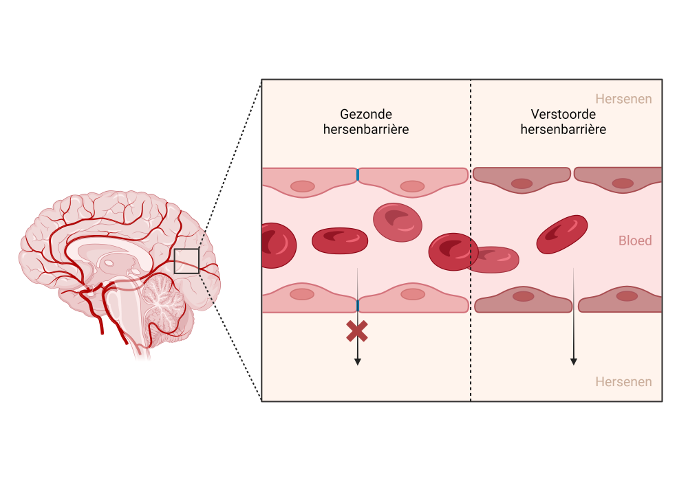
Een van de belangrijkste barrières is de bloed-hersenbarrière, die zich bevindt bij de bloedvaten in onze hersenen. De cellen van deze bloedvaten zijn nauw met elkaar verbonden, waardoor ze een muur vormen tussen het bloed en de hersenen. Deze barrière voorkomt dat schadelijke stoffen, zoals virussen en gifstoffen, onze hersenen binnendringen en zorgt ervoor dat alleen nuttige stoffen, zoals zuurstof en voedingsstoffen, toegang krijgen tot onze hersenen.
Gehackte hersenen
Net zoals een gehackte firewall de kwetsbaarheid van een computer vergroot en crashes kan veroorzaken, kunnen verstoringen in de hersenbarrières bijdragen aan de ontwikkeling van neurologische aandoeningen. Dit soort 'neurologische crashes' komt voor bij ziektes zoals multiple sclerose. Bij deze aandoening verzwakken de hersenbarrières, waardoor immuuncellen toegang krijgen tot het centrale zenuwstelsel. Eenmaal binnen kunnen deze cellen de beschermende laag van zenuwcellen, bekend als myeline, aanvallen, wat leidt tot communicatieproblemen tussen de hersenen en de rest van het lichaam, resulterend in motorische problemen.
Het herstellen van de integriteit en functie van de hersenbarrières in aandoeningen zoals multiple sclerose kan van cruciaal belang zijn om ontstekingen in de hersenen te verminderen. Dit opent de deur naar nieuwe therapieën die niet alleen gericht zijn op het behandelen van symptomen, maar ook op het aanpakken van de onderliggende oorzaken. Kortom, het herstellen van de "firewall" van onze hersenen kan bijdragen aan het beschermen en behouden van een gezond zenuwstelsel.
Regulatorische T cellen to the rescue
In mijn thesis-onderzoek ben ik dieper ingegaan op de invloed van het immuunsysteem op de integriteit en sterkte van de hersenbarrières. We wilden ontdekken welke immuuncellen een positieve of negatieve invloed hebben op deze barrières, met de hoop nieuwe behandelingen te ontwikkelen voor neurologische aandoeningen. Tijdens ons onderzoek ontdekten we dat het ontbreken van T-cellen resulteerde in verzwakte barrières, wat we aantoonden door een fluorescerende stof in het bloed van muizen te injecteren en te meten hoeveel ervan in de hersenen lekte. Door in te zoomen op deze T-cellen, identificeerden we dat een specifieke groep genaamd 'regulatorische T-cellen' of Tregs, verantwoordelijk waren voor het dit effect. Tregs spelen een essentiële rol in het stoppen van een overactief immuunsysteem om schade aan ons eigen lichaam te voorkomen, en we toonden dus ook aan dat ze een positief effect hebben op het versterken van de hersenbarrières.
IL-34 als boodschapper tussen Tregs en hersenbarrières
In de volgende fase van ons onderzoek wilden we nog dieper graven om te begrijpen hoe Tregs de integriteit van de hersenbarrières behouden. Om dit te achterhalen, zijn we gaan kijken naar de cytokines die door Tregs worden geproduceerd. Cytokines dienen als boodschappers van het immuunsysteem en spelen een cruciale rol bij de communicatie tussen verschillende cellen. We ontdekten dat het cytokine IL-34 een belangrijke factor is voor het versterken van de barrières. Interessant genoeg was het feit dat muismodellen van multiple sclerose en de ziekte van Alzheimer een verminderde concentratie IL-34 in hun bloed vertoonden. Wanneer deze muizen behandeld werden met IL-34 via intraveneuze toediening, zagen we dat de hersenbarrières daadwerkelijk sterker werden. Deze bevindingen dragen bij aan een beter begrip van de complexe relatie tussen Tregs en de functie van de hersenbarrière, wat nieuwe perspectieven opent voor therapeutische behandelingen bij verschillende neurologische aandoeningen.
Bibliografie
2022 Alzheimer’s disease facts and figures (2022) Alzheimer’s & Dementia 18: 700–789
Abbott NJ, Rönnbäck L & Hansson E (2006) Astrocyte–endothelial interactions at the blood–brain barrier. Nat Rev Neurosci 7: 41–53
Alcolado R, Weller RO, Parrish EP & Garrod D (1988) The Cranial Arachnoid and Pia Mater in Man: Anatomical and Ultrastructural Observations. Neuropathology and Applied Neurobiology 14: 1–17
Alves S, Churlaud G, Audrain M, Michaelsen-Preusse K, Fol R, Souchet B, Braudeau J, Korte M, Klatzmann D & Cartier N (2017) Interleukin-2 improves amyloid pathology, synaptic failure and memory in Alzheimer’s disease mice. Brain : a journal of neurology 140: 826–842
Anderson CM & Nedergaard M (2003) Astrocyte-mediated control of cerebral microcirculation. Trends in Neurosciences 26: 340–344
Armulik A, Genové G & Betsholtz C (2011) Pericytes: Developmental, Physiological, and Pathological Perspectives, Problems, and Promises. Developmental Cell 21: 193–215
Armulik A, Genové G, Mäe M, Nisancioglu MH, Wallgard E, Niaudet C, He L, Norlin J, Lindblom P, Strittmatter K, et al (2010) Pericytes regulate the blood–brain barrier. Nature 468: 557–561
Ascherio A (2013) Environmental factors in multiple sclerosis. Expert Rev Neurother 13: 3–9
Baecher-Allan C, Kaskow BJ & Weiner HL (2018) Multiple Sclerosis: Mechanisms and Immunotherapy. Neuron 97: 742–768
Baek H, Ye M, Kang G-H, Lee C, Lee G, Choi D Bin, Jung J, Kim H, Lee S, Kim JS, et al (2016) Neuroprotective effects of CD4+CD25+Foxp3+ regulatory T cells in a 3xTg-AD Alzheimer’s disease model. Oncotarget 7: 69347–69357
Balasa R, Barcutean L, Mosora O & Manu D (2021) Reviewing the Significance of Blood–Brain Barrier Disruption in Multiple Sclerosis Pathology and Treatment. Int J Mol Sci 22: 8370
Balusu S, Brkic M, Libert C & Vandenbroucke RE (2016) The choroid plexus-cerebrospinal fluid interface in Alzheimer’s disease: more than just a barrier. Neural Regen Res 11: 534–537
Banks WA, Gray AM, Erickson MA, Salameh TS, Damodarasamy M, Sheibani N, Meabon JS, Wing EE, Morofuji Y, Cook DG, et al (2015) Lipopolysaccharide-induced blood-brain barrier disruption: roles of cyclooxygenase, oxidative stress, neuroinflammation, and elements of the neurovascular unit. J Neuroinflammation 12: 223
Barbier P, Zejneli O, Martinho M, Lasorsa A, Belle V, Smet-Nocca C, Tsvetkov PO, Devred F & Landrieu I (2019) Role of Tau as a Microtubule-Associated Protein: Structural and Functional Aspects. Frontiers in Aging Neuroscience 11
Barnett MH & Prineas JW (2004) Relapsing and remitting multiple sclerosis: Pathology of the newly forming lesion. Annals of Neurology 55: 458–468
Bartholomäus I, Kawakami N, Odoardi F, Schläger C, Miljkovic D, Ellwart JW, Klinkert WEF, Flügel-Koch C, Issekutz TB, Wekerle H, et al (2009) Effector T cell interactions with meningeal vascular structures in nascent autoimmune CNS lesions. Nature 462: 94–98
Baughman EJ, Mendoza JP, Ortega SB, Ayers CL, Greenberg BM, Frohman EM & Karandikar NJ (2011) Neuroantigen-specific CD8+ regulatory T-cell function is deficient during acute exacerbation of multiple sclerosis. Journal of Autoimmunity 36: 115–124
Begley DJ & Brightman MW (2003) Structural and functional aspects of the blood-brain barrier. In Peptide Transport and Delivery into the Central Nervous System, Prokai L & Prokai-Tatrai K (eds) pp 39–78. Basel: Birkhäuser
Bernfield M, Kokenyesi R, Kato M, Hinkes MT, Spring J, Gallo RL & Lose EJ (1992) Biology of the syndecans: a family of transmembrane heparan sulfate proteoglycans. Annu Rev Cell Biol 8: 365–393
Bézie S, Picarda E, Ossart J, Tesson L, Usal C, Renaudin K, Anegon I & Guillonneau C (2015) IL-34 is a Treg-specific cytokine and mediates transplant tolerance. J Clin Invest 125: 3952–3964
Bis JC, Jian X, Kunkle BW, Chen Y, Hamilton-Nelson KL, Bush WS, Salerno WJ, Lancour D, Ma Y, Renton AE, et al (2020) Whole exome sequencing study identifies novel rare and common Alzheimer’s-Associated variants involved in immune response and transcriptional regulation. Molecular Psychiatry 25: 1859–1875
Braak H & Braak E (1995) Staging of alzheimer’s disease-related neurofibrillary changes. Neurobiology of Aging 16: 271–278
Brandstadter R & Katz Sand I (2017) The use of natalizumab for multiple sclerosis. Neuropsychiatr Dis Treat 13: 1691–1702
Brioschi S, Wang W-L, Peng V, Wang M, Shchukina I, Greenberg ZJ, Bando JK, Jaeger N, Czepielewski RS, Swain A, et al (2021) Heterogeneity of meningeal B cells reveals a lymphopoietic niche at the CNS borders. Science 373: eabf9277
Brown LS, Foster CG, Courtney J-M, King NE, Howells DW & Sutherland BA (2019) Pericytes and Neurovascular Function in the Healthy and Diseased Brain. Frontiers in Cellular Neuroscience 13
Brunnström HR & Englund EM (2009) Cause of death in patients with dementia disorders. Eur J Neurol 16: 488–492
Bryson KJ & Lynch MA (2016) Linking T cells to Alzheimer’s disease: from neurodegeneration to neurorepair. Current Opinion in Pharmacology 26: 67–73
Burns A & Iliffe S (2009) Alzheimer’s disease. BMJ 338
Cai Z, Qiao P-F, Wan C-Q, Cai M, Zhou N-K & Li Q (2018) Role of Blood-Brain Barrier in Alzheimer’s Disease. J Alzheimers Dis 63: 1223–1234
Charabati M, Wheeler MA, Weiner HL & Quintana FJ (2023) Multiple sclerosis: Neuroimmune crosstalk and therapeutic targeting. Cell 186: 1309–1327
Cheng X, Yang Y-L, Yang H, Wang Y-H & Du G-H (2018) Kaempferol alleviates LPS-induced neuroinflammation and BBB dysfunction in mice via inhibiting HMGB1 release and down-regulating TLR4/MyD88 pathway. International Immunopharmacology 56: 29–35
Chihara T, Suzu S, Hassan R, Chutiwitoonchai N, Hiyoshi M, Motoyoshi K, Kimura F & Okada S (2010) IL-34 and M-CSF share the receptor Fms but are not identical in biological activity and signal activation. Cell Death Differ 17: 1917–1927
Ciccocioppo F, Lanuti P, Pierdomenico L, Simeone P, Bologna G, Ercolino E, Buttari F, Fantozzi R, Thomas A, Onofrj M, et al (2019) The Characterization of Regulatory T-Cell Profiles in Alzheimer’s Disease and Multiple Sclerosis. Sci Rep 9: 8788
Compston A & Coles A (2002) Multiple sclerosis. The Lancet 359: 1221–1231
Cousins O, Hodges A, Schubert J, Veronese M, Turkheimer F, Miyan J, Engelhardt B & Roncaroli F (2022) The blood–CSF–brain route of neurological disease: The indirect pathway into the brain. Neuropathology and Applied Neurobiology 48: e12789
Croxford AL, Kurschus FC & Waisman A (2011) Mouse models for multiple sclerosis: Historical facts and future implications. Biochimica et Biophysica Acta (BBA) - Molecular Basis of Disease 1812: 177–183
Cserr HF (1971) Physiology of the choroid plexus. Physiological Reviews 51: 273–311
Cugurra A, Mamuladze T, Rustenhoven J, Dykstra T, Beroshvili G, Greenberg ZJ, Baker W, Papadopoulos Z, Drieu A, Blackburn S, et al (2021) Skull and vertebral bone marrow are myeloid cell reservoirs for the meninges and CNS parenchyma. Science 373: eabf7844
Cummings J, Lee G, Nahed P, Kambar MEZN, Zhong K, Fonseca J & Taghva K (2022) Alzheimer’s disease drug development pipeline: 2022. Alzheimer’s & Dementia: Translational Research & Clinical Interventions 8
Curotto de Lafaille MA, Kutchukhidze N, Shen S, Ding Y, Yee H & Lafaille JJ (2008) Adaptive Foxp3+ Regulatory T Cell-Dependent and -Independent Control of Allergic Inflammation. Immunity 29: 114–126
Cushman J, Lo J, Huang Z, Wasserfall C & Petitto JM (2003) Neurobehavioral Changes Resulting from Recombinase Activation Gene 1 Deletion. Clinical and Vaccine Immunology 10: 13–18
Daneman R (2012) The blood–brain barrier in health and disease. Annals of Neurology 72: 648–672
Daneman R & Prat A (2015) The Blood–Brain Barrier. Cold Spring Harb Perspect Biol 7: a020412
Dansokho C, Ait Ahmed D, Aid S, Toly-Ndour C, Chaigneau T, Calle V, Cagnard N, Holzenberger M, Piaggio E, Aucouturier P, et al (2016) Regulatory T cells delay disease progression in Alzheimer-like pathology. Brain : a journal of neurology 139: 1237–1251
De Bock M, Vandenbroucke RE, Decrock E, Culot M, Cecchelli R & Leybaert L (2014) A new angle on blood–CNS interfaces: A role for connexins? FEBS Letters 588: 1259–1270
Dehouck M-P, Méresse S, Delorme P, Fruchart J-C & Cecchelli R (1990) An Easier, Reproducible, and Mass-Production Method to Study the Blood–Brain Barrier In Vitro. Journal of Neurochemistry 54: 1798–1801
Dejana E, Orsenigo F & Lampugnani MG (2008) The role of adherens junctions and VE-cadherin in the control of vascular permeability. Journal of Cell Science 121: 2115–2122
Dobson R & Giovannoni G (2019) Multiple sclerosis – a review. European Journal of Neurology 26: 27–40
Dombrowski Y, O’Hagan T, Dittmer M, Penalva R, Mayoral SR, Bankhead P, Fleville S, Eleftheriadis G, Zhao C, Naughton M, et al (2017) Regulatory T cells promote myelin regeneration in the central nervous system. Nat Neurosci 20: 674–680
Dominguez-Villar M & Hafler DA (2018) Regulatory T cells in autoimmune disease. Nat Immunol 19: 665–673
Dujardin P, Vandenbroucke RE & Van Hoecke L (2022) Fighting fire with fire: The immune system might be key in our fight against Alzheimer’s disease. Drug Discovery Today 27: 1261–1283
van Dyck CH, Swanson CJ, Aisen P, Bateman RJ, Chen C, Gee M, Kanekiyo M, Li D, Reyderman L, Cohen S, et al (2023) Lecanemab in Early Alzheimer’s Disease. N Engl J Med 388: 9–21
Dyment DA, Ebers GC & Sadovnick AD (2004) Genetics of multiple sclerosis. Lancet Neurol 3: 104–110
Elmore MRP, Lee RJ, West BL & Green KN (2015) Characterizing Newly Repopulated Microglia in the Adult Mouse: Impacts on Animal Behavior, Cell Morphology, and Neuroinflammation. PLOS ONE 10: e0122912
Elmore MRP, Najafi AR, Koike MA, Dagher NN, Spangenberg EE, Rice RA, Kitazawa M, Matusow B, Nguyen H, West BL, et al (2014) Colony-Stimulating Factor 1 Receptor Signaling Is Necessary for Microglia Viability, Unmasking a Microglia Progenitor Cell in the Adult Brain. Neuron 82: 380–397
Engelhardt B, Carare RO, Bechmann I, Flügel A, Laman JD & Weller RO (2016) Vascular, glial, and lymphatic immune gateways of the central nervous system. Acta Neuropathologica 132: 317–338
Engelhardt B, Comabella M & Chan A (2022) Multiple sclerosis: Immunopathological heterogeneity and its implications. European Journal of Immunology 52: 869–881
Engelhardt B & Sorokin L (2009) The blood–brain and the blood–cerebrospinal fluid barriers: function and dysfunction. Semin Immunopathol 31: 497–511
Engelhardt B, Vajkoczy P & Weller RO (2017) The movers and shapers in immune privilege of the CNS. Nat Immunol 18: 123–131
Fabry Z, Schreiber HA, Harris MG & Sandor M (2008) Sensing the microenvironment of the central nervous system: Immune cells in the central nervous system and their pharmacological manipulation. Curr Opin Pharmacol 8: 496–507
Faridar A, Thome AD, Zhao W, Thonhoff JR, Beers DR, Pascual B, Masdeu JC & Appel SH (2020) Restoring regulatory T-cell dysfunction in Alzheimer’s disease through ex vivo expansion. Brain Commun 2: fcaa112
Filiano AJ, Xu Y, Tustison NJ, Marsh RL, Baker W, Smirnov I, Overall CC, Gadani SP, Turner SD, Weng Z, et al (2016) Unexpected role of interferon-γ in regulating neuronal connectivity and social behaviour. Nature 535: 425–429
Fitzpatrick Z, Frazer G, Ferro A, Clare S, Bouladoux N, Ferdinand J, Tuong ZK, Negro-Demontel ML, Kumar N, Suchanek O, et al (2020) Gut-educated IgA plasma cells defend the meningeal venous sinuses. Nature 587: 472–476
Forrester JV, McMenamin PG & Dando SJ (2018) CNS infection and immune privilege. Nat Rev Neurosci 19: 655–671
Freuchet A, Salama A, Bézie S, Tesson L, Rémy S, Humeau R, Règue H, Flippe L, Peterson P, Vimond N, et al (2022) IL-34 deficiency impairs FOXP3+ Treg function and increases susceptibility to autoimmunity. 2022.02.04.479184 doi:10.1101/2022.02.04.479184 [PREPRINT]
Frohman EM, Racke MK & Raine CS (2006) Multiple Sclerosis — The Plaque and Its Pathogenesis. N Engl J Med 354: 942–955
Frost GR, Jonas LA & Li YM (2019) Friend, Foe or Both? Immune Activity in Alzheimer’s Disease. Frontiers in Aging Neuroscience 11: 1–20
Gate D, Saligrama N, Leventhal O, Yang AC, Unger MS, Middeldorp J, Chen K, Lehallier B, Channappa D, De Los Santos MB, et al (2020) Clonally expanded CD8 T cells patrol the cerebrospinal fluid in Alzheimer’s disease. Nature 577: 399–404
Ghasemi N, Razavi S & Nikzad E (2017) Multiple Sclerosis: Pathogenesis, Symptoms, Diagnoses and Cell-Based Therapy. Cell J 19: 1–10
Giannakopoulos P, Herrmann FR, Bussière T, Bouras C, Kövari E, Perl DP, Morrison JH, Gold G & Hof PR (2003) Tangle and neuron numbers, but not amyloid load, predict cognitive status in Alzheimer’s disease. Neurology 60: 1495–1500
Gloor SM, Wachtel M, Bolliger MF, Ishihara H, Landmann R & Frei K (2001) Molecular and cellular permeability control at the blood-brain barrier. Brain Res Brain Res Rev 36: 258–264
Greenfield AL & Hauser SL (2018) B-cell Therapy for Multiple Sclerosis: Entering an era. Annals of Neurology 83: 13–26
Guillot-Sestier M-V & Town T (2013) Innate immunity in Alzheimer’s disease: a complex affair. CNS & neurological disorders drug targets 12: 593–607
van de Haar HJ, Burgmans S, Jansen JFA, van Osch MJP, van Buchem MA, Muller M, Hofman PAM, Verhey FRJ & Backes WH (2016) Blood-Brain Barrier Leakage in Patients with Early Alzheimer Disease. Radiology 281: 527–535
Halliday GM, Double KL, Macdonald V & Kril JJ (2003) Identifying severely atrophic cortical subregions in Alzheimer’s disease. Neurobiol Aging 24: 797–806
Han J, Fan Y, Zhou K, Zhu K, Blomgren K, Lund H, Zhang X-M & Harris RA (2020) Underestimated Peripheral Effects Following Pharmacological and Conditional Genetic Microglial Depletion. Int J Mol Sci 21: 8603
Heneka MT, Carson MJ, Khoury J El, Landreth GE, Brosseron F, Feinstein DL, Jacobs AH, Wyss-Coray T, Vitorica J, Ransohoff RM, et al (2015) Neuroinflammation in Alzheimer’s disease. The Lancet Neurology 14: 388–405
Heneka MT & O’Banion MK (2007) Inflammatory processes in Alzheimer’s disease. Journal of Neuroimmunology 184: 69–91
Heppner FL, Ransohoff RM & Becher B (2015) Immune attack: the role of inflammation in Alzheimer disease. Nature Reviews Neuroscience 16: 358–372
Herisson F, Frodermann V, Courties G, Rohde D, Sun Y, Vandoorne K, Wojtkiewicz GR, Masson GS, Vinegoni C, Kim J, et al (2018) Direct vascular channels connect skull bone marrow and the brain surface enabling myeloid cell migration. Nat Neurosci 21: 1209–1217
Hummel A van, Bi M, Ippati S, Hoven J van der, Volkerling A, Lee WS, Tan DCS, Bongers A, Ittner A, Ke YD, et al (2016) No Overt Deficits in Aged Tau-Deficient C57Bl/6.Mapttm1(EGFP)Kit GFP Knockin Mice. PLOS ONE 11: e0163236
Hutchings M & Weller RO (1986) Anatomical relationships of the pia mater to cerebral blood vessels in man. J Neurosurg 65: 316–325
Iadecola C & Anrather J (2011) The immunology of stroke: from mechanisms to translation. Nat Med 17: 796–808
Iliff JJ, Wang M, Liao Y, Plogg BA, Peng W, Gundersen GA, Benveniste H, Vates GE, Deane R, Goldman SA, et al (2012) A paravascular pathway facilitates CSF flow through the brain parenchyma and the clearance of interstitial solutes, including amyloid β. Sci Transl Med 4: 147ra111
Iqbal K, Alonso A del C, Chen S, Chohan MO, El-Akkad E, Gong C-X, Khatoon S, Li B, Liu F, Rahman A, et al (2005) Tau pathology in Alzheimer disease and other tauopathies. Biochim Biophys Acta 1739: 198–210
Ishiwata I, Ishiwata C, Ishiwata E, Sato Y, Kiguchi K, Tachibana T, Hashimoto H & Ishikawa H (2005) Establishment and characterization of a human malignant choroids plexus papilloma cell line (HIBCPP). Hum Cell 18: 67–72
Iwatsubo T, Odaka A, Suzuki N, Mizusawa H, Nukina N & Ihara Y (1994) Visualization of A beta 42(43) and A beta 40 in senile plaques with end-specific A beta monoclonals: evidence that an initially deposited species is A beta 42(43). Neuron 13: 45–53
Jan A, Gokce O, Luthi-Carter R & Lashuel HA (2008) The Ratio of Monomeric to Aggregated Forms of Aβ40 and Aβ42 Is an Important Determinant of Amyloid-β Aggregation, Fibrillogenesis, and Toxicity. J Biol Chem 283: 28176–28189
Jarrett JT, Berger EP & Lansbury PT (1993) The carboxy terminus of the beta amyloid protein is critical for the seeding of amyloid formation: implications for the pathogenesis of Alzheimer’s disease. Biochemistry 32: 4693–4697
Jellinger KA (1998) The neuropathological diagnosis of Alzheimer disease. Journal of Neural Transmission, Supplement 5: 97–118
Jessen NA, Munk ASF, Lundgaard I & Nedergaard M (2015) The Glymphatic System: A Beginner’s Guide. Neurochem Res 40: 2583–2599
Jin S, Sonobe Y, Kawanokuchi J, Horiuchi H, Cheng Y, Wang Y, Mizuno T, Takeuchi H & Suzumura A (2014) Interleukin-34 Restores Blood–Brain Barrier Integrity by Upregulating Tight Junction Proteins in Endothelial Cells. PLOS ONE 9: e115981
Johnston M, Zakharov A, Papaiconomou C, Salmasi G & Armstrong D (2004) Evidence of connections between cerebrospinal fluid and nasal lymphatic vessels in humans, non-human primates and other mammalian species. Cerebrospinal Fluid Res 1: 2
Kadry H, Noorani B & Cucullo L (2020) A blood–brain barrier overview on structure, function, impairment, and biomarkers of integrity. Fluids and Barriers of the CNS 17: 69
Kamboh MI, Demirci FY, Wang X, Minster RL, Carrasquillo MM, Pankratz VS, Younkin SG, Saykin AJ, Jun G, Baldwin C, et al (2012) Genome-wide association study of Alzheimer’s disease. Translational Psychiatry 2: e117–e117
Kent SA, Spires-Jones TL & Durrant CS (2020) The physiological roles of tau and Aβ: implications for Alzheimer’s disease pathology and therapeutics. Acta Neuropathol 140: 417–447
Korin B, Ben-Shaanan TL, Schiller M, Dubovik T, Azulay-Debby H, Boshnak NT, Koren T & Rolls A (2017) High-dimensional, single-cell characterization of the brain’s immune compartment. Nat Neurosci 20: 1300–1309
Koutrolos M, Berer K, Kawakami N, Wekerle H & Krishnamoorthy G (2014) Treg cells mediate recovery from EAE by controlling effector T cell proliferation and motility in the CNS. Acta Neuropathol Commun 2: 163
Kratzer I, Vasiljevic A, Rey C, Fevre-Montange M, Saunders N, Strazielle N & Ghersi-Egea J-F (2012) Complexity and developmental changes in the expression pattern of claudins at the blood-CSF barrier. Histochem Cell Biol 138: 861–879
LaFerla FM, Green KN & Oddo S (2007) Intracellular amyloid-beta in Alzheimer’s disease. Nat Rev Neurosci 8: 499–509
Lassmann H & Bradl M (2017) Multiple sclerosis: experimental models and reality. Acta Neuropathol 133: 223–244
Li P, Gan Y, Sun B-L, Zhang F, Lu B, Gao Y, Liang W, Thomson AW, Chen J & Hu X (2013) Adoptive regulatory T-cell therapy protects against cerebral ischemia. Ann Neurol 74: 458–471
Li R, Patterson KR & Bar-Or A (2018) Reassessing B cell contributions in multiple sclerosis. Nat Immunol 19: 696–707
Li Y-F, Zhang S-X, Ma X-W, Xue Y-L, Gao C, Li X-Y & Xu A-D (2019) The proportion of peripheral regulatory T cells in patients with Multiple Sclerosis: A meta-analysis. Multiple Sclerosis and Related Disorders 28: 75–80
Lin H, Lee E, Hestir K, Leo C, Huang M, Bosch E, Halenbeck R, Wu G, Zhou A, Behrens D, et al (2008) Discovery of a Cytokine and Its Receptor by Functional Screening of the Extracellular Proteome. Science 320: 807–811
Louveau A, Harris TH & Kipnis J (2015a) Revisiting the Mechanisms of CNS Immune Privilege. Trends Immunol 36: 569–577
Louveau A, Smirnov I, Keyes TJ, Eccles JD, Rouhani SJ, Peske JD, Derecki NC, Castle D, Mandell JW, Lee KS, et al (2015b) Structural and functional features of central nervous system lymphatic vessels. Nature 523: 337–341
Machado-Santos J, Saji E, Tröscher AR, Paunovic M, Liblau R, Gabriely G, Bien CG, Bauer J & Lassmann H (2018) The compartmentalized inflammatory response in the multiple sclerosis brain is composed of tissue-resident CD8+ T lymphocytes and B cells. Brain 141: 2066–2082
Maier M, Peng Y, Jiang L, Seabrook TJ, Carroll MC & Lemere CA (2008) Complement C3 Deficiency Leads to Accelerated Amyloid β Plaque Deposition and Neurodegeneration and Modulation of the Microglia/Macrophage Phenotype in Amyloid Precursor Protein Transgenic Mice. The Journal of Neuroscience 28: 6333 LP – 6341
Mandel LJ, Bacallao R & Zampighi G (1993) Uncoupling of the molecular ‘fence’ and paracellular ‘gate’ functions in epithelial tight junctions. Nature 361: 552–555
Marsh SE, Abud EM, Lakatos A, Karimzadeh A, Yeung ST, Davtyan H, Fote GM, Lau L, Weinger JG, Lane TE, et al (2016) The adaptive immune system restrains Alzheimer’s disease pathogenesis by modulating microglial function. Proceedings of the National Academy of Sciences of the United States of America 113: E1316–E1325
Mastorakos P & McGavern D (2019) The anatomy and immunology of vasculature in the central nervous system. Sci Immunol 4: eaav0492
Mazzitelli JA, Smyth LCD, Cross KA, Dykstra T, Sun J, Du S, Mamuladze T, Smirnov I, Rustenhoven J & Kipnis J (2022) Cerebrospinal fluid regulates skull bone marrow niches via direct access through dural channels. Nat Neurosci 25: 555–560
McAllister MS, Krizanac-Bengez L, Macchia F, Naftalin RJ, Pedley KC, Mayberg MR, Marroni M, Leaman S, Stanness KA & Janigro D (2001) Mechanisms of glucose transport at the blood-brain barrier: an in vitro study. Brain Res 904: 20–30
McAlpine FE & Tansey MG (2008) Neuroinflammation and tumor necrosis factor signaling in the pathophysiology of Alzheimer’s disease. Journal of inflammation research 1: 29–39
McCandless EE, Wang Q, Woerner BM, Harper JM & Klein RS (2006) CXCL12 Limits Inflammation by Localizing Mononuclear Infiltrates to the Perivascular Space during Experimental Autoimmune Encephalomyelitis1. The Journal of Immunology 177: 8053–8064
Medawar PB (1948) Immunity to homologous grafted skin; the fate of skin homografts transplanted to the brain, to subcutaneous tissue, and to the anterior chamber of the eye. Br J Exp Pathol 29: 58–69
Meng W & Takeichi M (2009) Adherens Junction: Molecular Architecture and Regulation. Cold Spring Harb Perspect Biol 1: a002899
Møllgård K, Beinlich FRM, Kusk P, Miyakoshi LM, Delle C, Plá V, Hauglund NL, Esmail T, Rasmussen MK, Gomolka RS, et al (2023) A mesothelium divides the subarachnoid space into functional compartments. Science 379: 84–88
Montagne A, Barnes SR, Sweeney MD, Halliday MR, Sagare AP, Zhao Z, Toga AW, Jacobs RE, Liu CY, Amezcua L, et al (2015) Blood-brain barrier breakdown in the aging human hippocampus. Neuron 85: 296–302
Montalban X, Hauser SL, Kappos L, Arnold DL, Bar-Or A, Comi G, de Seze J, Giovannoni G, Hartung H-P, Hemmer B, et al (2017) Ocrelizumab versus Placebo in Primary Progressive Multiple Sclerosis. New England Journal of Medicine 376: 209–220
Morita S, Furube E, Mannari T, Okuda H, Tatsumi K, Wanaka A & Miyata S (2016) Heterogeneous vascular permeability and alternative diffusion barrier in sensory circumventricular organs of adult mouse brain. Cell Tissue Res 363: 497–511
Mrdjen D, Pavlovic A, Hartmann FJ, Schreiner B, Utz SG, Leung BP, Lelios I, Heppner FL, Kipnis J, Merkler D, et al (2018) High-Dimensional Single-Cell Mapping of Central Nervous System Immune Cells Reveals Distinct Myeloid Subsets in Health, Aging, and Disease. Immunity 48: 380-395.e6
Murphy JB & Sturm E (1923) CONDITIONS DETERMINING THE TRANSPLANTABILITY OF TISSUES IN THE BRAIN. J Exp Med 38: 183–197
Nandi S, Cioce M, Yeung Y-G, Nieves E, Tesfa L, Lin H, Hsu AW, Halenbeck R, Cheng H-Y, Gokhan S, et al (2013) Receptor-type protein-tyrosine phosphatase ζ is a functional receptor for interleukin-34. J Biol Chem 288: 21972–21986
Neumann H, Medana IM, Bauer J & Lassmann H (2002) Cytotoxic T lymphocytes in autoimmune and degenerative CNS diseases. Trends in Neurosciences 25: 313–319
Nilsson P, Saito T & Saido TC (2014) New Mouse Model of Alzheimer’s. ACS Chem Neurosci 5: 499–502
Nitta T, Hata M, Gotoh S, Seo Y, Sasaki H, Hashimoto N, Furuse M & Tsukita S (2003) Size-selective loosening of the blood-brain barrier in claudin-5-deficient mice. J Cell Biol 161: 653–660
O’Brien RJ & Wong PC (2011) Amyloid Precursor Protein Processing and Alzheimer’s Disease. Annual Review of Neuroscience 34: 185–204
Pardo-Moreno T, González-Acedo A, Rivas-Domínguez A, García-Morales V, García-Cozar FJ, Ramos-Rodríguez JJ & Melguizo-Rodríguez L (2022) Therapeutic Approach to Alzheimer’s Disease: Current Treatments and New Perspectives. Pharmaceutics 14: 1117
Pardridge WM (2005) The blood-brain barrier: bottleneck in brain drug development. NeuroRx 2: 3–14
Pasciuto E, Burton OT, Roca CP, Lagou V, Rajan WD, Theys T, Mancuso R, Tito RY, Kouser L, Callaerts-Vegh Z, et al (2020) Microglia Require CD4 T Cells to Complete the Fetal-to-Adult Transition. Cell 182: 625-640.e24
Pauwels MJ, Xie J, Ceroi A, Balusu S, Castelein J, Van Wonterghem E, Van Imschoot G, Ward A, Menheniott TR, Gustafsson O, et al (2022) Choroid plexus-derived extracellular vesicles exhibit brain targeting characteristics. Biomaterials 290: 121830
Pearson HA & Peers C (2006) Physiological roles for amyloid β peptides. J Physiol 575: 5–10
Pette M, Fujita K, Kitze B, Whitaker JN, Albert E, Kappos L & Wekerle H (1990) Myelin basic protein‐specific T lymphocyte lines from MS patients and healthy individuals. Neurology 40: 1770–1770
Pietronigro E, Zenaro E & Constantin G (2016) Imaging of Leukocyte Trafficking in Alzheimer’s Disease. Frontiers in Immunology 7
Profaci CP, Munji RN, Pulido RS & Daneman R (2020) The blood–brain barrier in health and disease: Important unanswered questions. Journal of Experimental Medicine 217: e20190062
Pulous FE, Cruz-Hernández JC, Yang C, Kaya Ζ, Paccalet A, Wojtkiewicz G, Capen D, Brown D, Wu JW, Schloss MJ, et al (2022) Cerebrospinal fluid can exit into the skull bone marrow and instruct cranial hematopoiesis in mice with bacterial meningitis. Nat Neurosci 25: 567–576
Raichle ME & Gusnard DA (2002) Appraising the brain’s energy budget. Proceedings of the National Academy of Sciences 99: 10237–10239
Rattazzi L, Cariboni A, Poojara R, Shoenfeld Y & D’acquisto F (2015) Impaired sense of smell and altered olfactory system in RAG-1−∕− immunodeficient mice. Frontiers in Neuroscience 9
Rattazzi L, Piras G, Ono M, Deacon R, Pariante CM & D’Acquisto F (2013) CD4+ but not CD8+ T cells revert the impaired emotional behavior of immunocompromised RAG-1-deficient mice. Transl Psychiatry 3: e280–e280
Reardon S (2023a) FDA approves Alzheimer’s drug lecanemab amid safety concerns. Nature 613: 227–228
Reardon S (2023b) Alzheimer’s drug donanemab: what promising trial means for treatments. Nature 617: 232–233
Reboldi A, Coisne C, Baumjohann D, Benvenuto F, Bottinelli D, Lira S, Uccelli A, Lanzavecchia A, Engelhardt B & Sallusto F (2009) C-C chemokine receptor 6–regulated entry of TH-17 cells into the CNS through the choroid plexus is required for the initiation of EAE. Nat Immunol 10: 514–523
Ribeiro M, Brigas HC, Temido-Ferreira M, Pousinha PA, Regen T, Santa C, Coelho JE, Marques-Morgado I, Valente CA, Omenetti S, et al (2019) Meningeal γδ T cell-derived IL-17 controls synaptic plasticity and short-term memory. Sci Immunol 4: eaay5199
Robinson AP, Harp CT, Noronha A & Miller SD (2014) Chapter 8 - The experimental autoimmune encephalomyelitis (EAE) model of MS: utility for understanding disease pathophysiology and treatment. In Handbook of Clinical Neurology, Goodin DS (ed) pp 173–189. Elsevier
Rogers J, Luber-Narod J, Styren SD & Civin WH (1988) Expression of immune system-associated antigens by cells of the human central nervous system: Relationship to the pathology of Alzheimer’s disease. Neurobiology of Aging 9: 339–349
Rustenhoven J, Drieu A, Mamuladze T, de Lima KA, Dykstra T, Wall M, Papadopoulos Z, Kanamori M, Salvador AF, Baker W, et al (2021) Functional characterization of the dural sinuses as a neuroimmune interface. Cell 184: 1000-1016.e27
Saito T, Matsuba Y, Mihira N, Takano J, Nilsson P, Itohara S, Iwata N & Saido TC (2014) Single App knock-in mouse models of Alzheimer’s disease. Nat Neurosci 17: 661–663
Saito Y & Wright EM (1984) Regulation of bicarbonate transport across the brush border membrane of the bull-frog choroid plexus. J Physiol 350: 327–342
Saitou M, Furuse M, Sasaki H, Schulzke J-D, Fromm M, Takano H, Noda T & Tsukita S (2000) Complex Phenotype of Mice Lacking Occludin, a Component of Tight Junction Strands. MBoC 11: 4131–4142
Segaliny AI, Brion R, Mortier E, Maillasson M, Cherel M, Jacques Y, Le Goff B & Heymann D (2015) Syndecan-1 regulates the biological activities of interleukin-34. Biochim Biophys Acta 1853: 1010–1021
Seok SM, Kim JM, Park TY, Baik EJ & Lee SH (2013) Fructose-1,6-bisphosphate ameliorates lipopolysaccharide-induced dysfunction of blood–brain barrier. Arch Pharm Res 36: 1149–1159
Serrano-Pozo A, Frosch MP, Masliah E & Hyman BT (2011) Neuropathological Alterations in Alzheimer Disease. Cold Spring Harb Perspect Med 1: a006189
Shinkai Y, Rathbun G, Lam KP, Oltz EM, Stewart V, Mendelsohn M, Charron J, Datta M, Young F & Stall AM (1992) RAG-2-deficient mice lack mature lymphocytes owing to inability to initiate V(D)J rearrangement. Cell 68: 855–867
Sierksma A, Lu A, Mancuso R, Fattorelli N, Thrupp N, Salta E, Zoco J, Blum D, Buée L, De Strooper B, et al (2020) Novel Alzheimer risk genes determine the microglia response to amyloid-β but not to TAU pathology. EMBO Molecular Medicine 12: e10606
Solár P, Zamani A, Kubíčková L, Dubový P & Joukal M (2020) Choroid plexus and the blood–cerebrospinal fluid barrier in disease. Fluids and Barriers of the CNS 17: 35
Song J, Choi S-M, Whitcomb DJ & Kim BC (2017) Adiponectin controls the apoptosis and the expression of tight junction proteins in brain endothelial cells through AdipoR1 under beta amyloid toxicity. Cell Death Dis 8: e3102
Starr JM, Farrall AJ, Armitage P, McGurn B & Wardlaw J (2009) Blood–brain barrier permeability in Alzheimer’s disease: a case–control MRI study. Psychiatry Research: Neuroimaging 171: 232–241
Strazielle N & Ghersi-Egea JF (2013) Physiology of Blood–Brain Interfaces in Relation to Brain Disposition of Small Compounds and Macromolecules. Mol Pharmaceutics 10: 1473–1491
Stys PK, Zamponi GW, van Minnen J & Geurts JJG (2012) Will the real multiple sclerosis please stand up? Nat Rev Neurosci 13: 507–514
Tampi RR, Forester BP & Agronin M (2021) Aducanumab: evidence from clinical trial data and controversies. Drugs Context 10: 2021-7–3
Tarasoff-Conway JM, Carare RO, Osorio RS, Glodzik L, Butler T, Fieremans E, Axel L, Rusinek H, Nicholson C, Zlokovic BV, et al (2015) Clearance systems in the brain-implications for Alzheimer disease. Nat Rev Neurol 11: 457–470
Tejada-Vera B (2013) Mortality from Alzheimer’s disease in the United States: data for 2000 and 2010. NCHS Data Brief: 1–8
Tiruppathi C, Shimizu J, Miyawaki-Shimizu K, Vogel SM, Bair AM, Minshall RD, Predescu D & Malik AB (2008) Role of NF-κB-dependent Caveolin-1 Expression in the Mechanism of Increased Endothelial Permeability Induced by Lipopolysaccharide *. Journal of Biological Chemistry 283: 4210–4218
Togo T, Akiyama H, Iseki E, Kondo H, Ikeda K, Kato M, Oda T, Tsuchiya K & Kosaka K (2002) Occurrence of T cells in the brain of Alzheimer’s disease and other neurological diseases. Journal of neuroimmunology 124: 83–92
Trillo-Contreras JL, Toledo-Aral JJ, Echevarría M & Villadiego J (2019) AQP1 and AQP4 Contribution to Cerebrospinal Fluid Homeostasis. Cells 8: 197
Underlying Cause of Death, 1999-2020 Results Form
Unger MS, Li E, Scharnagl L, Poupardin R, Altendorfer B, Mrowetz H, Hutter-Paier B, Weiger TM, Heneka MT, Attems J, et al (2020) CD8(+) T-cells infiltrate Alzheimer’s disease brains and regulate neuronal- and synapse-related gene expression in APP-PS1 transgenic mice. Brain, behavior, and immunity 89: 67–86
Vandenbroucke RE, Dejonckheere E, Lint PV, Demeestere D, Wonterghem EV, Vanlaere I, Puimège L, Hauwermeiren FV, Rycke RD, Guire CM, et al (2012) Matrix Metalloprotease 8-Dependent Extracellular Matrix Cleavage at the Blood–CSF Barrier Contributes to Lethality during Systemic Inflammatory Diseases. J Neurosci 32: 9805–9816
Venken K, Hellings N, Thewissen M, Somers V, Hensen K, Rummens J-L, Medaer R, Hupperts R & Stinissen P (2008) Compromised CD4+ CD25high regulatory T-cell function in patients with relapsing-remitting multiple sclerosis is correlated with a reduced frequency of FOXP3-positive cells and reduced FOXP3 expression at the single-cell level. Immunology 123: 79–89
Vermunt L, Sikkes SAM, van den Hout A, Handels R, Bos I, van der Flier WM, Kern S, Ousset P-J, Maruff P, Skoog I, et al (2019) Duration of preclinical, prodromal, and dementia stages of Alzheimer’s disease in relation to age, sex, and APOE genotype. Alzheimer’s & Dementia 15: 888–898
Veszelka S, Pásztói M, Farkas AE, Krizbai I, Dung NTK, Niwa M, Ábrahám CS & Deli MA (2007) Pentosan polysulfate protects brain endothelial cells against bacterial lipopolysaccharide-induced damages. Neurochemistry International 50: 219–228
Vignali DAA, Collison LW & Workman CJ (2008) How regulatory T cells work. Nat Rev Immunol 8: 523–532
Wagner CA, Roqué PJ, Mileur TR, Liggitt D & Goverman JM (2020) Myelin-specific CD8+ T cells exacerbate brain inflammation in CNS autoimmunity. J Clin Invest 130: 203–213
Walker KA, Ficek BN & Westbrook R (2019) Understanding the Role of Systemic Inflammation in Alzheimer’s Disease. ACS Chem Neurosci 10: 3340–3342
Walton C, King R, Rechtman L, Kaye W, Leray E, Marrie RA, Robertson N, La Rocca N, Uitdehaag B, van der Mei I, et al (2020) Rising prevalence of multiple sclerosis worldwide: Insights from the Atlas of MS, third edition. Mult Scler 26: 1816–1821
Wang Y, Szretter KJ, Vermi W, Gilfillan S, Rossini C, Cella M, Barrow AD, Diamond MS & Colonna M (2012) IL-34 is a tissue-restricted ligand of CSF1R required for the development of Langerhans cells and microglia. Nat Immunol 13: 753–760
Watanabe T, Dohgu S, Takata F, Nishioku T, Nakashima A, Futagami K, Yamauchi A & Kataoka Y (2013) Paracellular barrier and tight junction protein expression in the immortalized brain endothelial cell lines bEND.3, bEND.5 and mouse brain endothelial cell 4. Biol Pharm Bull 36: 492–495
Weiner HL (2008) A shift from adaptive to innate immunity: a potential mechanism of disease progression in multiple sclerosis. J Neurol 255 Suppl 1: 3–11
Wekerle H (2006) Breaking ignorance: the case of the brain. Curr Top Microbiol Immunol 305: 25–50
Weller RO (2005) Microscopic morphology and histology of the human meninges. Morphologie 89: 22–34
Wichmann TO, Damkier HH & Pedersen M (2022) A Brief Overview of the Cerebrospinal Fluid System and Its Implications for Brain and Spinal Cord Diseases. Frontiers in Human Neuroscience 15
Wolburg H & Lippoldt A (2002) Tight junctions of the blood–brain barrier: development, composition and regulation. Vascular Pharmacology 38: 323–337
World Alzheimer Report 2022: Life after diagnosis: Navigating treatment, care and support (2022)
Wyss-Coray T & Rogers J (2012) Inflammation in Alzheimer disease-a brief review of the basic science and clinical literature. Cold Spring Harbor perspectives in medicine 2: a006346
Xie J, Gorlé N, Vandendriessche C, Van Imschoot G, Van Wonterghem E, Van Cauwenberghe C, Parthoens E, Van Hamme E, Lippens S, Van Hoecke L, et al (2021) Low-grade peripheral inflammation affects brain pathology in the AppNL-G-Fmouse model of Alzheimer’s disease. Acta Neuropathologica Communications 9: 163
Yshii L, Mascali L, Kouser L, Lemaitre P, Marino M, Dooley J, Burton O, Haughton J, Callaerts-Vegh Z, Strooper BD, et al (2022) The AppNL-G-F mouse model of Alzheimer’s disease is refractory to regulatory T cell treatment. 2022.03.11.483903 doi:10.1101/2022.03.11.483903 [PREPRINT]
Yu ASL, McCarthy KM, Francis SA, McCormack JM, Lai J, Rogers RA, Lynch RD & Schneeberger EE (2005) Knockdown of occludin expression leads to diverse phenotypic alterations in epithelial cells. Am J Physiol Cell Physiol 288: C1231-1241
Zamvil SS & Hauser SL (2021) Antigen Presentation by B Cells in Multiple Sclerosis. N Engl J Med 384: 378–381


