
Hoe diep ga je snijden, dokter?
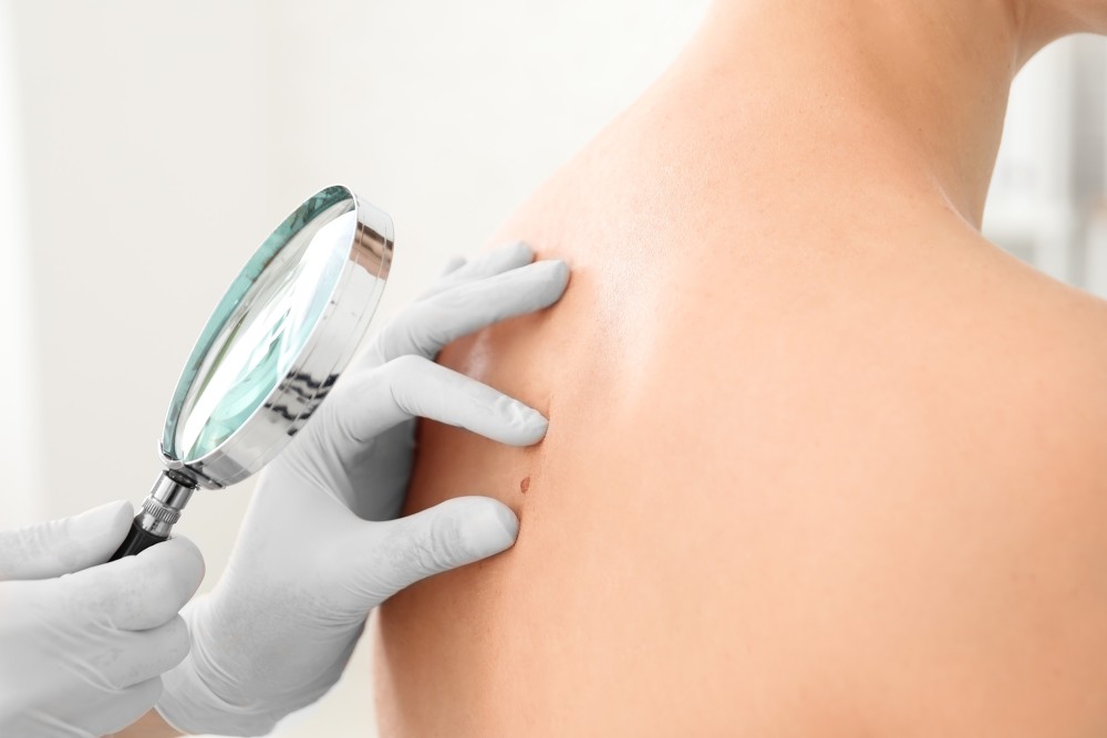
Je hebt een zorgwekkend vlekje op je huid. Je raadpleegt een dokter en die vermoedt dat het om huidkanker gaat. Er wordt een klein stukje van de mogelijke tumor weggesneden voor een controle op kankercellen, en die blijken aanwezig te zijn … Dit wordt voor steeds meer mensen werkelijkheid. Reden te over voor mij om -als student industrieel ingenieur elektromechanica- in mijn scriptie een nieuwe methode te introduceren om huidtumoren te detecteren. Daarbij komen voor het eerst onderhuidse eigenschappen van de tumor aan het licht en kunnen het aantal onnodige operaties aanzienlijk verminderen.
Bruinen, tegen welke kost?
De ‘perfecte’ zongebruinde huid kan een duistere keerzijde hebben. Terwijl we genieten van de warme zonnestralen, dringen UV-stralen diep door in de huid en richten onzichtbare schade aan. Huidkanker is onder andere daardoor uitgegroeid tot een wereldwijde zorg. Detectiesystemen optimaliseren is daarom geen luxe en mijn drijfveer tijdens het schrijven van mijn eindwerk. Momenteel wordt de ziekte opgespoord op basis van visuele kenmerken, waarbij interpretatie een grote rol speelt. De nieuw ontwikkelde methode op basis van nagebootste warmteverdeling van de huid, vergroot de objectivering van metingen aanzienlijk.
Kanker zichzelf laten vinden
Kankercellen zijn als supercellen: ze zijn hyperactief en hebben een verhoogd metabolisme. De ongecontroleerde celgroei vereist een grotere bloedtoevoer, wat resulteert in een hogere warmteproductie. Traditionele methoden voor het detecteren van huidtumoren maken gebruik van visuele kenmerken, zoals de vorm en kleur van de huidvlek. Maar wat als we de warmte die door de tumor wordt gegenereerd, kunnen gebruiken om deze kwaadaardige groei te identificeren en zelfs de diepte ervan te bepalen?
Computers vervangen patiënten
Thermografie, waarbij we werken met warmtebeelden, is een innovatieve methode voor het detecteren van huidtumoren. Om onderzoek te doen met deze methode zijn er data nodig. Die data kunnen gehaald worden uit testen bij patiënten. Dat kost veel tijd en geld. Een snellere, goedkopere optie is de data zelf creëren met hulp van computerkracht. Tijdens mijn scriptieonderzoek creëerde ik een geavanceerd 3D-FEM-huidmodel dat de warmteverdeling van de menselijke huid nabootst.
Afkoelen en opwarmen
Tijdens de simulaties koelen we het huidmodel af met koude lucht. Daarna warmt het huidmodel zich op een natuurlijke manier op en keert het terug naar zijn oorspronkelijke staat. De temperatuurveranderingen tijdens de afkoeling en opwarming zijn opmerkelijk. Door gebruik te maken van geavanceerde algoritmes en analyses van de thermogrammen, kunnen de vormeigenschappen van de tumor achterhaald worden. Niet alleen wordt de vorm van de tumor aan het huidoppervlak vastgesteld, ook de onderhuidse vormeigenschappen worden in beeld gebracht. Hierdoor is het mogelijk om bijvoorbeeld de richting te voorspellen waarin een onderhuidse tumor afwijkt.
Eén keer snijden
Eén van de belangrijkste bevindingen van mijn scriptieonderzoek is de mogelijkheid om de diepte van de tumor met een hoge nauwkeurigheid van 0,05 mm te bepalen. Dit heeft belangrijke implicaties voor de behandeling van huidkanker. Wanneer we de diepte van de tumor op voorhand kunnen bepalen, kan het aantal operaties verminderen. In plaats van telkens verschillende lagen van de tumor apart weg te snijden, naar het laboratorium te sturen en te onderzoeken, kan de tumor in één keer volledig worden verwijderd. Dit zorgt voor minder ongemak voor de patiënt en voor een efficiëntere en goedkopere behandeling.
Waar blijft AI?
Artificiële intelligentie die huidkanker opspoort en scherp in beeld brengt! Klinkt veraf maar komt door mijn scriptie heel dichtbij. In het gepresenteerde 3D-FEM-huidmodel is de tumor volledig aanpasbaar, waardoor er een eindeloze reeks thermische simulaties met verschillende tumoren kan worden uitgevoerd. Dat resulteert in een overvloed aan waardevolle data, wat de perfecte basis is voor het trainen van een AI-model.
Bibliografie
[1] N. R. Abbasi et al., “Early Diagnosis of Cutaneous Melanoma Revisiting the ABCD Criteria Downloaded From: https://jamanetwork.com/ by a Universiteit Antwerpen User on 12/07/2022,” 2004. [Online]. Available: www.jama.comwww.jama.com
[2] Dr. N. Kukutsch, “Huidkanker,” Sep. 2018. https://www.kanker.nl/kankersoorten/huidkanker (accessed Dec. 07, 2022).
[3] M. C. F. Simões, J. J. S. Sousa, and A. A. C. C. Pais, “Skin cancer and new treatment perspectives: A review,” Cancer Letters, vol. 357, no. 1. Elsevier Ireland Ltd, pp. 8–42, Feb. 01, 2015. doi: 10.1016/j.canlet.2014.11.001.
[4] J. Cook and J. A. Zitelli, “Mohs micrographic surgery: A cost analysis,” 1998.
[5] M. A. Marchetti et al., “Number needed to biopsy ratio and diagnostic accuracy for melanoma detection,” J Am Acad Dermatol, vol. 83, no. 3, pp. 780–787, Sep. 2020, doi: 10.1016/J.JAAD.2020.04.109.
[6] A. Nault, C. Zhang, K. M. Kim, S. Saha, D. D. Bennett, and Y. G. Xu, “Biopsy Use in Skin Cancer Diagnosis: Comparing Dermatology Physicians and Advanced Practice Professionals,” JAMA Dermatol, vol. 151, no. 8, pp. 899–902, Aug. 2015, doi: 10.1001/JAMADERMATOL.2015.0173.
[7] “Medische thermografie borsten en infraroodscreening lichaam - kosten in Nederland?” https://www.borstscreening.nl/ (accessed May 19, 2023).
[8] I. Peate, “The skin: largest organ of the body,” https://doi.org/10.12968/bjha.2021.15.9.446, vol. 15, no. 9, pp. 446–451, Oct. 2021, doi: 10.12968/BJHA.2021.15.9.446.
[9] F. J. G. Ebling, “human skin | Definition, Layers, Types, & Facts | Britannica.” https://www.britannica.com/science/human-skin (accessed Mar. 24, 2022).
[10] “Het stadium bij melanoom.” https://www.kanker.nl/kankersoorten/melanoom/diagnose/het-stadium-bij-m… (accessed Mar. 25, 2022).
[11] K. M. Zepon, M. S. Marques, M. M. da Silva Paula, F. D. P. Morisso, and L. A. Kanis, “Facile, green and scalable method to produce carrageenan-based hydrogel containing in situ synthesized AgNPs for application as wound dressing,” Int J Biol Macromol, vol. 113, pp. 51–58, 2018, doi: 10.1016/j.ijbiomac.2018.02.096.
[12] C. Vink, “Huid anatomie en fysiologie - ppt download.” https://slideplayer.nl/slide/15520850/ (accessed Mar. 25, 2022).
[13] A. L. Rippa, E. P. Kalabusheva, and E. A. Vorotelyak, “Regeneration of Dermis: Scarring and Cells Involved,” Cells, vol. 8, no. 6, p. 607, 2019, doi: 10.3390/cells8060607.
[14] M. Pirtini Çetingül and C. Herman, “A heat transfer model of skin tissue for the detection of lesions: Sensitivity analysis,” Phys Med Biol, vol. 55, no. 19, pp. 5933–5951, Oct. 2010, doi: 10.1088/0031-9155/55/19/020.
[15] “Anatomie van de huid - Eduwond.” https://www.eduwond.be/nl/digiwond/1968/anatomie-van-de-huid (accessed Mar. 24, 2022).
[16] D. Brokken and F. M. Hendriks, “Mechanical properties of human skin layers,” no. December 2014, 2012.
[17] H. Wang, X. X. Dong, J. C. Yang, H. Huang, Y. X. Li, and H. X. Zhang, “Finite element method simulating temperature distribution in skin induced by 980-nm pulsed laser based on pain stimulation,” Lasers Med Sci, vol. 32, no. 5, 2017, doi: 10.1007/s10103-017-2223-9.
[18] Queen Lauren, “SKIN CANCER 2 Acceptance of Senior Honors Thesis,” 2017.
[19] R. S. R. A. K. S. Ashish Dwivedi · Anurag Tripathi, Skin Cancer: Pathogenesis and Diagnosis. Springer Singapore, 2021. doi: 10.1007/978-981-16-0364-8.
[20] D. L. Cummins, J. M. Cummins, H. Pantle, M. A. Silverman, A. L. Leonard, and A. Chanmugam, “Cutaneous malignant melanoma,” Mayo Clinic Proceedings, vol. 81, no. 4. Elsevier Ltd, pp. 500–507, 2006. doi: 10.4065/81.4.500.
[21] D. E. Godar, “Worldwide Increasing Incidences of Cutaneous Malignant Melanoma,” J Skin Cancer, vol. 2011, pp. 1–6, 2011, doi: 10.1155/2011/858425.
[22] K. Kandala, D. Daxiang, and C. Herman, “Simulation of Discrete Blood Vessel Effects on the,” 2014, doi: 10.1115/IMECE2013-64451.SIMULATION.
[23] J. Iljaž, L. C. Wrobel, M. Hriberšek, and J. Marn, “Numerical modelling of skin tumour tissue with temperature-dependent properties for dynamic thermography,” Comput Biol Med, vol. 112, Sep. 2019, doi: 10.1016/j.compbiomed.2019.103367.
[24] Z. Apalla, A. Lallas, E. Sotiriou, E. Lazaridou, and D. Ioannides, “Epidemiological trends in skin cancer,” Dermatol Pract Concept, vol. 7, no. 2, Apr. 2017, doi: 10.5826/DPC.0702A01.
[25] M. Finoulst, P. Vankrunkelsven, and M. Garmyn, “Uit de pers gelicht De stijgende incidentie van huidkanker onder de loep,” 2019, doi: 10.2143/TVG.75.09.2002845.
[26] T.L.DIEPGEN and V.MAHLER, “The epidemiology of skin cancer,” British Journal of Dermatology 2002; 146 (Suppl. 61): 1–6., 2002.
[27] “NKR Cijfers.” https://iknl.nl/nkr-cijfers (accessed Feb. 27, 2023).
[28] “Melanoma Treatment Options at Each Stage.” https://www.healthcentral.com/slideshow/melanoma-treatment-by-stage (accessed May 19, 2023).
[29] “Oppervlakkig groeiend basaalcelcarcinoom – Da Vinci Clinic.” https://www.davinciclinic.be/aandoeningen/oppervlakkig-basaalcelcarcino… (accessed May 19, 2023).
[30] “Plaveiselcelcarcinoom - Huidkanker Stichting.” https://www.hukas.nl/soortenhuidkanker/plaveiselcelcarcinoom (accessed May 19, 2023).
[31] F. Liu-Smith, J. Jia, and Y. Zheng, “UV-Induced Molecular Signaling Differences in Melanoma and Non-melanoma Skin Cancer,” Adv Exp Med Biol, vol. 996, pp. 27–40, 2017, doi: 10.1007/978-3-319-56017-5_3.
[32] Y. Nartey and M. J. Sneyd, “The presenting features of melanoma in New Zealand: implications for earlier detection,” Aust N Z J Public Health, vol. 42, no. 6, pp. 567–571, Dec. 2018, doi: 10.1111/1753-6405.12815.
[33] D. L. Morton, D. G. Davtyan, L. A. Wanek, L. J. Foshag, and A. J. Cochran, “Multivariate analysis of the relationship between survival and the microstage of primary melanoma by clark level and breslow thickness,” Cancer, vol. 71, no. 11, pp. 3737–3743, 1993, doi: 10.1002/1097-0142(19930601)71:11<3737::AID-CNCR2820711143>3.0.CO;2-7.
[34] A. Scope et al., “The ‘ugly duckling’ sign: agreement between observers,” Arch Dermatol, vol. 144, no. 1, pp. 58–64, Jan. 2008, doi: 10.1001/ARCHDERMATOL.2007.15.
[35] J. E. Gershenwald et al., “Melanoma staging: Evidence-based changes in the American Joint Committee on Cancer eighth edition cancer staging manual,” CA Cancer J Clin, vol. 67, no. 6, pp. 472–492, Nov. 2017, doi: 10.3322/CAAC.21409.
[36] Kevin Wevers, Lukas Been, and Harald Hoekstra, “Chirurgische behandeling melanomen,” huisarts & wetenschap, 2013.
[37] E. Crocetti, L. Fancelli, A. Caldarella, and C. Buzzoni, “Thickness and diameter in melanoma: Is there a relation?,” Tumori, vol. 102, no. 1, pp. e1–e3, Jan. 2016, doi: 10.5301/tj.5000369.
[38] G. Ferrara, C. Tomasini, G. Argenziano, I. Zalaudek, and C. M. Stefanato, “Small-diameter melanoma: Toward a conceptual and practical reappraisal,” Journal of Cutaneous Pathology, vol. 39, no. 7. pp. 721–723, Jul. 2012. doi: 10.1111/j.1600-0560.2012.01884.x.
[39] “Skin Cancer,” Aug. 2022. https://www.mayoclinic.org/diseases-conditions/skin-cancer/diagnosis-tr… (accessed Dec. 07, 2022).
[40] “Mohs Micrographic Surgery - American Family Physician,” 2005. [Online]. Available: www.aafp.org/afp.
[41] R. J. Friedman, D. S. Rigel, and A. W. Kopf, “Early detection of malignant melanoma: the role of physician examination and self-examination of the skin,” CA Cancer J Clin, vol. 35, no. 3, pp. 130–151, May 1985, doi: 10.3322/CANJCLIN.35.3.130.
[42] H. Tsao et al., “Early detection of melanoma: Reviewing the ABCDEs American Academy of Dermatology Ad Hoc Task Force for the ABCDEs of Melanoma,” J Am Acad Dermatol, vol. 72, no. 4, pp. 717–723, Apr. 2015, doi: 10.1016/j.jaad.2015.01.025.
[43] S. Goldsmith, “A unifying approach to the clinical diagnosis of melanoma including ‘D’ for ‘Dark’ in the ABCDE criteria,” Dermatol Pract Concept, vol. 4, no. 5, Oct. 2014, doi: 10.5826/dpc.0404a16.
[44] S. M. Goldsmith and A. B. Cognetta, “Time to move forward after the report of the AAD Task Force for the ABCDEs of Melanoma,” Journal of the American Academy of Dermatology, vol. 73, no. 4. Mosby Inc., pp. e149–e150, Oct. 01, 2015. doi: 10.1016/j.jaad.2015.05.043.
[45] P. Carli et al., “Improvement of malignant/benign ratio in excised melanocytic lesions in the ‘dermoscopy era’: a retrospective study 1997-2001,” Br J Dermatol, vol. 150, no. 4, pp. 687–692, Apr. 2004, doi: 10.1111/J.0007-0963.2004.05860.X.
[46] G. Argenziano et al., “Dermoscopy of pigmented skin lesions: results of a consensus meeting via the Internet,” J Am Acad Dermatol, vol. 48, no. 5, pp. 679–693, May 2003, doi: 10.1067/MJD.2003.281.
[47] I. Zalaudek et al., “Clinically equivocal melanocytic skin lesions with features of regression: a dermoscopic-pathological study,” Br J Dermatol, vol. 150, no. 1, pp. 64–71, Jan. 2004, doi: 10.1111/J.1365-2133.2004.05657.X.
[48] H. Kittler, H. Pehamberger, K. Wolff, and M. Binder, “Diagnostic accuracy of dermoscopy,” Lancet Oncology, vol. 3, no. 3, pp. 159–165, Mar. 2002, doi: 10.1016/S1470-2045(02)00679-4.
[49] C. Tavares, M. Melo, J. M. Cameselle-Teijeiro, P. Soares, and M. Sobrinho-Simões, “ENDOCRINE TUMOURS: Genetic predictors of thyroid cancer outcome,” Eur J Endocrinol, vol. 174, no. 4, pp. R117–R126, Apr. 2016, doi: 10.1530/EJE-15-0605.
[50] Z. Ostrowski, P. Buliński, W. Adamczyk, P. Kozołub, and A. Nowak, “Numerical model of heat transfer in skin lesions,” Scientific Letters of University of Rzeszow Technology - Mechanics, vol. 32, no. 87(1/2015), pp. 55–62, 2015, doi: 10.7862/rm.2015.6.
[51] T. Y. Cheng and C. Herman, “Analysis of skin cooling for quantitative dynamic infrared imaging of near-surface lesions,” International Journal of Thermal Sciences, vol. 86, pp. 175–188, 2014, doi: 10.1016/j.ijthermalsci.2014.06.033.
[52] A. Bhowmik, R. Repaka, and S. C. Mishra, “Thermographic evaluation of early melanoma within the vascularized skin using combined non-Newtonian blood flow and bioheat models,” Comput Biol Med, vol. 53, pp. 206–219, 2014, doi: 10.1016/j.compbiomed.2014.08.002.
[53] M. Bonmarin and F. A. Le Gal, “Lock-in thermal imaging for the early-stage detection of cutaneous melanoma: A feasibility study,” Comput Biol Med, vol. 47, no. 1, pp. 36–43, Apr. 2014, doi: 10.1016/j.compbiomed.2014.01.008.
[54] M. Goyal, T. Knackstedt, S. Yan, and S. Hassanpour, “Artificial intelligence-based image classification methods for diagnosis of skin cancer: Challenges and opportunities,” Computers in Biology and Medicine, vol. 127. Elsevier Ltd, Dec. 01, 2020. doi: 10.1016/j.compbiomed.2020.104065.
[55] A. Antoniou and C. Damianou, “MR relaxation properties of tissue-mimicking phantoms,” Ultrasonics, vol. 119, p. 106600, Feb. 2022, doi: 10.1016/J.ULTRAS.2021.106600.
[56] T. Gomboc, J. Iljaž, L. C. Wrobel, M. Hriberšek, and J. Marn, “Design of constant temperature cooling device for melanoma screening by dynamic thermography,” Eng Anal Bound Elem, vol. 125, pp. 66–79, Apr. 2021, doi: 10.1016/J.ENGANABOUND.2021.01.009.
[57] J. E. Bischoff, E. M. Arruda, and K. Grosh, “Finite element modeling of human skin using an isotropic, nonlinear elastic constitutive model,” J Biomech, vol. 33, no. 6, pp. 645–652, Jun. 2000, doi: 10.1016/S0021-9290(00)00018-X.
[58] D. Ratovoson, V. Huon, V. Costalat, and F. Jourdan, “Combined model of human skin - Heat transfer in the vein and tissue: Experimental and numerical study1,” Quant Infrared Thermogr J, vol. 8, no. 2, pp. 165–186, 2011, doi: 10.3166/qirt.8.165-186.
[59] H. H. Pennes, “Applied physiology.,” Suom Laakaril, vol. 17, pp. 1865–1871, 1962, doi: 10.5005/jp/books/12678_10.
[60] L. L. Ferrás, N. J. Ford, M. L. Morgado, J. M. Nóbrega, and M. S. Rebelo, “Fractional Pennes’ Bioheat Equation: Theoretical and Numerical Studies,” Fract Calc Appl Anal, vol. 18, no. 4, pp. 1080–1106, 2015, doi: 10.1515/fca-2015-0062.
[61] A. Bhowmik, R. Singh, R. Repaka, and S. C. Mishra, “Conventional and newly developed bioheat transport models in vascularized tissues: A review,” J Therm Biol, vol. 38, no. 3, pp. 107–125, 2013, doi: 10.1016/j.jtherbio.2012.12.003.
[62] M. A. Ezzat, N. S. Alsowayan, Z. I. A. Al-Muhiameed, and S. M. Ezzat, “Fractional modelling of Pennes’ bioheat transfer equation,” Heat and Mass Transfer/Waerme- und Stoffuebertragung, vol. 50, no. 7, pp. 907–914, 2014, doi: 10.1007/s00231-014-1300-x.
[63] S. Andersson, “The Bio-heat Equation,” Medical Optics, pp. 1–14.
[64] “Alle informatie over verwarming die je nodig hebt - Gastrion Air Solutions - Gastrion.” https://gastrion.com/heating/verwarming-informatie/ (accessed Feb. 27, 2023).
[65] D. A. Torvi and J. D. Dale, “A finite element model of skin subjected to a flash fire,” J Biomech Eng, vol. 116, no. 3, pp. 250–255, 1994, doi: 10.1115/1.2895727.
[66] J. Werner and M. Buse, “Temperature profiles with respect to inhomogeneity and geometry of the human body,” J Appl Physiol (1985), vol. 65, no. 3, pp. 1110–1118, 1988, doi: 10.1152/JAPPL.1988.65.3.1110.
[67] S. B. Wilson and V. A. Spence, “A tissue heat transfer model for relating dynamic skin temperature changes to physiological parameters,” Phys Med Biol, vol. 33, no. 8, pp. 895–912, 1988, doi: 10.1088/0031-9155/33/8/001.
[68] T. Y. Cheng and C. Herman, “Optimization of Skin Cooling for Thermographic Imaging of Near-Surface Lesions,” ASME 2011 International Mechanical Engineering Congress and Exposition, IMECE 2011, vol. 2, pp. 351–360, Aug. 2012, doi: 10.1115/IMECE2011-65221.
[69] C. W. Song, A. Lokshina, J. G. Rhee, M. Patten, and S. H. Levitt, “Implication of Blood Flow in Hyperthermic Treatment of Tumors,” IEEE Trans Biomed Eng, vol. BME-31, no. 1, pp. 9–16, 1984, doi: 10.1109/TBME.1984.325364.
[70] M. Agrawal, K. R. Pardasani, and N. Adlakha, “Finite element model to study the thermal effect of tumors in dermal regions of irregular tapered shaped human limbs,” 2015, doi: 10.1016/j.ijthermalsci.2015.07.010.
[71] A. S. Ahuja, K. N. Prasad, W. R. Hendee, and P. L. Carson, “Thermal conductivity and diffusivity of neuroblastoma tumor cells,” Med Phys, vol. 5, no. 5, pp. 418–421, Sep. 1978, doi: 10.1118/1.594434.
[72] T. Latunde and O. M. Bamigbola, “Parameter Estimation and Sensitivity Analysis of an Optimal Control Model for Capital Asset Management,” Advances in Fuzzy Systems, vol. 2018, 2018, doi: 10.1155/2018/4756520.
[73] D. V. Likhachev, “Parametric sensitivity analysis as an essential ingredient of spectroscopic ellipsometry data modeling: An application of the Morris screening method,” J Appl Phys, vol. 126, no. 18, Nov. 2019, doi: 10.1063/1.5126074.
[74] A. Bhowmik and R. Repaka, “Estimation of growth features and thermophysical properties of melanoma within 3-D human skin using genetic algorithm and simulated annealing,” Int J Heat Mass Transf, vol. 98, pp. 81–95, Jul. 2016, doi: 10.1016/j.ijheatmasstransfer.2016.03.020.
[75] “NX software including CAD and CAM | Siemens Software.” https://plm.sw.siemens.com/en-US/nx/ (accessed May 08, 2023).
[76] “Optics calculator for Optris thermal imaging cameras.” https://www.optris.global/optics-calculator (accessed Dec. 08, 2022).
[77] N. M. Williams, K. D. Rojas, J. M. Reynolds, D. Kwon, J. Shum-Tien, and N. Jaimes, “Assessment of Diagnostic Accuracy of Dermoscopic Structures and Patterns Used in Melanoma Detection: A Systematic Review and Meta-analysis,” JAMA Dermatol, vol. 157, no. 9, p. 1, Sep. 2021, doi: 10.1001/JAMADERMATOL.2021.2845.
[78] “What is a Mesh? | SimWiki Documentation | SimScale.” https://www.simscale.com/docs/simwiki/preprocessing/what-is-a-mesh/ (accessed May 08, 2023).
[79] A. Katz and V. Sankaran, “Mesh quality effects on the accuracy of CFD solutions on unstructured meshes,” J Comput Phys, vol. 230, no. 20, pp. 7670–7686, Aug. 2011, doi: 10.1016/J.JCP.2011.06.023.
[80] Shashikant T. More and R. S. Bindu, “Effect of Mesh Size on Finite Element Analysis of Plate Structure”.
[81] A. Dutt, “Effect of Mesh Size on Finite Element Analysis of Beam,” SSRG International Journal of Mechanical Engineering, vol. 2, p. 12, 2015, Accessed: May 25, 2023. [Online]. Available: www.internationaljournalssrg.org
[82] “simulate | Etymology, origin and meaning of simulate by etymonline.” https://www.etymonline.com/word/simulate (accessed Feb. 27, 2023).
[83] “Simcenter HEEDS | Siemens Software.” https://plm.sw.siemens.com/en-US/simcenter/integration-solutions/heeds/ (accessed Feb. 27, 2023).
[84] “Cryo 6 - Zimmer MedizinSystems Aesthetic Division - Chillers.” https://aesthetic.zimmerusa.com/z-cryo-2/cryo-6/ (accessed Feb. 27, 2023).
[85] H. Chen, K. Wang, Z. Du, W. Liu, and Z. Liu, “Predicting the thermophysical properties of skin tumor based on the surface temperature and deep learning,” Int J Heat Mass Transf, vol. 180, Dec. 2021, doi: 10.1016/j.ijheatmasstransfer.2021.121804.


