
Een nieuwe kijk op erosieve artrose: AI als hulpmiddel voor radiologen
Je wordt op een ochtend wakker met gezwollen, pijnlijke vingers. Eerst lijkt het op overbelasting of de tekenen van ouderdom. Maar naarmate de dagen verstrijken, neemt de pijn toe, en wordt het uitvoeren van eenvoudige handelingen zoals het vasthouden van een pen of het dichtmaken van een knoop een dagelijkse strijd. Voor veel mensen met erosieve artrose is dit de realiteit. En het verontrustende? Zonder vroege interventie wordt deze aandoening alleen maar erger. Daarom is er een dringende behoefte aan innovatieve hulpmiddelen, zoals artificiële intelligentie (AI), die radiologen ondersteunen bij het diagnosticeren en monitoren van erosieve artrose. Een proces dat voorheen tijdrovend was, kan nu in enkele seconden worden uitgevoerd.
De onzichtbare strijd in onze handen
Artrose is een chronische gewrichtsaandoening die wereldwijd miljoenen mensen treft en het is een van de belangrijkste oorzaken van invaliditeit. Een bijzonder agressieve vorm van deze ziekte, erosieve artrose, is een meedogenloze aanvaller van de kleine vingergewrichten. In enkele maanden tijd kunnen de aangetaste gewrichten volledig vernietigd worden. Het verlies van handfunctie bedreigt niet alleen de zelfredzaamheid, maar ook iemands identiteit en levenskwaliteit. De psychologische impact hiervan mag niet worden onderschat.
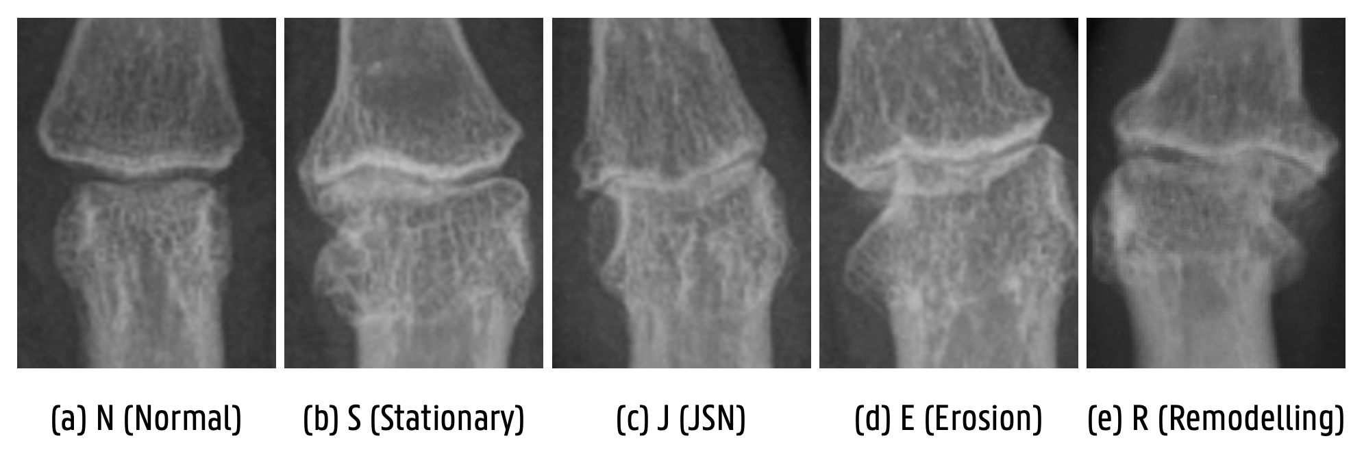
Radiologen gebruiken momenteel röntgenfoto’s om erosieve artrose op te sporen en op te volgen, waarbij correcte interpretatie een grote rol speelt. Hiervoor gebruiken ze het Verbruggen-Veys scoresysteem dat het verloop van de ziekte opdeelt in vijf fases of scores: normaal, zonder artrose (N); stationaire artrose (S); gewrichtsvernauwing (J); erosieve fase (E); en remodelering (R). Elke fase toont specifieke kenmerken. Radiologen moeten elk vingergewricht zorgvuldig beoordelen en een score toekennen, een tijdrovend en foutgevoelig proces.
De kracht van AI: een geautomatiseerd scoresysteem
Hier komt AI in beeld, in het bijzonder Convolutional Neural Networks (CNN’s). Deze AI-modellen, geïnspireerd door de werking van neuronen in het menselijk brein, zijn gespecialiseerd in beeldherkenning en kunnen complexe patronen in medische afbeeldingen detecteren. CNN’s worden al jaren onderzocht voor het opsporen van ziekten zoals reuma en hersentumoren, en nu helpen ze ook bij het diagnosticeren van erosieve artrose.
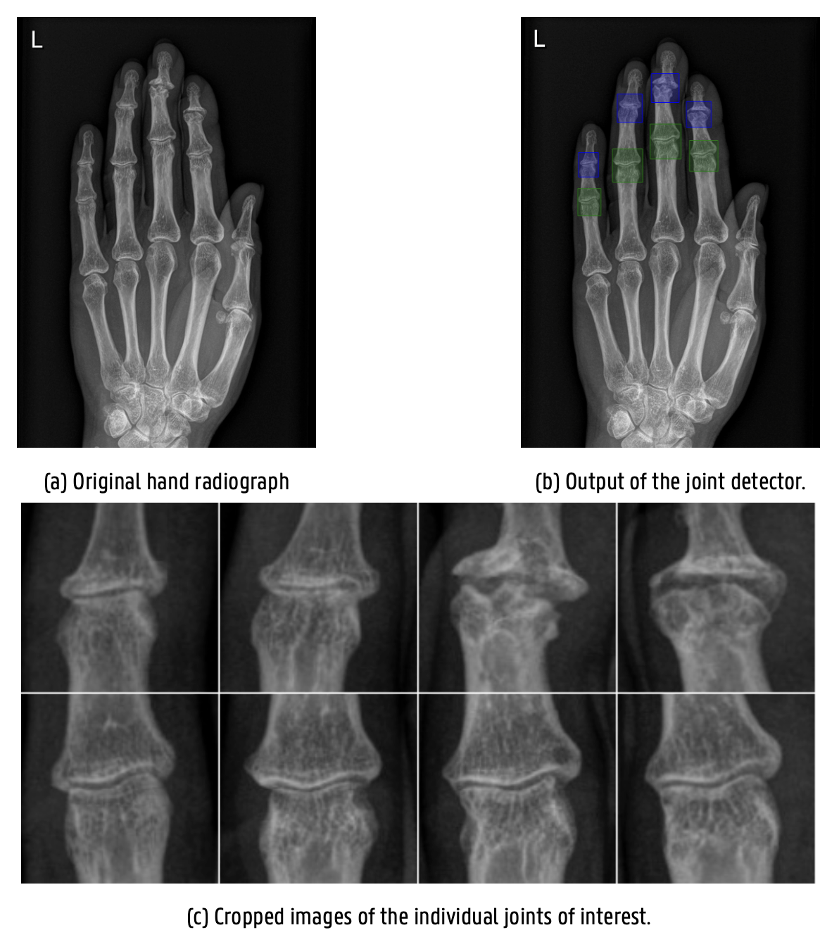
Tijdens mijn scriptieonderzoek ontwikkelde ik een geautomatiseerd scoresysteem dat in enkele seconden een score toekent. Dit systeem bestaat uit twee stappen:
- De Verkenner: deze AI detecteert en isoleert de vingergewrichten op röntgenfoto’s van mensenhanden.
- De Beoordelaar: deze AI beoordeelt de geïsoleerde gewrichten en kent ze de juiste score toe. Hierbij wordt rekening gehouden met de ernst en afwijking van eventuele foutieve voorspellingen: een gezond gewricht (N) verkeerd scoren als een licht aangetast gewricht (S), is minder erg dan hetzelfde gewricht scoren als een zwaar aangetast gewricht (E).
Met deze tweeledige aanpak kunnen andere scoresystemen, zoals GUSS (Ghent University Score System), in de toekomst geïntegreerd worden om een nog completere diagnostiek te bieden.
Van koekjes bakken naar medische doorbraken
Maar hoe train je een AI-systeem met beperkte medische data? Hier komt transfer learning om de hoek kijken. Dit concept werkt als volgt: stel je voor dat je goed bent in koekjes bakken. Als je vervolgens taarten leert bakken, begin je niet vanaf nul. Je weet al hoe je ingrediënten mengt en een oven bedient. AI werkt vergelijkbaar - we nemen een model dat getraind is op miljoenen alledaagse afbeeldingen en finetunen het voor onze specifieke medische taak. Net zoals jij je bakkennis van koekjes overdraagt naar taarten bakken, draagt het AI-model zijn kennis over van de ene taak naar de andere. Deze aanpak lost niet alleen het probleem van beperkte data op, maar versnelt ook het leerproces aanzienlijk.
Tijdens mijn onderzoek was dit echter niet de enige uitdaging. Een andere hindernis was de ondervertegenwoordiging van gewrichten in een gevorderde ziektestadia. De meest cruciale fasen van erosieve artrose (J en E), maakten elk maar 5% uit van de beschikbare afbeeldingen waarop de AI werd getraind. Dit weerspiegelt de realiteit in de klinische praktijk, waar we vaker gezonde gewrichten zien dan zieke. Hierdoor had het AI-model moeite om voldoende te leren van deze belangrijke, maar schaarse voorbeelden.
Om dit probleem te omzeilen, werd een slimme techniek genaamd data-augmentatie toegepast. Hierbij manipuleerde ik voorzichtig de bestaande afbeeldingen, bijvoorbeeld met kleine rotaties, om nieuwe variaties te creëren, zonder de accurate weergave van het gewricht aan te tasten. Dit gaf de AI de indruk dat het meer nieuwe afbeeldingen te zien kreeg dan er daadwerkelijk waren. Deze aanpak bleek succesvol: het geautomatiseerd systeem kon uiteindelijk 70% van nieuwe, ongeziene gewrichten een correcte score toekennen.
Meer dan cijfers: wat AI-fouten ons kunnen leren
Hoewel 70% misschien niet spectaculair lijkt, schuilt er een belangrijk inzicht achter dit cijfer. In plaats van alleen te focussen op correcte voorspellingen, is het minstens zo waardevol om te kijken waar de AI-fouten maakt. Bij het analyseren van deze foutieve scores, zag ik dat de AI voorspellingen maakte die een betere fit waren dan de voorziene score. Vanwege dit inzicht liet ik een ervaren radioloog, gespecialiseerd in erosieve artrose, een steekproef van deze foutieve voorspellingen zelf scoren. De radioloog kende voor deze foutieve voorspellingen dezelfde score toe als de AI in 50% van de gevallen.
Deze overeenkomst toonde aan dat bepaalde criteria van het scoresysteem eerder voor interpretatie vatbaar zijn. Deze analyse leverde inzichten om het Verbruggen-Veys systeem te herzien en objectievere regels op te stellen. Dit is precies waarom ik zo enthousiast ben over AI in medisch onderzoek: het helpt ons niet alleen bij het ontwikkelen van nieuwe diagnostische tools, maar het biedt ook nieuwe inzichten om bestaande methoden te optimaliseren.
What’s next?
AI-systemen kunnen het diagnostisch proces versnellen, wat cruciaal is voor tijdige behandeling en betere zorg. Daarnaast kan AI de werkdruk van zorgverleners verminderen en de efficiëntie in de klinische praktijk verhogen. Deze studie toont veelbelovende resultaten en opent de weg voor toekomstig onderzoek. Een geautomatiseerd scoresysteem biedt radiologen de kans voor grootschalige populatiestudies die ons begrip van de ziekte en de onderliggende risicofactoren verder kan vergroten.
Uiteindelijk zijn onze handen meer dan slechts gereedschappen. Ze zijn een weerspiegeling van onze onafhankelijkheid, en het is de moeite waard om er alles aan te doen om ze te beschermen.
Bibliografie
[1] C. M. Bishop, Pattern Recognition and Machine Learning (Information Science and Statistics). Berlin, Heidelberg: Springer-Verlag, Jul. 2006.
[2] A. Krenker, J. Bešter, A. Kos, A. Krenker, J. Bešter, and A. Kos, “Introduction to the Artificial Neural Networks,” in Artificial Neural Networks - Methodological Advances and Biomedical Applications. IntechOpen, Apr. 2011. [Online]. Available: https://www.intechopen.com/chapters/14881
[3] “Neural network diagram.” [Online]. Available: https://www.sciencelearn.org.nz/images/5156-neural-network-diagram
[4] “Multilayer Perceptrons — Knet. jl 0.7.2 documentation.” [Online]. Available: https://knet.readthedocs.io/en/v0.7.3/mlp.html
[5] B. Boehmke and B. M. Greenwell, Hands-On Machine Learning with R. New York: Chapman and Hall/CRC, Nov. 2019.
[6] A. Dertat, “Applied Deep Learning - Part 4: Convolutional Neural Networks,” Nov. 2017. [Online]. Available: https://towardsdatascience.com/applied-deep-learning-part-4-convolutional-neural-networks-584bc134c1e2
[7] S. Chaudhary, “A High-Level Guide to Autoencoders,” Aug. 2019. [Online]. Available: https://towardsdatascience.com/a-high-level-guide-to-autoencoders-b103ccd45924
[8] X. Shi, W. Cao, and S. Raschka, “Deep Neural Networks for Rank-Consistent Ordinal Regression Based On Conditional Probabilities,” Pattern Analysis and Applications, vol. 26, no. 3, pp. 941–955, Aug. 2023, arXiv:2111.08851 [cs, stat]. [Online]. Available: http://arxiv.org/abs/2111.08851
[9] W. Cao, V. Mirjalili, and S. Raschka, “Rank consistent ordinal regression for neural networks with application to age estimation,” Pattern Recognition Letters, vol. 140, pp. 325–331, Dec. 2020, arXiv:1901.07884 [cs, stat]. [Online]. Available: http://arxiv.org/abs/1901.07884
[10] V. Nanayakkara, G. Cotugno, N. Vitzilaios, D. Venetsanos, T. Nanayakkara, and M. Sahinkaya, “The Role of Morphology of the Thumb in Anthropomorphic Grasping: A Review,” Frontiers in Mechanical Engineering, vol. 3, Jun. 2017.
[11] G. Verbruggen, R. Wittoek, B. Cruyssen, and D. Elewaut, “Tumour necrosis factor blockade for the treatment of erosive osteoarthritis of the interphalangeal finger joints: A double blind, randomised trial on structure modification,” Annals of the rheumatic diseases, vol. 71, pp. 891–8, Nov. 2011.
[12] G. Verbruggen, R. Wittoek, B. Vander Cruyssen, and D. Elewaut, “Morbid anatomy of ’erosive osteoarthritis’ of the interphalangeal finger joints: an optimised scoring system to monitor disease progression in affected joints,” Annals of the Rheumatic Diseases, vol. 69, no. 5, pp. 862–867, May 2010.
[13] R. D. Sipio, “A Quick Guide to AUC-ROC in Machine Learning Models,” Aug. 2021. [Online]. Available: https://towardsdatascience.com/a-quick-guide-to-auc-roc-in-machine-learning-models-f0aedb78fbad
[14] Committee on Diagnostic Error in Health Care, Board on Health Care Services, Institute of Medicine, and The National Academies of Sciences, Engineering, and Medicine, Improving Diagnosis in Health Care, E. P. Balogh, B. T. Miller, and J. R. Ball, Eds. Washington (DC): National Academies Press (US), 2015. [Online]. Available: http://www.ncbi.nlm.nih.gov/books/NBK338596/
[15] P. H. Meyers, C. M. Nice, H. C. Becker, W. J. Nettleton, J. W. Sweeney, and G. R. Meckstroth, “AUTOMATED COMPUTER ANALYSIS OF RADIOGRAPHIC IMAGES,” Radiology, vol. 83, pp. 1029–1034, Dec. 1964.
[16] R. P. Kruger, J. R. Townes, D. L. Hall, S. J. Dwyer, and G. S. Lodwick, “Automated Radiographic Diagnosis via Feature Extraction and Classification of Cardiac Size and Shape Descriptors,” IEEE Transactions on Biomedical Engineering, vol. BME-19, no. 3, pp. 174–186, May 1972. [Online]. Available: https://ieeexplore.ieee.org/abstract/document/4120508
[17] J. -I. Toriwaki, Y. Suenaga, T. Negoro, and T. Fukumura, “Pattern recognition of chest X-ray images,” Computer Graphics and Image Processing, vol. 2, no. 3, pp. 252–271, Dec. 1973. [Online]. Available: https://www.sciencedirect.com/science/article/pii/0146664X73900051
[18] K. Doi, “Computer-aided diagnosis in medical imaging: historical review, current status and future potential,” Computerized Medical Imaging and Graphics: The Official Journal of the Computerized Medical Imaging Society, vol. 31, no. 4-5, pp. 198–211, 2007.
[19] G. Verbruggen and E. M. Veys, “Numerical scoring systems for the anatomic evolution of osteoarthritis of the finger joints,” Arthritis and Rheumatism, vol. 39, no. 2, pp. 308–320, Feb. 1996.
[20] A. M. Turing, “Computing Machinery and Intelligence,” Mind, vol. 59, no. October, pp. 433–60, 1950.
[21] S. J. Russell and P. Norvig, Artificial intelligence: A modern approach, 4th ed. Pearson, 2021.
[22] P. Ongsulee, “Artificial intelligence, machine learning, and deep learning,
International Conference on ICT and Knowledge Engineering, 2017.
[23] T. Panch, P. Szolovits, and R. Atun, “Artificial intelligence, machine learning and health systems,” Journal of Global Health, vol. 8, no. 2, p. 020303. [Online]. Available: https://www.ncbi.nlm.nih.gov/pmc/articles/PMC6199467/
[24] C. Janiesch, P. Zschech, and K. Heinrich, “Machine learning and deep learning,” Electronic Markets, vol. 31, no. 3, pp. 685–695, Sep. 2021, arXiv:2104.05314 [cs]. [Online]. Available: http://arxiv.org/abs/2104.05314
[25] M. I. Jordan and T. M. Mitchell, “Machine learning: Trends, perspectives, and prospects,” Science, Jul. 2015. [Online]. Available: https://www.science.org/doi/10.1126/science.aaa8415
[26] T. M. Mitchell, Machine Learning. McGraw-Hill, 1997, google-Books-ID: EoYBngEACAAJ.
[27] I. Goodfellow, Y. Bengio, and C. Aaron, Deep Learning. MIT Press, 2016. [Online]. Available: http://www.deeplearningbook.org
[28] Y. LeCun, Y. Bengio, and G. Hinton, “Deep learning,” Nature, vol. 521, no. 7553, pp. 436–444, May 2015. [Online]. Available: https://www.nature.com/articles/nature14539
[29] M. Naeem, S. Rizvi, and A. Coronato, “A Gentle Introduction to Reinforcement Learning and its Application in Different Fields,” IEEE Access, vol. 8, pp. 209 320–209 344, Jan. 2020.
[30] P. Domingos, “A few useful things to know about machine learning,” Communications of the ACM, vol. 55, no. 10, pp. 78–87, 2012. [Online]. Available: https://dl.acm.org/doi/10.1145/2347736.2347755
[31] A. Jain, R. Duin, and J. Mao, “Statistical pattern recognition: a review,” IEEE Transactions on Pattern Analysis and Machine Intelligence, vol. 22, no. 1, pp. 4–37, Jan. 2000. [Online]. Available: https://ieeexplore.ieee.org/abstract/document/824819
[32] W. S. McCulloch and W. Pitts, “A logical calculus of the ideas immanent in nervous activity,” The Bulletin of Mathematical Biophysics, vol. 5, no. 4, pp. 115–133, Dec. 1943. [Online]. Available: https://doi.org/10.1007/BF02478259
[33] D. O. Hebb, The Organization of Behavior: A Neuropsychological Theory. New York: Psychology Press, May 2002.
[34] B. W. White and F. Rosenblatt, “Principles of Neurodynamics: Perceptrons and the Theory of Brain Mechanisms,” The American Journal of Psychology, vol. 76, no. 4, p. 705, Dec. 1963. [Online]. Available: https://www.jstor.org/stable/1419730?origin=crossref
[35] M. Minsky and S. Papert, Perceptrons; an Introduction to Computational Geometry. MIT Press, 1969, Google-Books-ID: Ow1OAQAAIAAJ.
[36] S. Ruder, “An overview of gradient descent optimization algorithms,” Jun. 2017, arXiv:1609.04747 [cs]. [Online]. Available: http://arxiv.org/abs/1609.04747
[37] S. Albawi, T. A. Mohammed, and S. Al-Zawi, “Understanding of a convolutional neural network,” in 2017 International Conference on Engineering and Technology (ICET), Aug. 2017, pp. 1–6. [Online]. Available: https://ieeexplore.ieee.org/document/8308186
[38] A. Krizhevsky, I. Sutskever, and G. E. Hinton, “ImageNet classification with deep convolutional neural networks,” in Proceedings of the 25th International Conference on Neural Information Processing Systems - Volume 1, ser. NIPS’12. Red Hook, NY, USA: Curran Associates Inc., Dec. 2012, pp. 1097–1105.
[39] K. He, X. Zhang, S. Ren, and J. Sun, “Deep Residual Learning for Image Recognition,” Dec. 2015, arXiv:1512.03385 [cs]. [Online]. Available: http://arxiv.org/abs/1512.03385
[40] J. Yosinski, J. Clune, Y. Bengio, and H. Lipson, “How transferable are features in deep neural networks?” Nov. 2014, arXiv:1411.1792 [cs]. [Online]. Available: http://arxiv.org/abs/1411.1792
[41] S. J. Pan and Q. Yang, “A Survey on Transfer Learning,” IEEE Transactions on Knowledge and Data Engineering, vol. 22, no. 10, pp. 1345–1359, Oct. 2010. [Online]. Available: https://ieeexplore.ieee.org/document/5288526
[42] N. Tajbakhsh, J. Y. Shin, S. R. Gurudu, R. T. Hurst, C. B. Kendall, M. B. Gotway, and J. Liang, “Convolutional Neural Networks for Medical Image Analysis: Full Training or Fine Tuning?” IEEE Transactions on Medical Imaging, vol. 35, no. 5, pp. 1299–1312, May 2016. [Online]. Available: http://www.scopus.com/inward/record.url?scp=84968649810&partnerID=8YFLogxK
[43] Y. Bengio, A. C. Courville, and P. Vincent, “Unsupervised Feature Learning and Deep Learning: A Review and New Perspectives,” ArXiv, Jun. 2012. [Online]. Available: https://www.semanticscholar.org/paper/Unsupervised-Feature-Learning-and-Deep-Learning%3A-A-Bengio-Courville/f8c8619ea7d68e604e40b814b40c72888a755e95
[44] E. Frank and M. Hall, “A Simple Approach to Ordinal Classification,” in Machine Learning: ECML 2001, L. De Raedt and P. Flach, Eds. Berlin, Heidelberg: Springer, 2001, pp. 145–156.
[45] S. S. Stevens, “On the Theory of Scales of Measurement,” Science, vol. 103, no. 2684, pp. 677–680, Jun. 1946. [Online]. Available: https://www.science.org/doi/10.1126/science.103.2684.677
[46] R. Likert, “A technique for the measurement of attitudes,” Archives of Psychology, vol. 22 140, pp. 55–55, 1932.
[47] G. Sullivan and A. Artino, “Analyzing and Interpreting Data From Likert-Type Scales,” Journal of Graduate Medical Education, vol. 5, pp. 541–2, Dec. 2013.
[48] P. McCullagh, “Regression Models for Ordinal Data,” Journal of the Royal Statistical Society: Series B (Methodological), vol. 42, no. 2, pp. 109–127, Jan. 1980. [Online]. Available: https://rss.onlinelibrary.wiley.com/doi/10.1111/j.2517-6161.1980.tb01109.x
[49] L. Li and H. t. Lin, “Ordinal Regression by Extended Binary Classification,” in Advances in Neural Information Processing Systems, vol. 19. MIT Press, 2006. [Online]. Available: https://papers.nips.cc/paper_files/paper/2006/hash/019f8b946a256d9357eadc5ace2c8678-Abstract.html
[50] K. Crammer and Y. Singer, “Pranking with Ranking,” in Advances in Neural Information Processing Systems, vol. 14. MIT Press, 2001. [Online]. Available: https://papers.nips.cc/paper_files/paper/2001/hash/5531a5834816222280f20d1ef9e95f69-Abstract.html
[51] A. Shashua and A. Levin, “Ranking with Large Margin Principle: Two Approaches,” in Advances in Neural Information Processing Systems, vol. 15. MIT Press, 2002. [Online]. Available: https://papers.nips.cc/paper_files/paper/2002/hash/51de85ddd068f0bc787691d356176df9-Abstract.html
[52] S. Rajaram, A. Garg, X. S. Zhou, and T. S. Huang, “Classification Approach towards Ranking and Sorting Problems,” in Machine Learning: ECML 2003, N. Lavrač, D. Gamberger, H. Blockeel, and L. Todorovski, Eds. Berlin, Heidelberg: Springer, 2003, pp. 301–312.
[53] W. Chu and S. S. Keerthi, “New approaches to support vector ordinal regression,” Proceedings of the 22nd international conference on Machine learning - ICML ’05, pp. 145–152, 2005. [Online]. Available: http://portal.acm.org/citation.cfm?doid=1102351.1102370
[54] J. Cheng, “A neural network approach to ordinal regression,” Apr. 2007, arXiv:0704.1028 [cs]. [Online]. Available: http://arxiv.org/abs/0704.1028
[55] Z. Niu, M. Zhou, L. Wang, X. Gao, and G. Hua, “Ordinal Regression with Multiple Output CNN for Age Estimation,” in 2016 IEEE Conference on Computer Vision and Pattern Recognition (CVPR), Jun. 2016, pp. 4920–4928, iSSN: 1063-6919. [Online]. Available: https://ieeexplore.ieee.org/document/7780901
[56] D. G. Arias, A. C. Black, and M. Varacallo, “Anatomy, Shoulder and Upper Limb, Hand Bones,” in StatPearls. Treasure Island (FL): StatPearls Publishing, 2023. [Online]. Available: http://www.ncbi.nlm.nih.gov/books/NBK547684/
[57] S. Panchal – Kildare and K. Malone, “Skeletal anatomy of the hand” Hand Clinics, vol. 29, no. 4, pp. 459–471, Nov. 2013.
[58] E. R. Vina and C. K. Kwoh, “Epidemiology of osteoarthritis: literature update,” Current Opinion in Rheumatology, vol. 30, no. 2, pp. 160–167, Mar. 2018.
[59] M. Cross, E. Smith, D. Hoy, S. Nolte, I. Ackerman, M. Fransen, L. Bridgett, S. Williams, F. Guillemin, C. L. Hill, L. L. Laslett, G. Jones, F. Cicuttini, R. Osborne, T. Vos, R. Buchbinder, A. Woolf, and L. March, “The global burden of hip and knee osteoarthritis: estimates from the global burden of disease 2010 study,” Annals of the Rheumatic Diseases, vol. 73, no. 7, pp. 1323–1330, Jul. 2014.
[60] J. Martel-Pelletier, A. J. Barr, F. M. Cicuttini, P. G. Conaghan, C. Cooper, M. B. Goldring, S. R. Goldring, G. Jones, A. J. Teichtahl, and J. -P. Pelletier, “Osteoarthritis,” Nature Reviews Disease Primers, vol. 2, no. 1, pp. 1–18, Oct. 2016. [Online]. Available: https://www.nature.com/articles/nrdp201672
[61] D. J. Hunter and S. Bierma-Zeinstra, “Osteoarthritis,” The Lancet, vol. 393, no. 10182, pp. 1745–1759, Apr. 2019. [Online]. Available: https://www.thelancet.com/journals/lancet/article/PIIS0140-6736(19)30417-9/fulltext
[62] M. Marshall, F. E. Watt, T. L. Vincent, and K. Dziedzic, “Hand osteoarthritis: clinical phenotypes, molecular mechanisms and disease management,” Nature Reviews. Rheumatology, vol. 14, no. 11, pp. 641–656, Nov. 2018.
[63] D. C. Crain, “Interphalangeal Osteoarthritis: Characterized by Painful, Inflammatory Episodes Resulting in Deformity of the Proximal and Distal Articulations,” JAMA, vol. 175, no. 12, pp. 1049–1053, Mar. 1961. [Online]. Available: https://doi.org/10.1001/jama.1961.03040120011003
[64] G. E. Ehrlich, “Osteoarthritis beginning with inflammation. Definitions and correlations,” JAMA, vol. 232, no. 2, pp. 157–159, Apr. 1975.
[65] R. M. Stecher and H. Hauser, “Heberden’s nodes; the roentgenological and clinical appearance of degenerative joint disease of the fingers,” The American Journal of Roentgenology and Radium Therapy, vol. 59, no. 3, pp. 326–337, Mar. 1948.
[66] W. Zhang, M. Doherty, B. F. Leeb, L. Alekseeva, N. K. Arden, J. W. Bijlsma, F. Dincer, K. Dziedzic, H. J. Hauselmann, P. Kaklamanis, M. Kloppenburg, L. S. Lohmander, E. Maheu, E. Martin-Mola, K. Pavelka, L. Punzi, S. Reiter, J. Smolen, G. Verbruggen, I. Watt, I. Zimmermann-Gorska, and ESCISIT, “EULAR evidence-based recommendations for the diagnosis of hand osteoarthritis: report of a task force of ESCISIT,” Annals of the Rheumatic Diseases, vol. 68, no. 1, pp. 8–17, Jan. 2009.
[67] M. Favero, E. Belluzzi, A. Ortolan, M. Lorenzin, F. Oliviero, A. Doria, C. R. Scanzello, and R. Ramonda, “Erosive hand osteoarthritis: latest findings and outlook,” Nature Reviews Rheumatology, vol. 18, no. 3, pp. 171–183, Mar. 2022. [Online]. Available: https://www.nature.com/articles/s41584-021-00747-3
[68] L. Punzi, R. Ramonda, and P. Sfriso, “Erosive osteoarthritis,” Best Practice & Research. Clinical Rheumatology, vol. 18, no. 5, pp. 739–758, Oct. 2004.
[69] I. K. Haugen, M. Englund, P. Aliabadi, J. Niu, M. Clancy, T. K. Kvien, and D. T. Felson, “Prevalence, incidence and progression of hand osteoarthritis in the general population: the Framingham Osteoarthritis Study,” Annals of the Rheumatic Diseases, vol. 70, no. 9, pp. 1581–1586, Sep. 2011.
[70] M. C. Kortekaas, W. -Y. Kwok, M. Reijnierse, T. W. J. Huizinga, and M. Kloppenburg, “In erosive hand osteoarthritis more inflammatory signs on ultrasound are found than in the rest of hand osteoarthritis,” Annals of the Rheumatic Diseases, vol. 72, no. 6, pp. 930–934, Jun. 2013.
[71] I. K. Haugen, A. Mathiessen, B. Slatkowsky-Christensen, K. Magnusson, P. Bøyesen, S. Sesseng, D. van der Heijde, T. K. Kvien, and H. B. Hammer, “Synovitis and radiographic progression in non-erosive and erosive hand osteoarthritis: is erosive hand osteoarthritis a separate inflammatory phenotype?” Osteoarthritis and Cartilage, vol. 24, no. 4, pp. 647–654, Apr. 2016.
[72] L. R. Belhorn and E. V. Hess, “Erosive osteoarthritis,” Seminars in Arthritis and Rheumatism, vol. 22, no. 5, pp. 298–306, Apr. 1993.
[73] W. Y. Kwok, M. Kloppenburg, F. R. Rosendaal, J. B. vanMeurs, A. Hofman, and S. M. A. Bierma-Zeinstra, “Erosive hand osteoarthritis: its prevalence and clinical impact in the general population and symptomatic hand osteoarthritis,” Annals of the Rheumatic Diseases, vol. 70, no. 7, pp. 1238–1242, Jul. 2011.
[74] R. Wittoek, B. V. Cruyssen, and G. Verbruggen, “Predictors of functional impairment and pain in erosive osteoarthritis of the interphalangeal joints: comparison with controlled inflammatory arthritis,” Arthritis and Rheumatism, vol. 64, no. 5, pp. 1430–1436, May 2012.
[75] C. Duarte-Salazar, N. Marín-Arriaga, and A. Miranda-Duarte, “The High Clinical Burden of Erosive Hand Osteoarthritis is Associated with Clinical Findings, Pain, and Radiographic Severity,” Reumatologia Clinica, pp. S1699–258X(21)00 087–5, Apr. 2021.
[76] A. W. Visser, P. Bøyesen, I. K. Haugen, J. W. Schoones, D. M. v. d. Heijde, F. R. Rosendaal, and M. Kloppenburg, “Radiographic scoring methods in hand osteoarthritis – a systematic literature search and descriptive review,” Osteoarthritis and Cartilage, vol. 22, no. 10, pp. 1710–1723, Oct. 2014. [Online]. Available: https://www.oarsijournal.com/article/S1063-4584(14)01108-X/fulltext
[77] L. Punzi, M. Frigato, P. Frallonardo, and R. Ramonda, “Inflammatory osteoarthritis of the hand, ”Best Practice & Research. Clinical Rheumatology, vol. 24, no. 3, pp. 301–312, Jun. 2010.
[78] E. Maheu, R. D. Altman, D. A. Bloch, M. Doherty, M. Hochberg, A. Mannoni, L. Punzi, T. Spector, and G. Verbruggen, “Design and conduct of clinical trials in patients with osteoarthritis of the hand: recommendations from a task force of the Osteoarthritis Research Society International,” Osteoarthritis and Cartilage, vol. 14, no. 4, pp. 303–322, Apr. 2006. [Online]. Available: https://www.oarsijournal.com/article/S1063-4584(06)00040-9/fulltext#secd55771695e1398
[79] J. Kellgren and J. Lawrence, The epidemiology of chronic rheumatism in Atlas of Standard Radiographs. Philadelphia: FA Davis, 1963, vol. 10–11.
[80] D. A. Kallman, F. M. Wigley, W. W. Scott, M. C. Hochberg, and J. D. Tobin, “New radiographic grading scales for osteoarthritis of the hand. Reliability for determining prevalence and progression, ” Arthritis and Rheumatism, vol. 32, no. 12, pp. 1584–1591, Dec. 1989.
[81] G. Verbruggen, S. Goemaere, and E. M. Veys, “Systems to assess the progression of finger joint osteoarthritis and the effects of disease modifying osteoarthritis drugs,” Clinical Rheumatology, vol. 21, no. 3, pp. 231–243, Jun. 2002.
[82] W. Grassi, E. Filippucci, A. Farina, and C. Cervini, “Sonographic imaging of the distal phalanx,” Seminars in Arthritis and Rheumatism, vol. 29, no. 6, pp. 379–384, Jun. 2000.
[83] R. Ramonda, P. Frallonardo, E. Musacchio, S. Vio, and L. Punzi, “Joint and bone assessment in hand osteoarthritis,” Clinical Rheumatology, vol. 33, no. 1, pp. 11–19, Jan. 2014.
[84] A. Iagnocco, E. Filippucci, A. Ossandon, A. Ciapetti, F. Salaffi, S. Basili, W. Grassi, and G. Valesini, “High resolution ultrasonography in detection of bone erosions in patients with hand osteoarthritis,” The Journal of Rheumatology, vol. 32, no. 12, pp. 2381–2383, Dec. 2005.
[85] A. J. Grainger, J. M. Farrant, P. J. O’Connor, A. L. Tan, S. Tanner, P. Emery, and D. McGonagle, “MR imaging of erosions in interphalangeal joint osteoarthritis: is all osteoarthritis erosive?” Skeletal Radiology, vol. 36, no. 8, pp. 737–745, Aug. 2007.
[86] M. Kloppenburg, “Hand osteoarthritis-nonpharmacological and pharmacological treatments,” Nature Reviews. Rheumatology, vol. 10, no. 4, pp. 242–251, Apr. 2014.
[87] F. P. B. Kroon, L. Carmona, J. W. Schoones, and M. Kloppenburg, “Efficacy and safety of non-pharmacological, pharmacological and surgical treatment for hand osteoarthritis: a systematic literature review informing the 2018 update of the EULAR recommendations for the management of hand osteoarthritis,” RMD open, vol. 4, no. 2, p. e000734, 2018.
[88] M. Kloppenburg, C. Peterfy, I. K. Haugen, F. Kroon, S. Chen, L. Wang, W. Liu, G. Levy, R. M. Fleischmann, F. Berenbaum, D. van der Heijde, P. Bansal, R. Wittoek, S. Feng, Y. Fang, M. Saltarelli, J. K. Medema, and M. C. Levesque, “Phase IIa, placebo-controlled, randomised study of lutikizumab, an anti-interleukin-1α and anti-interleukin-1β dual variable domain immunoglobulin, in patients with erosive hand osteoarthritis,” Annals of the Rheumatic Diseases, vol. 78, no. 3, pp. 413–420, Mar. 2019.
[89] M. Kloppenburg, R. Ramonda, K. Bobacz, W. -Y. Kwok, D. Elewaut, T. W. J. Huizinga, F. P. B. Kroon, L. Punzi, J. S. Smolen, B. V. Cruyssen, R. Wolterbeek, G. Verbruggen, and R. Wittoek, “Etanercept in patients with inflammatory hand osteoarthritis (EHOA): a multicentre, randomised, double-blind, placebo-controlled trial,” Annals of the Rheumatic Diseases, vol. 77, no. 12, pp. 1757–1764, Dec. 2018. [Online]. Available: https://ard.bmj.com/content/77/12/1757
[90] R. Wittoek, G. Verbruggen, T. Vanhaverbeke, and D. Elewaut, “OP0071 EFFECT OF DENOSUMAB ON STRUCTURE MODIFICATION IN EROSIVE HAND OSTEOARTHRITIS: RESULTS OF A 48-WEEKS, MONOCENTRIC, RANDOMIZED, PLACEBO-CONTROLLED, DOUBLE-BLIND PHASE 2 STUDY AND OPEN LABEL EXTENSION PHASE,” Annals of the Rheumatic Diseases, vol. 82, no. Suppl 1, pp. 48–49, Jun. 2023. [Online]. Available: https://ard.bmj.com/content/82/Suppl_1/48.1
[91] R. Wittoek, G. Verbruggen, T. Vanhaverbeke, R. Colman, and D. Elewaut, “RANKL blockade for erosive hand osteoarthritis: a randomized placebo-controlled phase 2a trial,” Nature Medicine, pp. 1–8, Feb. 2024. [Online]. Available: https://www.nature.com/articles/s41591-024-02822-0
[92] M. Binvignat, V. Pedoia, A. J. Butte, K. Louati, D. Klatzmann, F. Berenbaum, E. Mariotti-Ferrandiz, and J. Sellam, “Use of machine learning in osteoarthritis research: a systematic literature review,” RMD Open, vol. 8, no. 1, p. e001998, Mar. 2022. [Online]. Available: https://rmdopen.bmj.com/content/8/1/e001998
[93] B. S. Overgaard, A. B. H. Christensen, L. Terslev, T. R. Savarimuthu, and S. A. Just, “Artificial intelligence model for segmentation and severity scoring of osteophytes in hand osteoarthritis on ultrasound images,” Frontiers in Medicine, vol. 11, Mar. 2024. [Online]. Available: https://www.frontiersin.org/articles/10.3389/fmed.2024.1297088
[94] Z. Zhou, M. M. R. Siddiquee, N. Tajbakhsh, and J. Liang, “UNet++: A Nested U-Net Architecture for Medical Image Segmentation,” Jul. 2018, arXiv:1807.10165 [cs, eess, stat]. [Online]. Available: http://arxiv.org/abs/1807.10165
[95] R. Ponnusamy, M. Zhang, Z. Chang, Y. Wang, C. Guida, S. Kuang, X. Sun, J. Blackadar, J. B. Driban, T. McAlindon, J. Duryea, L. Schaefer, C. B. Eaton, I. K. Haugen, and J. Shan, “Automatic measuring of finger joint space width on hand radiograph using deep learning and conventional computer vision methods,” Biomedical Signal Processing and Control, vol. 84, p. 104713, Jul. 2023. [Online]. Available: https://www.sciencedirect.com/science/article/pii/S1746809423001465
[96] Y. Xue, R. Zhang, Y. Deng, K. Chen, and T. Jiang, “A preliminary examination of the diagnostic value of deep learning in hip osteoarthritis,” PLOS ONE, vol. 12, no. 6, p. e0178992, Jun. 2017. [Online]. Available: https://journals.plos.org/plosone/article?id=10.1371/journal.pone.0178992
[97] A. Tiulpin, S. Klein, S. M. A. Bierma-Zeinstra, J. Thevenot, E. Rahtu, J. v. Meurs, E. H. G. Oei, and S. Saarakkala, “Multimodal Machine Learning-based Knee Osteoarthritis Progression Prediction from Plain Radiographs and Clinical Data,” Scientific Reports, vol. 9, no. 1, p. 20038, Dec. 2019. [Online]. Available: https://www.nature.com/articles/s41598-019-56527-3
[98] S. Olsson, E. Akbarian, A. Lind, A. S. Razavian, and M. Gordon, “Automating classification of osteoarthritis according to Kellgren-Lawrence in the knee using deep learning in an unfiltered adult population,” BMC Musculoskeletal Disorders, vol. 22, no. 1, p. 844, Oct. 2021. [Online]. Available: https://doi.org/10.1186/s12891-021-04722-7
[99] K. Üreten and H. H. Maraş, “Automated Classification of Rheumatoid Arthritis, Osteoarthritis, and Normal Hand Radiographs with Deep Learning Methods,” Journal of Digital Imaging, vol. 35, no. 2, pp. 193–199, Apr. 2022. [Online]. Available: https://doi.org/10.1007/s10278-021-00564-w
[100] K. Maziarz, A. Krason, and Z. Wojna, “Deep Learning for Rheumatoid Arthritis: Joint Detection and Damage Scoring in X-rays,” Nov. 2022, arXiv:2104.13915 [cs]. [Online] Available: http://arxiv.org/abs/2104.13915
[101] Y. Hioki, K. Makino, K. Koyama, H. Haro, and H. Terada, “Evaluation Method of Rheumatoid Arthritis by the X-ray Photograph using Deep Learning,” 2021 IEEE 3rd Global Conference on Life Sciences and Technologies (LifeTech), pp. 444–447, Mar. 2021. [Online]. Available: https://ieeexplore.ieee.org/document/9391953/
[102] T. Hirano, M. Nishide, N. Nonaka, J. Seita, K. Ebina, K. Sakurada, and A. Kumanogoh, “Development and validation of a deep-learning model for scoring of radiographic finger joint destruction in rheumatoid arthritis,” Rheumatology Advances in Practice, vol. 3, no. 2, p. rkz047, Nov. 2019. [Online]. Available: https://www.ncbi.nlm.nih.gov/pmc/articles/PMC6921374/
[103] N. Chaturvedi, “Deep RA: Predicting Joint Damage From Radiographs Using CNN with Attention,” May 2022, arXiv:2102.06982 [cs]. [Online]. Available: http://arxiv.org/abs/2102.06982
[104] S. Lee, M. Choi, H. -s. Choi, M. S. Park, and S. Yoon, “FingerNet: Deep learning-based robust finger joint detection from radiographs,” in 2015 IEEE Biomedical Circuits and Systems Conference (BioCAS), Oct. 2015, pp. 1–4. [Online]. Available: https://ieeexplore.ieee.org/document/7348440
[105] J. Rohrbach, T. Reinhard, B. Sick, and O. Dürr, “Bone erosion scoring for rheumatoid arthritis with deep convolutional neural networks,” Computers & Electrical Engineering, vol. 78, pp. 472–481, Sep. 2019. [Online]. Available: https://www.sciencedirect.com/science/article/pii/S0045790618329409
[106] S. Boini and F. Guillemin, “Radiographic scoring methods as outcome measures in rheumatoid arthritis: properties and advantages,” Annals of the Rheumatic Diseases, vol. 60, no. 9, pp. 817–827, Sep. 2001. [Online]. Available: https://www.ncbi.nlm.nih.gov/pmc/articles/PMC1753828/
[107] H. E. Kim, A. Cosa-Linan, N. Santhanam, M. Jannesari, M. E. Maros, and T. Ganslandt, “Transfer learning for medical image classification: a literature review,” BMC Medical Imaging, vol. 22, no. 1, p. 69, Apr. 2022. [Online]. Available: https://doi.org/10.1186/s12880-022-00793-7
[108] B. van Ginneken, A. A. A. Setio, C. Jacobs, and F. Ciompi, “Off-the-shelf convolutional neural network features for pulmonary nodule detection in computed tomography scans,” in 2015 IEEE 12th International Symposium on Biomedical Imaging (ISBI), Apr. 2015, pp. 286–289, iSSN: 1945-8452. [Online]. Available: https://ieeexplore.ieee.org/document/7163869
[109] Y. Bar, I. Diamant, L. Wolf, and H. Greenspan, “Deep learning with non-medical training used for chest pathology identification,” vol. 9414, p. 94140V, Mar. 2015, aDS Bibcode: 2015SPIE.9414E..0VB. [Online]. Available: https://ui.adsabs.harvard.edu/abs/2015SPIE.9414E..0VB
[110] O. A. B. Penatti, K. Nogueira, and J. A. dos Santos, “Do deep features generalize from everyday objects to remote sensing and aerial scenes domains?” in 2015 IEEE Conference on Computer Vision and Pattern Recognition Workshops (CVPRW), Jun. 2015, pp. 44–51, iSSN: 2160-7516. [Online]. Available: https://ieeexplore.ieee.org/document/7301382
[111] A. S. Razavian, H. Azizpour, J. Sullivan, and S. Carlsson, “CNN Features off-the-shelf: an Astounding Baseline for Recognition,” May 2014, arXiv:1403.6382 [cs]. [Online]. Available: http://arxiv.org/abs/1403.6382
[112] G. Carneiro, J. Nascimento, and A. P. Bradley, “Unregistered Multiview Mammogram Analysis with Pre- trained Deep Learning Models,” in Medical Image Computing and Computer-Assisted Intervention – MICCAI 2015, N. Navab, J. Hornegger, W. M. Wells, and A. F. Frangi, Eds. Cham: Springer International Publishing, 2015, pp. 652–660.
[113] T. Schlegl, J. Ofner, and G. Langs, “Unsupervised Pretraining Across Image Domains Improves Lung Tissue Classification,” in Medical Computer Vision: Algorithms for Big Data, B. Menze, G. Langs, A. Montillo, M. Kelm, H. Müller, S. Zhang, W. T. Cai, and D. Metaxas, Eds. Cham: Springer International Publishing, 2014, pp. 82–93.
[114] H. Azizpour, A. S. Razavian, J. Sullivan, A. Maki, and S. Carlsson, “From generic to specific deep representations for visual recognition,” in 2015 IEEE Conference on Computer Vision and Pattern Recognition Workshops (CVPRW), Jun. 2015, pp. 36–45, iSSN: 2160-7516. [Online]. Available: https://ieeexplore.ieee.org/document/7301270
[115] H. Chen, D. Ni, J. Qin, S. Li, X. Yang, T. Wang, and P. A. Heng, “Standard Plane Localization in Fetal Ultrasound via Domain Transferred Deep Neural Networks,” IEEE Journal of Biomedical and Health Informatics, vol. 19, no. 5, pp. 1627–1636, Sep. 2015. [Online]. Available: https://ieeexplore.ieee.org/document/7090943
[116] A. Paszke, S. Gross, F. Massa, A. Lerer, J. Bradbury, G. Chanan, T. Killeen, Z. Lin, N. Gimelshein, L. Antiga, A. Desmaison, A. Köpf, E. Yang, Z. DeVito, M. Raison, A. Tejani, S. Chilamkurthy, B. Steiner, L. Fang, J. Bai, and S. Chintala, “PyTorch: An Imperative Style, High-Performance Deep Learning Library,” Dec. 2019, arXiv:1912.01703 [cs, stat]. [Online]. Available: http://arxiv.org/abs/1912.01703
[117] W. Falcon, J. Borovec, A. Wälchli, N. Eggert, J. Schock, J. Jordan, N. Skafte, Ir1dXD, V. Bereznyuk, E. Harris, Tullie Murrell, P. Yu, S. Præsius, T. Addair, J. Zhong, D. Lipin, S. Uchida, Shreyas Bapat, H. Schröter, B. Dayma, A. Karnachev, Akshay Kulkarni, Shunta Komatsu, Martin. B, Jean-Baptiste SCHIRATTI, H. Mary, D. Byrne, Cristobal Eyzaguirre, Cinjon, and A. Bakhtin, “PyTorchLightning/pytorch-lightning: 0.7.6 release,” May 2020. [Online]. Available: https://zenodo.org/record/3828935
[118] G. Jocher, A. Chaurasia, and Jing Qiu, “Ultralytics YOLOv8,” 2023. [Online]. Available: https://github.com/ultralytics/ultralytics
[119] R. Wightman, “PyTorch Image Models,” 2019. [Online]. Available: https://github.com/rwightman/pytorch- image-models
[120] “HumanSignal/label-studio,” Apr. 2024, original-date: 2019-06-19T02:00:44Z. [Online]. Available: https://github.com/HumanSignal/label-studio
[121] “Research Infrastructure(iLab. t).” [Online]. Available: https://idlab.technology/infrastructure/
[122] T. p. d. team, “pandas-dev/pandas: Pandas, ” Apr. 2024. [Online]. Available: https://zenodo.org/records/10957263
[123] D. L. Fung, Q. Liu, S. Islam, L. Lac, L. O’Neil, C. A. Hitchon, and P. Hu, “Deep learning-based joint detection in Rheumatoid arthritis hand radiographs, ” AMIA Summits on Translational Science Proceedings, vol. 2023, pp. 206–215, Jun. 2023. [Online]. Available: https://www.ncbi.nlm.nih.gov/pmc/articles/PMC10283127/
[124] Y. Ma, I. Pan, S. Y. Kim, G. G. Wieschhoff, K. P. Andriole, and J. C. Mandell, “Deep learning discrimination of rheumatoid arthritis from osteoarthritis on hand radiography,” Skeletal Radiology, vol. 53, no. 2, pp. 377–383, Feb. 2024. [Online]. Available: https://doi.org/10.1007/s00256-023-04408-2
[125] J. Redmon, S. Divvala, R. Girshick, and A. Farhadi, “You Only Look Once: Unified, Real-Time Object Detection,” May 2016, arXiv:1506.02640 [cs]. [Online]. Available: http://arxiv.org/abs/1506.02640
[126] K. Simonyan and A. Zisserman, “Very Deep Convolutional Networks for Large-Scale Image Recognition,” Apr. 2015, arXiv:1409.1556 [cs]. [Online]. Available: http://arxiv.org/abs/1409.1556
[127] R. Wightman, H. Touvron, and H. Jégou, “ResNet strikes back: An improved training procedure in timm,” Oct. 2021, arXiv:2110.00476 [cs]. [Online]. Available: http://arxiv.org/abs/2110.00476
[128] C. Szegedy, V. Vanhoucke, S. Ioffe, J. Shlens, and Z. Wojna, “Rethinking the Inception Architecture for Computer Vision,” Dec. 2015, arXiv:1512.00567 [cs]. [Online]. Available: http://arxiv.org/abs/1512.00567
[129] C. Szegedy, S. Ioffe, V. Vanhoucke, and A. Alemi, “Inception-v4, Inception-ResNet and the Impact of Residual Connections on Learning,” Aug. 2016, arXiv:1602.07261 [cs]. [Online]. Available: http://arxiv.org/abs/1602.07261
[130] S. M. Pizer, E. P. Amburn, J. D. Austin, R. Cromartie, A. Geselowitz, T. Greer, B. ter Haar Romeny, J. B. Zimmerman, and K. Zuiderveld, “Adaptive histogram equalization and its variations,” Computer Vision, Graphics, and Image Processing, vol. 39, no. 3, pp. 355–368, Sep. 1987. [Online]. Available: https://www.sciencedirect.com/science/article/pii/S0734189X8780186X
[131] S. Pizer, R. Johnston, J. Ericksen, B. Yankaskas, and K. Muller, “Contrast-limited adaptive histogram equalization: speed and effectiveness,” in [1990] Proceedings of the First Conference on Visualization in Biomedical Computing, May 1990, pp. 337–345. [Online]. Available: https://ieeexplore.ieee.org/document/109340
[132] E. A. Tjoa, I. P. Yowan Nugraha Suparta, R. Magdalena, and N. Kumalasari Cp, “The use of CLAHE for improving an accuracy of CNN architecture for detecting pneumonia,” SHS Web of Conferences, vol. 139, p. 03026, 2022. [Online]. Available: https://www.shs-conferences.org/10.1051/shsconf/202213903026
[133] E. Goceri, “Medical image data augmentation: techniques, comparisons and interpretations,” Artificial Intelligence Review, vol. 56, no. 11, pp. 12 561–12 605, Nov. 2023. [Online]. Available: https://doi.org/10.1007/s10462-023-10453-z
[134] N. Salem, H. Malik, and A. Shams, “Medical image enhancement based on histogram algorithms,” Procedia Computer Science, vol. 163, pp. 300–311, Jan. 2019. [Online]. Available: https://www.sciencedirect.com/science/article/pii/S1877050919321519
[135] A. Buslaev, A. Parinov, E. Khvedchenya, V. I. Iglovikov, and A. A. Kalinin, “Albumentations: fast and flexible image augmentations,” Information, vol. 11, no. 2, p. 125, Feb. 2020, arXiv:1809.06839 [cs]. [Online]. Available: http://arxiv.org/abs/1809.06839
[136] M. Talo, O. Yildirim, U. B. Baloglu, G. Aydin, and U. R. Acharya, “Convolutional neural networks for multi-class brain disease detection using MRI images,” Computerized Medical Imaging and Graphics: The Official Journal of the Computerized Medical Imaging Society, vol. 78, p. 101673, Dec. 2019.
[137] “Raschka-research-group/coral-pytorch,” May 2024, original-date: 2020-11-07T16:49:16Z. [Online]. Available: https://github.com/Raschka-research-group/coral-pytorch
[138] H. Ferreira, “Confusion matrix and other metrics in machine learning | by Hugo Ferreira | Hugo Ferreira’s blog | Medium.” [Online]. Available: https://medium.com/hugo-ferreiras-blog/confusion-matrix-and-other-metrics-in-machine-learning-894688cb1c0a
[139] B. Snyder and R. Barzilay, “Multiple Aspect Ranking Using the Good Grief Algorithm,” in Human Language Technologies 2007: The Conference of the North American Chapter of the Association for Computational Linguistics; Proceedings of the Main Conference, C. Sidner, T. Schultz, M. Stone, and C. Zhai, Eds. Rochester, New York: Association for Computational Linguistics, Apr. 2007, pp. 300–307. [Online]. Available: https://aclanthology.org/N07-1038
[140] S. Baccianella, A. Esuli, and F. Sebastiani, “Evaluation Measures for Ordinal Regression,” in 2009 Ninth International Conference on Intelligent Systems Design and Applications, Nov. 2009, pp. 283–287, iSSN: 2164-7151. [Online]. Available: https://ieeexplore.ieee.org/document/5364825
[141] A. Odena, V. Dumoulin, and C. Olah, “Deconvolution and Checkerboard Artifacts,” Distill, vol. 1, no. 10, p. 10. 23915/distill.00003, Oct. 2016. [Online]. Available: http://distill.pub/2016/deconv-checkerboard
[142] D. P. Kingma and J. Ba, “Adam: A Method for Stochastic Optimization,” Jan. 2017, arXiv:1412.6980 [cs]. [Online]. Available: http://arxiv.org/abs/1412.6980
[143] M. Buda, A. Maki, and M. A. Mazurowski, “Asystematic study of the class imbalance problem in convolutional neural networks,” Neural Networks, vol. 106, pp. 249–259, Oct. 2018, arXiv:1710.05381 [cs, stat]. [Online]. Available: http://arxiv.org/abs/1710.05381
[144] J. M. Johnson and T. M. Khoshgoftaar, “Survey on deep learning with class imbalance,” Journal of Big Data, vol. 6, no. 1, p. 27, Mar. 2019. [Online]. Available: https://doi.org/10.1186/s40537-019-0192-5


