
Beïnvloeden metalen de werking van jouw DNA?
Metalen zijn onmisbaar voor ons lichaam—ze spelen een sleutelrol in onze groei, ontwikkeling en algehele gezondheid. Maar wat als deze essentiële moleculen ook invloed hebben op de meest fundamentele blauwdruk van ons bestaan: ons DNA? In dit onderzoek trachten we een antwoord te vinden op de fascinerende vraag of ijzer kan binden aan histonen, de eiwitten die onze genen aan- en uitzetten. Hoe beïnvloedt dit onze genen en hoe we zijn?
Een bibliotheek vol genen
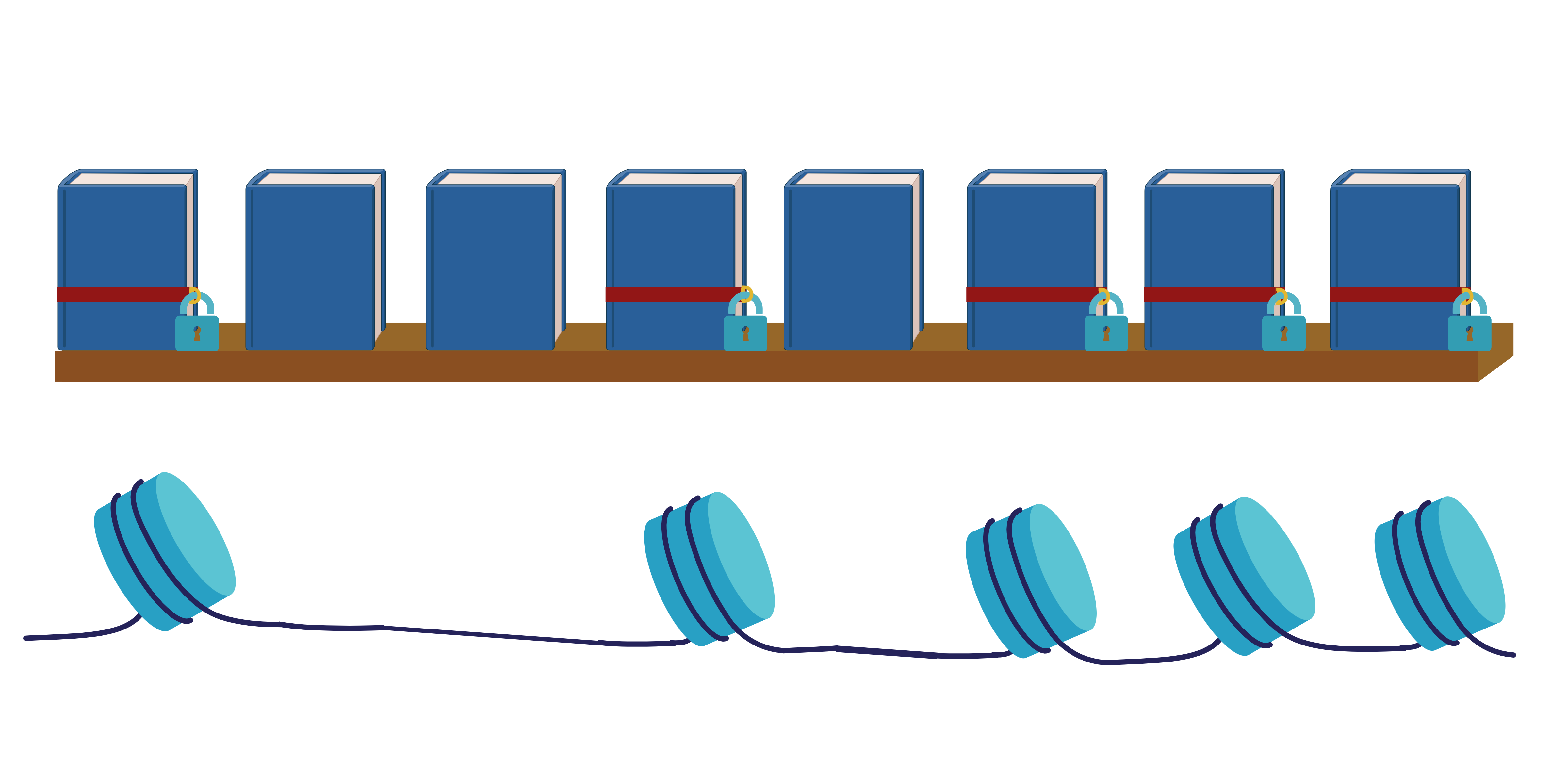
Ons DNA kan voorgesteld worden als een grote bibliotheek vol boeken, waarin elk boek een gen vertegenwoordigt. Deze boeken geven de instructies voor het maken van eiwitten, de bouwstenen van ons lichaam. Deze eiwitten bepalen veel van onze eigenschappen, zoals je huidskleur, de kleur van je ogen en de werking van je lichaam. Ons DNA draagt als het ware het ontwerp voor je bestaan. Toch kunnen twee personen met exact hetzelfde DNA er heel verschillend uitzien of zich anders gedragen. Neem bijvoorbeeld eeneiige tweelingen: waarom kan één helft van een eeneiige tweeling ten prooi vallen aan een hartaandoening op jongere leeftijd, terwijl de andere zus kerngezond blijft? Dit fenomeen is te danken aan epigenetica, waarbij de invloed van omgevingsfactoren en levensstijl ervoor zullen zorgen dat bepaalde genen of boeken meer worden ‘uitgelezen’ dan andere. Hierdoor kunnen mensen met dezelfde genetische aanleg heel verschillende levens leiden.
Histonen als sloten
Niet alle genen hoeven continu actief te zijn. Ze worden alleen gebruikt als dat nodig is. Om deze beschikbaarheid te reguleren, kan het DNA worden opgerold rond histonen (een bepaald type eiwit) om de genetische informatie compact op te vouwen. Wanneer DNA zich oprolt rond een complex van acht histonen, noemen we dat ook wel een nucleosoom. In opgevouwen toestand is het DNA niet beschikbaar en kunnen er geen eiwitten worden aangemaakt - die genen staan dan als het ware ‘uit’. Je kunt histonen zien als de sloten op de boeken in onze genetische bibliotheek: wanneer ze op slot zijn, is de informatie binnenin ontoegankelijk. Welke genen 'open' of 'op slot' zijn, bepaalt voor een groot deel hoe we eruitzien en functioneren.
Metalen zijn van levensbelang
Naast DNA en eiwitten spelen metalen ook een belangrijke rol in het lichaam. We kunnen niet leven zonder metalen. Bekende mineralen zoals calcium en fosfor zorgen bijvoorbeeld voor sterke botten, natrium, kalium en chloride daarentegen helpen bij het reguleren van onze vochtbalans. Bovendien bevat ongeveer de helft van alle gekende eiwitten een metaal, wat essentieel is voor de werking van dat eiwit. Een goed voorbeeld hiervan is hemoglobine, een belangrijk eiwit in ons bloed dat dankzij ijzer zuurstof van de longen naar de rest van het lichaam transporteert.
Aan de andere kant hebben metalen ook een keerzijde. Ze kunnen oxidatieve stress veroorzaken, wat betekent dat ze ervoor zorgen dat zuurstof in een schadelijke vorm in de cellen komt. Dit kan ons DNA beschadigen, wat op zijn beurt kan leiden tot kanker. Daarom hebben veel onderzoeken gekeken naar hoe metalen bijdragen aan DNA-schade. De rol van deze metalen op onze ‘sloten’ is echter nog niet helemaal duidelijk.
Kunnen metalen onze genen reguleren?
Omdat metalen zo belangrijk zijn, rijst de vraag: spelen ze ook een rol in het reguleren van onze genen? Epigenetica is een complex proces waar veel factoren op elkaar inspelen en elkaar beïnvloeden. Naast histonen, die functioneren als 'sloten' op onze genen, kunnen andere moleculen en stoffen de genen verder vergrendelen of juist toegankelijk maken. Eén van deze factoren zijn acetylaties, die de sloten 'verzwakken' waardoor het DNA zich ontrolt en weer gelezen kan worden. Maar wat als metalen deze sloten ook kunnen beïnvloeden? Recente studies hebben aangetoond dat koper kan binden aan histonen. Aangezien ijzer in veel eiwitten een rol speelt, is de volgende vraag: kan ijzer ook aan histonen binden? En zou dit invloed hebben op welke genen actief zijn?
De zoektocht naar ijzerbinding aan histonen
Om te onderzoeken of ijzer zich daadwerkelijk aan histonen kan binden, moeten we eerst een 3D-model maken van het nucleosoom—het complex van DNA en 8 histonen. Om dit complex in grote hoeveelheden te produceren, maken we gebruik van verschillende stappen. Dit proces begint met genetisch gemodificeerde bacteriën, die de instructies krijgen om de benodigde eiwitten aan te maken. Vervolgens worden deze eiwitten gezuiverd uit de bacteriën en samengebracht om het complex te vormen. Dit proces is in realiteit niet evident. Het nucleosoom bestaat uit vier verschillende soorten histonen, wat betekent dat elke histon zijn eigen specifieke stappen nodig heeft om geoptimaliseerd te worden. Desondanks slaagden we erin om drie van de vier histonen aan te maken. Het onderzoek naar het vierde histon is nog in volle gang, maar binnenkort kunnen we wellicht vaststellen of ijzer in staat is om aan histonen te binden en zo onze genen te beïnvloeden.
Het begrijpen van hoe metalen zoals ijzer invloed kunnen hebben op onze genen opent nieuwe mogelijkheden in de geneeskunde. Als blijkt dat ijzer en andere metalen een rol spelen in het 'aanzetten' of 'uitzetten' van genen, zou dit kunnen leiden tot baanbrekende behandelingen voor genetische aandoeningen of ziekten zoals kanker. Bepaalde therapieën zouden juist de aanwezigheid van de metalen kunnen gebruiken om kankercellen onschadelijk te maken of hun ontwikkeling af te remmen.
Bibliografie
1. Schindelin, J., et al., Fiji: an open-source platform for biological-image analysis. Nature Methods, 2012. 9(7): p. 676-682.
2. Jurrus, E., et al., Improvements to the APBS biomolecular solvation software suite. Protein Science, 2018. 27(1): p. 112-128.
3. Gibney, E.R. and C.M. Nolan, Epigenetics and gene expression. Heredity, 2010. 105(1): p. 4-13.
4. Tronick, E. and R.G. Hunter, Waddington, Dynamic Systems, and Epigenetics. Front Behav Neurosci, 2016. 10: p. 107.
5. Dupont, C., D.R. Armant, and C.A. Brenner, Epigenetics: definition, mechanisms and clinical perspective. Semin Reprod Med, 2009. 27(5): p. 351-7.
6. Al Aboud, N.M., C. Tupper, and I. Jialal, Genetics, Epigenetic Mechanism, in StatPearls. 2024: Treasure Island (FL).
7. Hamilton, J.P., Epigenetics: principles and practice. Dig Dis, 2011. 29(2): p. 130-5.
8. Jin, B., Y. Li, and K.D. Robertson, DNA methylation: superior or subordinate in the epigenetic hierarchy? Genes Cancer, 2011. 2(6): p. 607-17.
9. Jang, H.S., et al., CpG and Non-CpG Methylation in Epigenetic Gene Regulation and Brain Function. Genes (Basel), 2017. 8(6).
10. Pontecorvo, G., B. De Felice, and M. Carfagna, Novel methylation at GpC dinucleotide in the fish Sparus aurata genome. Molecular Biology Reports, 2000. 27(4): p. 225-230.
11. van der Wijst, M.G., et al., Experimental mitochondria-targeted DNA methylation identifies GpC methylation, not CpG methylation, as potential regulator of mitochondrial gene expression. Sci Rep, 2017. 7(1): p. 177.
12. Theys, C., et al., Mitochondrial GpC and CpG DNA Hypermethylation Cause Metabolic Stress-Induced Mitophagy and Cholestophagy. International Journal of Molecular Sciences, 2023. 24(22): p. 16412.
13. Moore, L.D., T. Le, and G. Fan, DNA Methylation and Its Basic Function. Neuropsychopharmacology, 2013. 38(1): p. 23-38.
14. Kriukienė, E., Z. Liutkevičiūtė, and S. Klimašauskas, 5-Hydroxymethylcytosine--the elusive epigenetic mark in mammalian DNA. Chem Soc Rev, 2012. 41(21): p. 6916-30.
15. Dhar, G.A., et al., DNA methylation and regulation of gene expression: Guardian of our health. The Nucleus, 2021. 64(3): p. 259-270.
16. Suzuki, M.M. and A. Bird, DNA methylation landscapes: provocative insights from epigenomics. Nature Reviews Genetics, 2008. 9(6): p. 465-476.
17. Bommarito, P.A. and R.C. Fry, Chapter 2-1 - The Role of DNA Methylation in Gene Regulation, in Toxicoepigenetics, S.D. McCullough and D.C. Dolinoy, Editors. 2019, Academic Press. p. 127-151.
18. Sadakierska-Chudy, A. and M. Filip, A Comprehensive View of the Epigenetic Landscape. Part II: Histone Post-translational Modification, Nucleosome Level, and Chromatin Regulation by ncRNAs. Neurotoxicity Research, 2015. 27(2): p. 172-197.
19. Shahid, Z., et al., Genetics, Histone Code, in StatPearls. 2024: Treasure Island (FL).
20. Dai, H. and Z. Wang, Histone Modification Patterns and Their Responses to Environment. Current Environmental Health Reports, 2014. 1(1): p. 11-21.
21. Albini, S., V. Zakharova, and S. Ait-Si-Ali, Chapter 3 - Histone Modifications, in Epigenetics and Regeneration, D. Palacios, Editor. 2019, Academic Press. p. 47-72.
22. Karmodiya, K., et al., H3K9 and H3K14 acetylation co-occur at many gene regulatory elements, while H3K14ac marks a subset of inactive inducible promoters in mouse embryonic stem cells. BMC Genomics, 2012. 13(1): p. 424.
23. DesJarlais, R. and P.J. Tummino, Role of Histone-Modifying Enzymes and Their Complexes in Regulation of Chromatin Biology. Biochemistry, 2016. 55(11): p. 1584-1599.
24. Miller, J.L. and P.A. Grant, The role of DNA methylation and histone modifications in transcriptional regulation in humans. Subcell Biochem, 2013. 61: p. 289-317.
25. Greer, E.L. and Y. Shi, Histone methylation: a dynamic mark in health, disease and inheritance. Nat Rev Genet, 2012. 13(5): p. 343-57.
26. He, S., et al., Chapter 33 - Epigenetic Mechanisms of Retinal Disease, in Retina (Fifth Edition), S.J. Ryan, et al., Editors. 2013, W.B. Saunders: London. p. 642-651.
27. Liu, R., et al., Post-translational modifications of histones: Mechanisms, biological functions, and therapeutic targets. MedComm, 2023. 4(3): p. e292.
28. Yang, X., et al., Epigenetic modulations of noncoding RNA: a novel dimension of Cancer biology. Molecular Cancer, 2020. 19(1): p. 64.
29. Wei, J.-W., et al., Non-coding RNAs as regulators in epigenetics (Review). Oncol Rep, 2017. 37(1): p. 3-9.
30. Wu, Y.-L., et al., Epigenetic regulation in metabolic diseases: mechanisms and advances in clinical study. Signal Transduction and Targeted Therapy, 2023. 8(1): p. 98.
31. Choudhuri, S., Y. Cui, and C.D. Klaassen, Molecular targets of epigenetic regulation and effectors of environmental influences. Toxicol Appl Pharmacol, 2010. 245(3): p. 378-93.
32. Collins, L.J., B. Schönfeld, and X.S. Chen, Chapter 4 - The Epigenetics of Non-coding RNA, in Handbook of Epigenetics, T. Tollefsbol, Editor. 2011, Academic Press: San Diego. p. 49-61.
33. Statello, L., et al., Gene regulation by long non-coding RNAs and its biological functions. Nature Reviews Molecular Cell Biology, 2021. 22(2): p. 96-118.
34. Li, J., et al., Long noncoding RNA XIST: Mechanisms for X chromosome inactivation, roles in sex-biased diseases, and therapeutic opportunities. Genes & Diseases, 2022. 9(6): p. 1478-1492.
35. Gong, C. and L.E. Maquat, lncRNAs transactivate STAU1-mediated mRNA decay by duplexing with 3' UTRs via Alu elements. Nature, 2011. 470(7333): p. 284-8.
36. Ana Luisa Pedroso, A., et al., The Function of lncRNAs as Epigenetic Regulators, in Non-Coding RNAs, T. Lütfi, A. Sümer, and T. Esen, Editors. 2019, IntechOpen: Rijeka. p. Ch. 9.
37. Lolak, S., P. Suwannarat, and R.H. Lipsky, Chapter Five - Epigenetics of Depression, in Progress in Molecular Biology and Translational Science, S. Akbarian and F. Lubin, Editors. 2014, Academic Press. p. 103-137.
38. Gan, J., et al., A unique glimpse into the crosstalk between different epigenetic mechanisms in porcine embryonic development†. Biology of Reproduction, 2022. 107(6): p. 1411-1424.
39. Cedar, H. and Y. Bergman, Linking DNA methylation and histone modification: patterns and paradigms. Nature Reviews Genetics, 2009. 10(5): p. 295-304.
40. Huang, W., et al., LncRNA-mediated DNA methylation: an emerging mechanism in cancer and beyond. Journal of Experimental & Clinical Cancer Research, 2022. 41(1): p. 100.
41. Kornberg, R.D., Structure of chromatin. Annu Rev Biochem, 1977. 46: p. 931-54.
42. Heitz, E., “Das” Heterochromatin der Moose. 1928: Bornträger.
43. Straub, T., Heterochromatin dynamics. PLoS Biol, 2003. 1(1): p. E14.
44. Cosgrove, M.S. and C. Wolberger, How does the histone code work? Biochem Cell Biol, 2005. 83(4): p. 468-76.
45. Luger, K., et al., Crystal structure of the nucleosome core particle at 2.8 A resolution. Nature, 1997. 389(6648): p. 251-60.
46. Szerlong, H.J. and J.C. Hansen, Nucleosome distribution and linker DNA: connecting nuclear function to dynamic chromatin structure. Biochem Cell Biol, 2011. 89(1): p. 24-34.
47. Woodcock, C.L.F., J.P. Safer, and J.E. Stanchfield, Structural repeating units in chromatin: I. Evidence for their general occurrence. Experimental Cell Research, 1976. 97(1): p. 101-110.
48. Zhu, P. and G. Li, Structural insights of nucleosome and the 30-nm chromatin fiber. Curr Opin Struct Biol, 2016. 36: p. 106-15.
49. McGhee, J.D., et al., Higher order structure of chromatin: orientation of nucleosomes within the 30 nm chromatin solenoid is independent of species and spacer length. Cell, 1983. 33(3): p. 831-41.
50. Li, G. and D. Reinberg, Chromatin higher-order structures and gene regulation. Curr Opin Genet Dev, 2011. 21(2): p. 175-86.
51. Maeshima, K., et al., Chromatin as dynamic 10-nm fibers. Chromosoma, 2014. 123(3): p. 225-37.
52. Hou, Z., et al., Structure of native chromatin fibres revealed by Cryo-ET in situ. Nature Communications, 2023. 14(1): p. 6324.
53. Hocher, A., et al., Histones with an unconventional DNA-binding mode in vitro are major chromatin constituents in the bacterium Bdellovibrio bacteriovorus. Nature Microbiology, 2023. 8(11): p. 2006-2019.
54. Luzhetskaya, O.P., S.E. Sedykh, and G.A. Nevinsky, How Human H1 Histone Recognizes DNA. Molecules, 2020. 25(19).
55. Workman, J. and S.M. Abmayr, Fundamentals of Chromatin. 2014. 1-587.
56. Ramaswamy, A. and I. Ioshikhes, Chapter Four - Dynamics of Modeled Oligonucleosomes and the Role of Histone Variant Proteins in Nucleosome Organization, in Advances in Protein Chemistry and Structural Biology, R. Donev, Editor. 2013, Academic Press. p. 119-149.
57. Zheng, C. and J.J. Hayes, Structures and interactions of the core histone tail domains. Biopolymers, 2003. 68(4): p. 539-46.
58. Sato, S., M. Dacher, and H. Kurumizaka, Nucleosome Structures Built from Highly Divergent Histones: Parasites and Giant DNA Viruses. Epigenomes, 2022. 6(3): p. 22.
59. McGinty, R.K. and S. Tan, Histone, Nucleosome, and Chromatin Structure, in Fundamentals of Chromatin, J.L. Workman and S.M. Abmayr, Editors. 2014, Springer New York: New York, NY. p. 1-28.
60. Zhang, H., et al., Mapping the electrostatic potential of the nucleosome acidic patch. Scientific Reports, 2021. 11(1): p. 23013.
61. Paul, S., Histone “acidic patch”: a hotspot in chromatin biology. The Nucleus, 2021. 64(3): p. 271-275.
62. McGinty, R.K. and S. Tan, Principles of nucleosome recognition by chromatin factors and enzymes. Current Opinion in Structural Biology, 2021. 71: p. 16-26.
63. Luger, K. and T.J. Richmond, The histone tails of the nucleosome. Curr Opin Genet Dev, 1998. 8(2): p. 140-6.
64. Iwasaki, W., et al., Contribution of histone N-terminal tails to the structure and stability of nucleosomes. FEBS Open Bio, 2013. 3: p. 363-9.
65. Li, Z. and H. Kono, Distinct Roles of Histone H3 and H2A Tails in Nucleosome Stability. Sci Rep, 2016. 6: p. 31437.
66. Peng, Y., et al., Histone tails as signaling antennas of chromatin. Curr Opin Struct Biol, 2021. 67: p. 153-160.
67. Luger, K., Dynamic nucleosomes. Chromosome Research, 2006. 14(1): p. 5-16.
68. Peterson, C.L. and M.-A. Laniel, Histones and histone modifications. Current Biology, 2004. 14(14): p. R546-R551.
69. Bure, I.V., M.V. Nemtsova, and E.B. Kuznetsova, Histone Modifications and Non-Coding RNAs: Mutual Epigenetic Regulation and Role in Pathogenesis. International Journal of Molecular Sciences, 2022. 23(10): p. 5801.
70. Strahl, B.D. and C.D. Allis, The language of covalent histone modifications. Nature, 2000. 403(6765): p. 41-45.
71. Lyubitelev, A.V., et al., Structure and functions of linker histones. Biochemistry (Moscow), 2016. 81(3): p. 213-223.
72. Buschbeck, M. and S.B. Hake, Variants of core histones and their roles in cell fate decisions, development and cancer. Nature Reviews Molecular Cell Biology, 2017. 18(5): p. 299-314.
73. Amatori, S., et al., The dark side of histones: genomic organization and role of oncohistones in cancer. Clinical Epigenetics, 2021. 13(1): p. 71.
74. Weber, C.M. and S. Henikoff, Histone variants: dynamic punctuation in transcription. Genes & Development, 2014. 28(7): p. 672-682.
75. Seal, R.L., et al., A standardized nomenclature for mammalian histone genes. Epigenetics & Chromatin, 2022. 15(1): p. 34.
76. Flaus, A., J.A. Downs, and T. Owen-Hughes, Histone isoforms and the oncohistone code. Current Opinion in Genetics & Development, 2021. 67: p. 61-66.
77. Martire, S. and L.A. Banaszynski, The roles of histone variants in fine-tuning chromatin organization and function. Nature Reviews Molecular Cell Biology, 2020. 21(9): p. 522-541.
78. Biterge, B. and R. Schneider, Histone variants: key players of chromatin. Cell and Tissue Research, 2014. 356(3): p. 457-466.
79. De Rop, V., A. Padeganeh, and P.S. Maddox, CENP-A: the key player behind centromere identity, propagation, and kinetochore assembly. Chromosoma, 2012. 121(6): p. 527-38.
80. Zhang, W., J. Feng, and Q. Li, The replisome guides nucleosome assembly during DNA replication. Cell & Bioscience, 2020. 10(1): p. 37.
81. Feng, S., et al., RIF1-ASF1-mediated high-order chromatin structure safeguards genome integrity. Nature Communications, 2022. 13(1): p. 957.
82. De Koning, L., et al., Histone chaperones: an escort network regulating histone traffic. Nature Structural & Molecular Biology, 2007. 14(11): p. 997-1007.
83. Serra-Cardona, A. and Z. Zhang, Replication-Coupled Nucleosome Assembly in the Passage of Epigenetic Information and Cell Identity. Trends Biochem Sci, 2018. 43(2): p. 136-148.
84. Ohtomo, H., et al., Dynamic Solution Structures of Whole Human NAP1 Dimer Bound to One and Two Histone H2A-H2B Heterodimers Obtained by Integrative Methods. Journal of Molecular Biology, 2023. 435(15): p. 168189.
85. Robert, F. and C. Jeronimo, Transcription-coupled nucleosome assembly. Trends in Biochemical Sciences, 2023. 48(11): p. 978-992.
86. Tyagi, M., et al., Chromatin remodelers: We are the drivers!! Nucleus, 2016. 7(4): p. 388-404.
87. Reyes, A.A., R.D. Marcum, and Y. He, Structure and Function of Chromatin Remodelers. Journal of Molecular Biology, 2021. 433(14): p. 166929.
88. Liang, H., et al., The role of SWI/SNF complexes in digestive system neoplasms. Medical Oncology, 2024. 41(5): p. 119.
89. Sahu, R.K., S. Singh, and R.S. Tomar, The mechanisms of action of chromatin remodelers and implications in development and disease. Biochemical Pharmacology, 2020. 180: p. 114200.
90. Permyakov, E.A., Metal Binding Proteins. Encyclopedia, 2021. 1(1): p. 261-292.
91. Yruela, I., Transition metals in plant photosynthesis. Metallomics, 2013. 5(9): p. 1090-109.
92. Thiele, D.J., Metal-regulated transcription in eukaryotes. Nucleic Acids Res, 1992. 20(6): p. 1183-91.
93. Smethurst, D.G.J. and N. Shcherbik, Interchangeable utilization of metals: New perspectives on the impacts of metal ions employed in ancient and extant biomolecules. J Biol Chem, 2021. 297(6): p. 101374.
94. Janisse, S.E., R.L. Fernandez, and M.C. Heffern, Characterizing metal–biomolecule interactions by mass spectrometry. Trends in Biochemical Sciences, 2023. 48(9): p. 815-825.
95. Ibers, J.A. and R.H. Holm, Modeling coordination sites in metallobiomolecules. Science, 1980. 209(4453): p. 223-35.
96. Chalkley, M.J., S.I. Mann, and W.F. DeGrado, De novo metalloprotein design. Nat Rev Chem, 2022. 6(1): p. 31-50.
97. Tainer, J.A., V.A. Roberts, and E.D. Getzoff, Metal-binding sites in proteins. Curr Opin Biotechnol, 1991. 2(4): p. 582-91.
98. Andreini, C., et al., The human iron-proteome†. Metallomics, 2018. 10(9): p. 1223-1231.
99. Dlouhy, A.C. and C.E. Outten, The Iron Metallome in Eukaryotic Organisms, in Metallomics and the Cell, L. Banci, Editor. 2013, Springer Netherlands: Dordrecht. p. 241-278.
100. Feig, A.L. and S.J. Lippard, Reactions of Non-Heme Iron(II) Centers with Dioxygen in Biology and Chemistry. Chemical Reviews, 1994. 94(3): p. 759-805.
101. Kaplan, J. and D.M. Ward, The essential nature of iron usage and regulation. Curr Biol, 2013. 23(15): p. R642-6.
102. Skrzypczak-Jankun, E., et al., Lipoxygenase: a molecular complex with a non-heme iron. Journal of Molecular Structure: THEOCHEM, 1996. 374(1): p. 47-52.
103. Wang, D.L., et al., Electrochemical properties of the diiron core of uteroferrin and its anion complexes. Biochemistry, 1991. 30(33): p. 8187-94.
104. Palmer, G. and J. Reedijk, Nonmenclature of electron-transfer proteins: Recommendations 1989. Biochimica et Biophysica Acta (BBA) - Bioenergetics, 1991. 1060(3): p. vii-xix.
105. Cyr, A.R. and F.E. Domann, The redox basis of epigenetic modifications: from mechanisms to functional consequences. Antioxid Redox Signal, 2011. 15(2): p. 551-89.
106. Wang, Y., et al., Iron Metabolism in Cancer. Int J Mol Sci, 2018. 20(1).
107. Logie, E., et al., Ferroptosis Induction in Multiple Myeloma Cells Triggers DNA Methylation and Histone Modification Changes Associated with Cellular Senescence. Int J Mol Sci, 2021. 22(22).
108. Mohideen, K., R. Muhammad, and C.A. Davey, Perturbations in nucleosome structure from heavy metal association. Nucleic Acids Res, 2010. 38(18): p. 6301-11.
109. Saavedra, R.A., Structure, function, and evolution of the metal-binding domain in the nucleosome. Bioessays, 2023. 45(5): p. e2200192.
110. Palma, F.R., et al., Histone H3.1 is a chromatin-embedded redox sensor triggered by tumor cells developing adaptive phenotypic plasticity and multidrug resistance. Cell Rep, 2024. 43(3): p. 113897.
111. Peana, M., et al., Nickel binding sites in histone proteins: Spectroscopic and structural characterization. Coordination Chemistry Reviews, 2013. 257(19): p. 2737-2751.
112. Bal, W., H. Kozłowski, and K.S. Kasprzak, Molecular models in nickel carcinogenesis. Journal of Inorganic Biochemistry, 2000. 79(1): p. 213-218.
113. Zoroddu, M.A., et al., Molecular mechanisms in nickel carcinogenesis: modeling Ni(II) binding site in histone H4. Environ Health Perspect, 2002. 110 Suppl 5(Suppl 5): p. 719-23.
114. Karavelas, T., et al., Coordination properties of Cu(ii) and Ni(ii) ions towards the C-terminal peptide fragment -TYTEHA- of histone H4. Dalton Transactions, 2008(9): p. 1215-1223.
115. Mylonas, M., et al., The binding of Ni(II) ions to terminally blocked hexapeptides derived from the metal binding -ESHH- motif of histone H2A. Journal of the Chemical Society-Dalton Transactions, 2002. 38: p. 4296-4306.
116. Nunes, A.M., et al., Coordination of Cu2+and Ni2+ with the histone model peptide of H2B N-terminal tail (1-31 residues): A spectroscopic study. Dalton Transactions, 2010. 39(18): p. 4369-4381.
117. Nunes, A.M., et al., Interaction of histone H2B (fragment 63-93) with Ni(ii). An NMR study. Dalton Trans, 2009(11): p. 1904-13.
118. Nunes, A.M., et al., The possible role of 94-125 peptide fragment of histone H2B in nickel-induced carcinogenesis. Inorganic chemistry, 2010. 49 12: p. 5658-68.
119. Midorikawa, K., M. Murata, and S. Kawanishi, Histone peptide AKRHRK enhances H(2)O(2)-induced DNA damage and alters its site specificity. Biochemical and biophysical research communications, 2005. 333 4: p. 1073-7.
120. Panagiotou, K., et al., Cu(II) and Ni(II) interactions with the terminally blocked hexapeptide Ac-Leu-Ala-His-Tyr-Asn-Lys-amide model of histone H2B (80-85). Bioinorg Chem Appl, 2008. 2008: p. 257038.
121. Zavitsanos, K., et al., Copper effective binding with 32–62 and 94–125 peptide fragments of histone H2B. Journal of Inorganic Biochemistry, 2011. 105(1): p. 102-110.
122. Zavitsanos, K., et al., Interaction of Cu(ii) and Ni(ii) with the 63–93 fragment of histone H2B. Dalton Transactions, 2008(44): p. 6179-6187.
123. Saavedra, R.A., Histones and Metal-Binding Domains. Science, 1986. 234(4783): p. 1589-1589.
124. Adamczyk, M., et al., A zinc-finger like metal binding site in the nucleosome. FEBS Lett, 2007. 581(7): p. 1409-16.
125. Mylonas, M., J.C. Plakatouras, and N. Hadjiliadis, Interactions of Ni(II) and Cu(II) ions with the hydrolysis products of the C-terminal -ESHH- motif of histone H2A model peptides. Association of the stability of the complexes formed with the cleavage of the -E-S- bond. Dalton Trans, 2004(24): p. 4152-60.
126. Daban, J.R. and C.R. Cantor, Use of fluorescent probes to study nucleosomes. Methods Enzymol, 1989. 170: p. 192-214.
127. Wang, B.C., et al., The octameric histone core of the nucleosome. Structural issues resolved. J Mol Biol, 1994. 236(1): p. 179-88.
128. Wiedemann, C., et al., Cysteines and Disulfide Bonds as Structure-Forming Units: Insights From Different Domains of Life and the Potential for Characterization by NMR. Frontiers in Chemistry, 2020. 8.
129. Camerini-Otero, R.D. and G. Felsenfeld, Histone H3 disulfide dimers and nucleosome structure. Proc Natl Acad Sci U S A, 1977. 74(12): p. 5519-23.
130. Blundell, T., et al., Insulin: The Structure in the Crystal and its Reflection in Chemistry and Biology by, in Advances in Protein Chemistry, C.B. Anfinsen, J.T. Edsall, and F.M. Richards, Editors. 1972, Academic Press. p. 279-402.
131. Yamashita, M.M., et al., Where metal ions bind in proteins. Proc Natl Acad Sci U S A, 1990. 87(15): p. 5648-52.
132. Bal, W., et al., Interactions of nickel(II) with histones. Stability and solution structure of complexes with CH3CO-Cys-Ala-Ile-His-NH2, a putative metal binding sequence of histone H3. Chem Res Toxicol, 1995. 8(5): p. 683-92.
133. Bal, W., et al., Interaction of Nickel(II) with histones: in vitro binding of nickel(II) to the core histone tetramer. Arch Biochem Biophys, 1999. 364(2): p. 161-6.
134. Attar, N., et al., The histone H3-H4 tetramer is a copper reductase enzyme. Science, 2020. 369(6499): p. 59-64.
135. Entezari, S., et al., Iron Chelators in Treatment of Iron Overload. Journal of Toxicology, 2022. 2022: p. 4911205.
136. Bryan, S.E., et al., Interactions of mercury and copper with constitutive heterochromatin and euchromatin in vivo and in vitro. Biochemistry, 1976. 15(8): p. 1667-76.
137. Schneider, C.A., W.S. Rasband, and K.W. Eliceiri, NIH Image to ImageJ: 25 years of image analysis. Nature Methods, 2012. 9(7): p. 671-675.
138. Longo, P.A., et al., Transient mammalian cell transfection with polyethylenimine (PEI). Methods Enzymol, 2013. 529: p. 227-40.
139. VanderWall, K., et al., Iron in multiple myeloma. Crit Rev Oncog, 2013. 18(5): p. 449-61.
140. Mobarra, N., et al., A Review on Iron Chelators in Treatment of Iron Overload Syndromes. Int J Hematol Oncol Stem Cell Res, 2016. 10(4): p. 239-247.
141. Tsvetkov, P., et al., Copper induces cell death by targeting lipoylated TCA cycle proteins. Science, 2022. 375(6586): p. 1254-1261.
142. Gerson, S.L., et al., Chapter 57 - Pharmacology and Molecular Mechanisms of Antineoplastic Agents for Hematologic Malignancies, in Hematology (Seventh Edition), R. Hoffman, et al., Editors. 2018, Elsevier. p. 849-912.
143. Rensvold, J.W., et al., Iron Deprivation Induces Transcriptional Regulation of Mitochondrial Biogenesis. J Biol Chem, 2016. 291(40): p. 20827-20837.
144. Luger, K., et al., Characterization of nucleosome core particles containing histone proteins made in bacteria. J Mol Biol, 1997. 272(3): p. 301-11.
145. Tanaka, Y., et al., Expression and purification of recombinant human histones. Methods, 2004. 33(1): p. 3-11.
146. Burnett, V.L. and D.L. Springer, High-Level Expression of Human Histone H4 in E. Coli. BioTechniques, 1999. 26(1): p. 30-34.
147. Gopal, G.J. and A. Kumar, Strategies for the Production of Recombinant Protein in Escherichia coli. The Protein Journal, 2013. 32(6): p. 419-425.
148. Mauro, V.P. and S.A. Chappell, A critical analysis of codon optimization in human therapeutics. Trends Mol Med, 2014. 20(11): p. 604-13.
149. Bonekamp, F. and K.F. Jensen, The AGG codon is translated slowly in E. coli even at very low expression levels. Nucleic Acids Research, 1988. 16(7): p. 3013-3024.
150. Wang, Y., et al., An Engineered Rare Codon Device for Optimization of Metabolic Pathways. Scientific Reports, 2016. 6(1): p. 20608.
151. Luger, K., T.J. Rechsteiner, and T.J. Richmond, Expression and purification of recombinant histones and nucleosome reconstitution. Methods Mol Biol, 1999. 119: p. 1-16.
152. Murby, M., M. Uhlén, and S. Ståhl, Upstream Strategies to Minimize Proteolytic Degradation upon Recombinant Production inEscherichia coli. Protein Expression and Purification, 1996. 7(2): p. 129-136.
153. Dong, W., et al., The Role of Non-Catalytic Domains of Hrp3 in Nucleosome Remodeling. International Journal of Molecular Sciences, 2021. 22: p. 1793.
154. Francis, D.M. and R. Page, Strategies to optimize protein expression in E. coli. Curr Protoc Protein Sci, 2010. Chapter 5(1): p. 5.24.1-5.24.29.
155. Lusser, A. and J.T. Kadonaga, Strategies for the reconstitution of chromatin. Nature Methods, 2004. 1(1): p. 19-26.
156. Yamaguchi, H. and M. Miyazaki, Refolding techniques for recovering biologically active recombinant proteins from inclusion bodies. Biomolecules, 2014. 4(1): p. 235-51.
157. Lercher, L., et al., Generation of a synthetic GlcNAcylated nucleosome reveals regulation of stability by H2A-Thr101 GlcNAcylation. Nature Communications, 2015. 6(1): p. 7978.
158. Dyer, P.N., et al., Reconstitution of nucleosome core particles from recombinant histones and DNA. Methods Enzymol, 2004. 375: p. 23-44.
159. Bhattacharya, S. and D.S. Gupta, A Simple Method to Produce Sub-Nucleosome Complexes of High Purity In Vitro. Advances in Bioscience and Biotechnology, 2016. 07: p. 133-141.
160. Singh, A., et al., Protein recovery from inclusion bodies of Escherichia coli using mild solubilization process. Microbial Cell Factories, 2015. 14(1): p. 41.
161. Mirhosseini, S., et al., The Efficient Solubilization and Refolding of Recombinant Organophosphorus Hydrolyses Inclusion Bodies Produced in Escherichia coli. Journal of Applied Biotechnology Reports, 2019. 6: p. 20-25.
162. Tessadori, F., et al., Recurrent de novo missense variants across multiple histone H4 genes underlie a neurodevelopmental syndrome. Am J Hum Genet, 2022. 109(4): p. 750-758.
163. Tagami, H., et al., Histone H3.1 and H3.3 complexes mediate nucleosome assembly pathways dependent or independent of DNA synthesis. Cell, 2004. 116(1): p. 51-61.
164. Zhang, J., et al., ACSL4: a double-edged sword target in multiple myeloma, promotes cell proliferation and sensitizes cell to ferroptosis. Carcinogenesis, 2023. 44(3): p. 242-251.
165. Yang, F., et al., The iron chelator deferoxamine decreases myeloma cell survival. J Int Med Res, 2021. 49(1): p. 300060520987396.
166. Pullarkat, V., et al., Iron chelators induce autophagic cell death in multiple myeloma cells. Leuk Res, 2014. 38(8): p. 988-96.
167. Feng, H. and B. Stockwell, Unsolved mysteries: How does lipid peroxidation cause ferroptosis? PLOS Biology, 2018. 16: p. e2006203.
168. Enright, H.U., W.J. Miller, and R.P. Hebbel, Nucleosomal histone protein protects DNA from iron-mediated damage. Nucleic Acids Research, 1992. 20(13): p. 3341-3346.
169. Liang, Q. and P.C. Dedon, Cu(II)/H2O2-induced DNA damage is enhanced by packaging of DNA as a nucleosome. Chem Res Toxicol, 2001. 14(4): p. 416-22.
170. Dragar, Č., S. Kralj, and P. Kocbek, Bioevaluation methods for iron-oxide-based magnetic nanoparticles. International Journal of Pharmaceutics, 2021. 597: p. 120348.
171. Murphy, M.P., et al., Guidelines for measuring reactive oxygen species and oxidative damage in cells and in vivo. Nature Metabolism, 2022. 4(6): p. 651-662.
172. Sharma, A., K. Singh, and A. Almasan, Histone H2AX phosphorylation: a marker for DNA damage. Methods Mol Biol, 2012. 920: p. 613-26.
173. Luger, K. and T.J. Richmond, DNA binding within the nucleosome core. Curr Opin Struct Biol, 1998. 8(1): p. 33-40.
174. Muthurajan, U.M., et al., Crystal structures of histone Sin mutant nucleosomes reveal altered protein-DNA interactions. Embo j, 2004. 23(2): p. 260-71.
175. Ordu, O., et al., Modification of the histone tetramer at the H3-H3 interface impacts tetrasome conformations and dynamics. J Chem Phys, 2018. 148(12): p. 123323.


