Heb jij een standaardlijf?
- EmmaDeceuninck
Hoe verschillend is elk lichaam? Open een anatomieboek en je ziet een menselijk lichaam met botten die op een vaste plaats zitten, organen met een vaste vorm en bloedvaten die netjes een uitgetekende route volgen. Maar bestaat er iets als een standaardlichaam? Het antwoord is eenvoudig: nee. En om ervoor te zorgen dat artsen niet voor verrassingen komen te staan tijdens een operatie, is het van belang om op voorhand te weten hoe anatomie kan variëren.

Anatomie van de arteriën zoals voorgesteld in een anatomieboek.
Dit is bijvoorbeeld belangrijk voor de anatomie van de arteriën in onze benen. Deze arteriën kunnen vernauwd of verstopt geraken, wat perifeer vasculair lijden wordt genoemd. Door deze vernauwingen bereikt er minder zuurstof het been, wat kan leiden tot pijn, krampen, vermoeidheid of moeilijke wondheling. De behandeling bestaat uit levensstijlaanpassingen en medicatie. Bij uitgesproken klachten is een ingreep nodig, waarbij de vernauwing of verstopping vaak wordt geopend met een ballonkatheter.
Al in 1928 toonde Adachi in ‘Das Arteriensystem der Japaner’ aan dat de arteriën, in tegenstelling tot wat anatomieboeken ons vertellen, niet bij iedereen dezelfde route volgen.
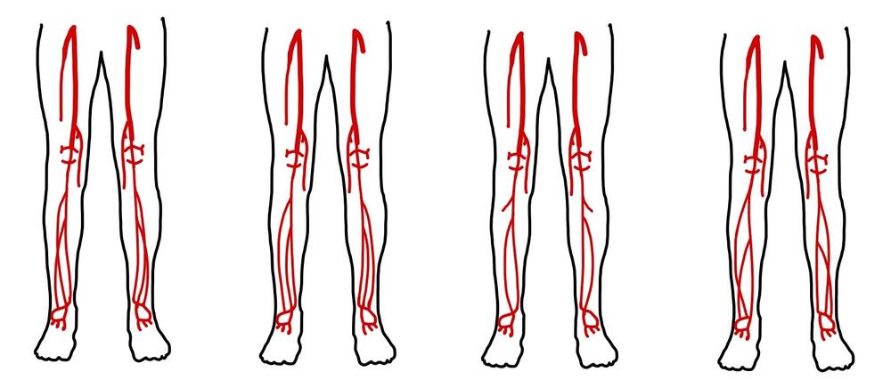
Vier voorbeelden van hoe het vertakkingspatroon van de arteriën in het onderbeen kan verschillen. Zoek de verschillen!
Voor een arts is het nuttig de routes van deze arteriën te kunnen volgen, hoe dik zijn ze, en is er een verschil tussen mannen en vrouwen? Om dat te achterhalen doken we een jaar lang in tientallen studies over dit onderwerp. Talrijke studies over de arteriën en hun routes en diameter zijn gepubliceerd maar een eenduidig overzicht ontbreekt door het gebrek aan gestandaardiseerde definities. Bovendien worden belangrijke kenmerken als geslacht of etniciteit vaak niet gerapporteerd. Een sluitend antwoord bleef dus uit.
Ook al is het duidelijk dat er een verschil is, er is geen manier om die verschillen duidelijk weer te geven. Bovendien zijn anatomische verschillen tussen mannen en vrouwen al lange tijd bekend, maar blijft het moeilijk om deze verschillen duidelijk in kaart te brengen en wordt dit in onderzoek niet altijd bestudeerd en gerapporteerd. Tot vandaag wordt de Kaukasische man vaker vertegenwoordigd in onderzoek. Deze resultaten worden gebruikt als basis voor de behandeling van alle patiënten. Maar is dit logisch? Nee. Het is een beetje alsof je shampoo enkel test op steil haar en verwacht dat het ook voor krullend haar werkt.
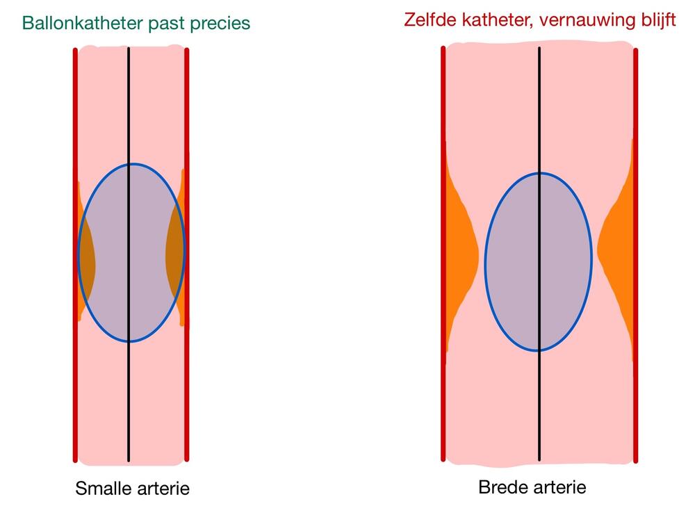
Eén ballonkatheter, twee verschillende diameters, dit illustreert dat dezelfde maat niet overal past.
Hoewel er dus veel anatomische verschillen bestaan, zijn deze in de huidige studies niet steeds zichtbaar. En die blinde vlek blijft jammer genoeg niet beperkt tot de wetenschappelijk artikels. Wanneer medische materialen ontworpen worden op basis van een beperkte populatie en vervolgens als maat voor alle patiënten gebruikt worden, kan dit grote gevolgen hebben voor de resultaten na een ingreep. Denk aan een ballonkatheter die net niet aansluit op de diameter van een bloedvat: dat kan de ingreep bemoeilijken of het risico op complicaties vergroten, terwijl dat net te vermijden was met betere kennis vooraf. Het is belangrijk dat behandelingen worden aangepast aan de individuele verschillen van elke patiënt. Daarom moeten we niet alleen afvragen: ‘Wat weten we?’ We moeten ook durven vragen: ‘Voor wie is de kennis van toepassing?’ Want als we echt medische zorg op maat van de patiënt willen maken, moeten we eerst zeker weten dat onze wetenschap niet langer uitgaat van één standaardmens.
Bibliografie
1. Mateus S, Paulo R, Coelho P, Rodrigues F, Marques V, Neiva HP, et al. Evaluation of Lower Limb Arteriovenous Diameters in Indoor Soccer Athletes: Arterial Doppler Ultrasound Study. Frontiers in Physiology. 2021;12.
2. Klein WM, Bartels LW, Bax L, van der Graaf Y, Mali W. Magnetic resonance imaging measurement of blood volume flow in peripheral arteries in healthy subjects. Journal of Vascular Surgery. 2003;38(5):1060-6.
3. Kil SW, Jung GS. Anatomical variations of the popliteal artery and its tibial branches: analysis in 1242 extremities. Cardiovasc Intervent Radiol. 2009;32(2):233-40.
4. Aragonés P, Rodríguez-Niedenführ M, Quinones S, de Blas CS, Konschake M, Sanudo JR, et al. Popliteal artery: Anatomical study and review of the literature. Ann Anat. 2021;234:151654.
5. Badagabettu Nayak S, Padur Aithal A, Kumar N, Regunathan D, Shetty P, Alathady Maloor P. A cadaveric study of variations of external iliac artery and its implication in trauma and radiology. Morphologie. 2019;103(341):24-31.
6. Nasr AY, Badawoud MH, Al-Hayani AA, Hussein AM. Origin of profunda femoris artery and its circumflex femoral branches: anatomical variations and clinical significance. Folia Morphol (Warsz). 2014;73(1):58-67.
7. Lorbeer R, Grotz A, Dörr M, Völzke H, Lieb W, Kühn JP, et al. Reference values of vessel diameters, stenosis prevalence, and arterial variations of the lower limb arteries in a male population sample using contrast-enhanced MR angiography. Plos One. 2018;13(6).
8. Adachi B, Hasebe K, Kyōto Teikoku D, Kenkyūsha, History of Medicine C, Paul Kligfield C. Das Arteriensystem der Japaner. Kyoto: Verlag der Kaiserlich-Japanischen Universität zu Kyoto; 1928.
9. Kim D, Orron DE, Skillman JJ. Surgical significance of popliteal arterial variants. A unified angiographic classification. Ann Surg. 1989;210(6):776-81.
10. Daniele A, Lucas SJE, Rendeiro C. Variability of flow-mediated dilation across lower and upper limb conduit arteries. European journal of applied physiology. 2024;124(11):3265-78.
11. Takahashi N, Higuchi T, Hirose Y, Yamanouchi H, Takatsuka H, Funayama K. Changes in aortic shape and diameters after death: comparison of early postmortem computed tomography with antemortem computed tomography. Forensic Sci Int. 2013;225(1-3):27-31.
12. Li X, Liu X, Li X, Xu L, Chen X, Liang F. Tortuosity of the superficial femoral artery and its influence on blood flow patterns and risk of atherosclerosis. Biomech Model Mechanobiol. 2019;18(4):883-96.
13. Wood NB, Zhao SZ, Zambanini A, Jackson M, Gedroyc W, Thom SA, et al. Curvature and tortuosity of the superficial femoral artery: a possible risk factor for peripheral arterial disease. J Appl Physiol (1985). 2006;101(5):1412-8.
14. Bruno A. Are There Differences in Vascular Disease Between Ethnic and Racial Groups? Stroke. 1998;29(1):2-3.
15. Chiam PT, Koh AS, Ewe SH, Sin YK, Chao VT, Ng CK, et al. Iliofemoral anatomy among Asians: implications for transcatheter aortic valve implantation. Int J Cardiol. 2013;167(4):1373-9.
16. Zhang T, Tsang W, Wijeysundera HC, Ko DT. Reporting and representation of ethnic minorities in cardiovascular trials: A systematic review. American Heart Journal. 2013;166(1):52-7.
17. Groman R, Ginsburg J. Racial and ethnic disparities in health care: a position paper of the American College of Physicians. Ann Intern Med. 2004;141(3):226-32.
18. Li JK. Arterial Wall Properties in Men and Women: Hemodynamic Analysis and Clinical Implications. Adv Exp Med Biol. 2018;1065:291-306.
19. Ji H, Kwan AC, Chen MT, Ouyang D, Ebinger JE, Bell SP, et al. Sex Differences in Myocardial and Vascular Aging. Circ Res. 2022;130(4):566-77.
20. Lee H, Kim H. How South Korea can build better gender diversity into research. Nature. 2024;632(8026):S13.
21. Gailloud P. Normal, accessory, and aberrant branches of the common iliac artery: angiographic prevalence and clinical implications. Surg Radiol Anat. 2024;46(11):1845-57.
22. Panagouli E, Antonopoulos I, Protogerou V, Troupis T. Anatomical study of the common iliac arteries. Folia Morphol (Warsz). 2021;80(4):845-9.
23. Kazim M, Razian SA, Zamani E, Varandani D, Shahbad R, Zolfaghari Sichani A, et al. Mechanical, structural, and morphological differences in the iliac arteries. J Mech Behav Biomed Mater. 2024;155:106535.
24. Tomaszewski KA, Henry BM, Vikse J, Pękala P, Roy J, Svensen M, et al. Variations in the origin of the deep femoral artery: A meta-analysis. Clinical Anatomy. 2017;30(1):106-13.
25. Massoud TF, Fletcher EWL. Anatomical variants of the profunda femoris artery: An angiographic study. Surgical and Radiologic Anatomy. 1997;19(2):99-103.
26. Demirtaş H, Değirmenci B, Çelik AO, Umul A, Kara M, Aktaş AR, et al. Anatomic variations of popliteal artery: Evaluation with 128-section CT-angiography in 1261 lower limbs. Diagn Interv Imaging. 2016;97(6):635-42.
27. Celtikci P, Ergun O, Durmaz HA, Conkbayir I, Hekimoglu B. Evaluation of popliteal artery branching patterns and a new subclassification of the 'usual' branching pattern. Surg Radiol Anat. 2017;39(9):1005-15.
28. Olewnik Ł, Łabętowicz P, Podgórski M, Polguj M, Ruzik K, Topol M. Variations in terminal branches of the popliteal artery: cadaveric study. Surg Radiol Anat. 2019;41(12):1473-82.
29. Shahid S, Saghir N, Cawley O, Saujani S. A Cadaveric Study of the Branching Pattern and Diameter of the Genicular Arteries: A Focus on the Middle Genicular Artery. J Knee Surg. 2015;28(5):417-24.
30. Jethwa B, Sharma R, Tanner J, Rahimi OB. Collateral circulation of the femoral and genicular systems in human lower limbs is highly uncommon. Journal of Anatomy. 2020;237(4):791-7.
31. Callese TE, Cusumano L, Redwood KD, Genshaft S, Plotnik A, Stewart J, et al. Classification of Genicular Artery Anatomic Variants Using Intraoperative Cone-Beam Computed Tomography. Cardiovascular and Interventional Radiology. 2023;46(5):628-34.
32. Kropman RH, Kiela G, Moll FL, de Vries JP. Variations in anatomy of the popliteal artery and its side branches. Vasc Endovascular Surg. 2011;45(6):536-40.
33. Çandir BN, Gayretli Ö, Gürses IA, Coskun O, Saglam L, Öztürk A, et al. MORPHOMETRIC AND MORPHOLOGIC EVALUATION OF ANTERIOR TIBIAL ARTERY. Journal of Istanbul Faculty of Medicine-Istanbul Tip Fakultesi Dergisi. 2023;86(4):360-7.
34. Karip B, Ertaş A, Balcıoğlu HA. Anatomical study of the distal course of the posterior tibial artery: A cadaver study. Acta orthopaedica et traumatologica turcica. 2023;57(3):92-8.
35. Ntuli S, Nalla S, Kiter A. Anatomical variation of the Dorsalis pedis artery in a South African population - A Cadaveric Study. Foot (Edinb). 2018;35:16-27.
36. Luckrajh JS, Lazarus L, Naidoo N, Rennie C, Satyapal KS. Anatomy of the Dorsalis Pedis Artery. International Journal of Morphology. 2018;36(2):730-6.
37. Jordan Z, Lockwood C, Munn Z, Aromataris E. The updated Joanna Briggs Institute Model of Evidence-Based Healthcare. Int J Evid Based Healthc. 2019;17(1):58-71.
38. Zeng X, Zhang Y, Kwong JS, Zhang C, Li S, Sun F, et al. The methodological quality assessment tools for preclinical and clinical studies, systematic review and meta-analysis, and clinical practice guideline: a systematic review. J Evid Based Med. 2015;8(1):2-10.
39. Moola S, Munn Z, Sears K, Sfetcu R, Currie M, Lisy K, et al. Conducting systematic reviews of association (etiology): The Joanna Briggs Institute's approach. Int J Evid Based Healthc. 2015;13(3):163-9.
40. Munn Z, Moola S, Lisy K, Riitano D, Tufanaru C. Methodological guidance for systematic reviews of observational epidemiological studies reporting prevalence and cumulative incidence data. Int J Evid Based Healthc. 2015;13(3):147-53.
41. George PP, DeCastro Molina JA, Heng BH. The methodological quality of systematic reviews comparing intravitreal bevacizumab and alternates for neovascular age related macular degeneration: A systematic review of reviews. Indian J Ophthalmol. 2014;62(7):761-7.
42. Başar R, Sargon MF, Cumhur M, Bayramoğlu A, Demiryürek D. Distinct intergender difference in the femoral artery ramification patterns found in the Turkish population: angiographic study. Anat Sci Int. 2002;77(4):250-3.
43. Lippert H. Arterial variation in the hand and foot. Handchirurgie Mikrochirurgie Plastische Chirurgie. 1984;16(4):254-8.
44. Vazquez MT, Murillo J, Maranillo E, Parkin I, Sanudo J. Patterns of the circumflex femoral arteries revisited. Clin Anat. 2007;20(2):180-5.
45. Łabętowicz P, Olewnik Ł, Podgórski M, Majos M, Stefańczyk L, Topol M, et al. A morphological study of the medial and lateral femoral circumflex arteries: a proposed new classification. Folia Morphol (Warsz). 2019;78(4):738-45.
46. Claassen H, Schmitt O, Schulze M, Wree A. Deep femoral artery: A new point of view based on cadaveric study. Annals of Anatomy. 2021;237.
47. Keen JA. A STUDY OF ARTERIAL VARIATIONS IN LIMBS, WITH SPECIAL REFERENCE TO SYMMETRY OF VASCULAR PATTERNS. American Journal of Anatomy. 1961;108(3):245-+.
48. Bardsley JL, Staple TW. Variations in branching of the popliteal artery. Radiology. 1970;94(3):581-7.
49. Calisir C, Simsek S, Tepe M. Variations in the popliteal artery branching in 342 patients studied with peripheral CT angiography using 64-MDCT. Jpn J Radiol. 2015;33(1):13-20.
50. Oner S, Oner Z. Popliteal Artery Branching Variations: A Study on Multidetector CT Angiography. Sci Rep. 2020;10(1):8147.
51. Tindall AJ, Shetty AA, James KD, Middleton A, Fernando KW. Prevalence and surgical significance of a high-origin anterior tibial artery. J Orthop Surg (Hong Kong). 2006;14(1):13-6.
52. Kulkarni V, Ramesh B. A Morphological Study of Dorsalis Pedis Artery and Its Clinical Correlation. International organisation of scientific research. 2012;2:2278-3008.
53. Patel D, Shinde A. Variations of branching pattern of the dorsalis pedis artery: A cadaveric study. National Journal of Clinical Anatomy. 2023;12(1):42-5.
54. Siddharth P, Smith NL, Mason RA, Giron F. Variational anatomy of the deep femoral artery. Anat Rec. 1985;212(2):206-9.
55. Samarawickrama MB, Nanayakkara BG, Wimalagunarathna KWR, Nishantha DG, Walawage UB. Branching pattern of the femoral artery at the femoral triangle: a cadaver study. Galle Medical Journal. 2009.
56. Prakash, Kumari J, Kumar Bhardwaj A, Jose BA, Kumar Yadav S, Singh G. Variations in the origins of the profunda femoris, medial and lateral femoral circumflex arteries: a cadaver study in the Indian population. Rom J Morphol Embryol. 2010;51(1):167-70.
57. Dixit D, Kubavat DM, Rathod SP, Patel MM, Singel TC. A study of variations in the origin of profunda femoris artery and its circumflex branches. Int J Biol Med Res. 2011;2(4):1084-9.
58. Thitilertdecha S, Rungruang T, Voraphattropas C. The origin of profunda femoris artery in Thais. 2012;64.
59. Rajani SJ, Ravat MK, Rajani JK, Bhedi AN. Cadaveric Study of Profunda Femoris Artery with Some Unique Variations. J Clin Diagn Res. 2015;9(5):Ac01-3.
60. Manjappa T, Prasanna LC. Anatomical variations of the profunda femoris artery and its branches-a cadaveric study in South Indian population. Indian J Surg. 2014;76(4):288-92.
61. Kulkarni S, Nikade V. A study of branching pattern of femoral artery in femoral triangle in cadavers. Int J Recent Trends Technol. 2013;6:53-5.
62. Sabnis A. Anatomical variations of profunda femoris artery. Journal of clinical research letters. 2013;4(1):54.
63. Suthar K, Patil D, Mehta C, Patel V, Prajapati B, Bhatt C. Cadaveric study: morphological study of branches of femoral artery in front of thigh. CIBTech Journal of Surgery. 2013;2(2):16-22.
64. Day CP, Orme R. Popliteal artery branching patterns - an angiographic study. Clinical Radiology. 2006;61(8):696-9.
65. Ozgur Z, Ucerler H, Aktan Ikiz ZA. Branching patterns of the popliteal artery and its clinical importance. Surg Radiol Anat. 2009;31(5):357-62.
66. Tomaszewski KA, Popieluszko P, Graves MJ, Pękala PA, Henry BM, Roy J, et al. The evidence-based surgical anatomy of the popliteal artery and the variations in its branching patterns. J Vasc Surg. 2017;65(2):521-9.e6.
67. Mavili E, Dönmez H, Kahriman G, Özaşlamacı A, Özcan N, Taşdemir K. Popliteal artery branching patterns detected by digital subtraction angiography. Diagn Interv Radiol. 2011;17(1):80-3.
68. Lappas D, Stavropoulos NA, Noussios G, Sakellariou V, Skandalakis P. Anatomic study of infrapopliteal vessels. Folia Morphol (Warsz). 2012;71(3):164-7.
69. Sandgren T, Sonesson B, Ahlgren ÅR, Länne T. The diameter of the common femoral artery in healthy human: influence of sex, age, and body size. Journal of vascular surgery. 1999;29(3):503-10.
70. Sandgren T, Sonesson B, Ahlgren ÅR, Länne T. Factors predicting the diameter of the popliteal artery in healthy humans. Journal of vascular surgery. 1998;28(2):284-9.
71. Tsai J, Brown T, van Herzeele I, Prendes C, Pouncey AL. Editor's Choice - Evaluation of Sex Specific Representation and Sex Disaggregated Reporting in European Society for Vascular Surgery 2024 Clinical Practice Guidelines on the Management of Abdominal Aorto-iliac Artery Aneurysms. Eur J Vasc Endovasc Surg. 2025;70(4):458-65.
72. Duarte-Mendes P, Paulo R, Coelho P, Rodrigues F, Marques V, Mateus S. Variability of Lower Limb Artery Systolic-Diastolic Velocities in Futsal Athletes and Non-Athletes: Evaluation by Arterial Doppler Ultrasound. International Journal of Environmental Research and Public Health. 2020;17(2).
73. Fonkoue L, Behets C, Steyaert A, Kouassi JEK, Detrembleur C, Cornu O. Anatomical study of the descending genicular artery and implications forimage-guidedinterventions for knee pain. Clinical Anatomy. 2021;34(4):634-43.
74. Fukuda H, Ashida M, Ishii R, Abe S, Ibukuro K. Anatomical variants of the lateral femoral circumflex artery: an angiographic study. Surg Radiol Anat. 2005;27(3):260-4.
75. Hemamalini, Manjunatha HN. Variations in the origin, course and branching pattern of dorsalis pedis artery with clinical significance. Sci Rep. 2021;11(1):1448.
76. Ziegler T, Kamolz LP, Vasilyeva A, Schintler M, Neuwirth M, Parvizi D. Descending genicular artery. Branching patterns and measuring parameters: A systematic review and meta-analysis of several anatomical studies. J Plast Reconstr Aesthet Surg. 2018;71(7):967-75.
77. García-Pumarino R, Franco JM. Anatomical variability of descending genicular artery. Ann Plast Surg. 2014;73(5):607-11.