Van kleuterklas naar laboratorium: het gaspedaal van leukemie uitschakelen
Een van mijn klasgenootjes in de kleuterschool moest vechten tegen leukemie, kort nadat ik mijn ‘Moeke’ aan een agressieve leukemie had verloren. Op dat moment wist ik: “ik wil later het verschil maken voor patiënten.” Die woorden zijn me altijd bijgebleven. Twintig jaar later stapte ik het laboratorium voor de Biologie van Leukemie binnen, niet als patiënt of familielid, maar als onderzoeker. Tijdens mijn thesis onderzocht ik negen nieuwe kandidaat-geneesmiddelen. Twee daarvan bleken bijzonder veelbelovend en worden vandaag al verder onderzocht. Dit kan een veelbelovende stap zijn richting gerichtere behandelingen met mogelijk minder afhankelijkheid van intensieve chemotherapie.
Leukemie is nog steeds de meest voorkomende kanker bij kinderen. De afgelopen decennia zijn de overlevingskansen sterk gestegen, dankzij steeds beter afgestemde intensieve chemotherapie. Toch hervalt 15% tot 20% van de kinderen, waardoor de overlevingskansen aanzienlijk dalen. Bovendien is chemotherapie gericht op alle snel delende cellen, inclusief gezonde cellen, wat zorgt voor heel wat bijwerkingen. Veel patiënten ervaren vermoeidheid, misselijkheid, haaruitval en een verminderde afweer, waardoor kinderen gevoeliger worden voor infecties. De nood aan meer gerichte behandelingen is voor mij geen abstract wetenschappelijk probleem. Ik heb van dichtbij gezien wat kanker met patiënten en hun omgeving doet.
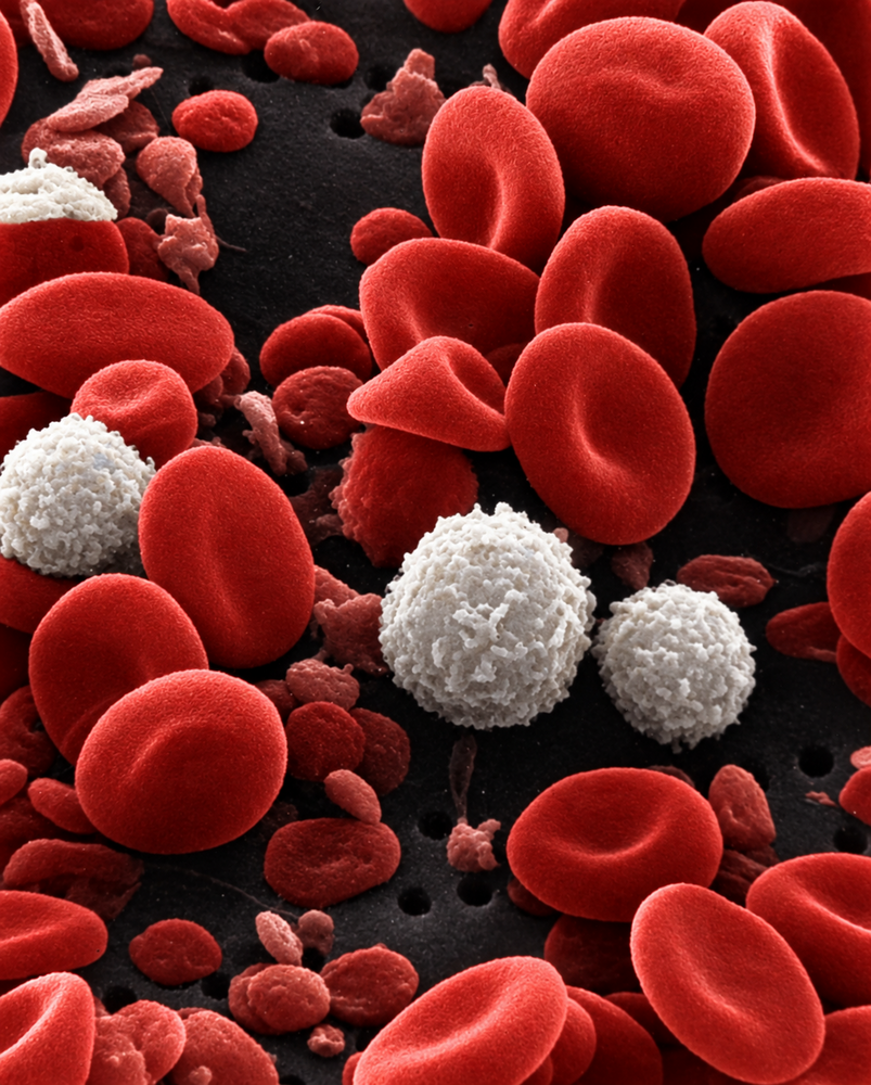
Rode en witte bloedcellen onder de microscoop. Kanker in de witte bloedcellen wordt leukemie genoemd.
Een gaspedaal dat blijft ingedrukt
Mijn onderzoek richtte zich op een specifieke vorm van kinderleukemie: hoog-hyperdiploïde B-cel acute lymfatische leukemie (HeH B-ALL). Deze vorm is de meest voorkomende vorm van B-ALL bij kinderen en treft ongeveer drie op de tien kinderen met B-ALL. Deze specifieke vorm van leukemie wordt gekenmerkt door extra chromosomen en dus extra genetisch materiaal (DNA) in de B-cellen. Extra genetisch materiaal verstoort de normale werking van de cel, waardoor veel cellen niet meer goed kunnen functioneren en zelfs afsterven. Cellen die bovenop het extra genetisch materiaal ook fouten opstapelen in hun groeisysteem, zoals het RAS-systeem, krijgen zo een voordeel. Zo’n fout in het RAS-systeem kan ervoor zorgen dat het gaspedaal van de cel als het ware blijft ingedrukt. Het eindresultaat is dat bloedcellen zich abnormaal ontwikkelen, hun normale functies niet meer uitoefenen en ongecontroleerd blijven delen. De vraag die mij daarom drijft, is eenvoudig, maar tegelijk complex: kunnen we de ongeremde leukemiecellen uitschakelen?
De voet van het gaspedaal halen
In mijn onderzoek maakte ik celmodellen in het labo die exact dezelfde fouten in het RAS-systeem bevatten. Nadien bekeek ik welke nieuwe geneesmiddelen (RAS-remmers) het meest geschikt waren om die ontspoorde groei weer te blokkeren. Ik testte negen veelbelovende kandidaat-geneesmiddelen, elk met een andere strategie om het ontspoorde RAS-systeem uit te schakelen. Sommige remmen het RAS-eiwit rechtstreeks af, andere zorgen ervoor dat het op de foute plaats in de cel terechtkomt of grijpen verderop in dezelfde signaalroute in. Daarnaast testte ik geneesmiddelen die het eiwit rechtstreeks naar de afvalcontainer van de cel sturen, waardoor het RAS-eiwit wordt afgebroken. Ik onderzocht bij elke strategie zowel hoe sterk de geneesmiddelen de groei van de leukemiecellen remden, als hun effect op het RAS-signaal zelf en hun toxiciteit.
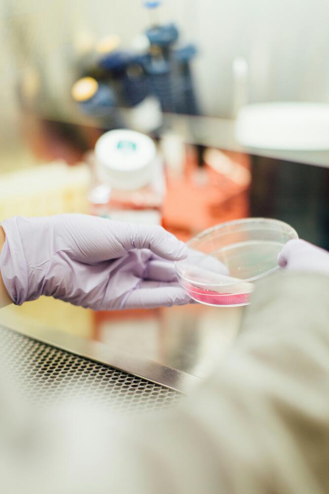
Cellen in het labo worden in leven gehouden in een plaat.
Verschillende geneesmiddelen slaagden erin het ontspoorde groeisignaal stil te leggen, zonder gezonde cellen ernstig te beschadigen. Zeven van de negen kandidaat-geneesmiddelen toonden veelbelovende resultaten. Uiteindelijk bleven twee duidelijke toppers over. De sterkste resultaten werden behaald met Daraxonrasib, dat het RAS-eiwit direct blokkeert, en Trametinib, dat het signaal verderop stopt. Opvallend was dat beide geneesmiddelen effectief waren, ongeacht welke fout aanwezig was in het RAS-systeem. Andere geneesmiddelen gaven ook veelbelovende resultaten, maar enkel wanneer bepaalde fouten in het RAS-systeem voorkwamen. Twee van de negen geneesmiddelen vielen af omdat ze toxische effecten hadden of omdat ze de celgroei niet konden blokkeren.
Van proefbuis naar patiënt
Tijdens mijn onderzoek testte ik deze geneesmiddelen in het labo. De volgende stap is testen in patiëntmodellen. Deze modellen zijn afgeleid van echte leukemiecellen. Zo kunnen we nagaan of onze resultaten uit het labo ook standhouden in een realistischer model van leukemie. Het mooiste resultaat? Daraxonrasib en Trametinib bleven niet beperkt tot mijn thesis. Intussen worden beide geneesmiddelen, op basis van deze resultaten, al getest in patiëntmodellen die in ons labo zijn ontwikkeld. Dat betekent dat mijn thesis niet eindigt op papier, maar vandaag al verder leeft in nieuw onderzoek.
Als de volgende resultaten positief zijn, kunnen klinische studies een volgende stap worden. Pas daarna kan worden onderzocht of deze geneesmiddelen uiteindelijk een plaats kunnen krijgen in de behandeling van patiënten. Omdat deze RAS-remmers zich richten op een specifiek groeisysteem van de leukemiecellen, hopen we dat ze in de toekomst een gerichtere behandeling met mogelijk minder bijwerkingen kunnen bieden dan klassieke chemotherapie. Dat mijn onderzoek vandaag al deel uitmaakt van vervolgonderzoek naar betere behandelingen voor kinderen met leukemie, betekent voor mij bijzonder veel.
Twintig jaar geleden ontstond mijn droom in een kleuterklas. Vandaag staat die droom tussen proefbuizen en petrischaaltjes. En morgen? Dankzij mijn doctoraatsopleiding aan de KU Leuven krijg ik de kans om die droom verder na te jagen en me verder te verdiepen in leukemieonderzoek. Want zolang kinderen met leukemie blijven vechten, blijf ik zoeken naar betere behandelingen.
Bibliografie
1. Cumano, A., & Godin, I. (2007). Ontogeny of the Hematopoietic System. Annual Review of Immunology, 25, 745–785. https://doi.org/10.1146/annurev.immunol.25.022106.141538
2. Kato, H., & Igarashi, K. (2019). To be red or white: Lineage commitment and maintenance of the hematopoietic system by the “inner myeloid.” Haematologica, 104(10), 1919–1927. https://doi.org/10.3324/haematol.2019.216861
3. Puhr, S., Lee, J., Zvezdova, E., et al. (2015). Dendritic Cell Development – History, Advances, and Open Questions. Seminars in Immunology, 27(6), 388–396. https://doi.org/10.1016/j.smim.2016.03.012
4. Rieger, M. A., & Schroeder, T. (2012). Hematopoiesis. Cold Spring Harbor Perspectives in Biology, 4(12), a008250. https://doi.org/10.1101/cshperspect.a008250
5. Orakpoghenor, O., Avazi, D. O., Markus, T. P., et al. (2019). Lymphocytes: A Brief Review. Scientific Journal of Immunology & Immunotherapy, 5.
6. Zlotoff, D. A., & Bhandoola, A. (2011). Hematopoietic progenitor migration to the adult thymus. Annals of the New York Academy of Sciences, 1217(1), 122–138. https://doi.org/10.1111/j.1749-6632.2010.05881.x
7. Lai, A. Y., & Kondo, M. (2008). T and B lymphocyte differentiation from hematopoietic stem cell. Seminars in Immunology, 20(4), 207–212. https://doi.org/10.1016/j.smim.2008.05.002
8. Rothenberg, E. V. (2011). T cell lineage commitment: Identity and renunciation. Journal of Immunology (Baltimore, Md. : 1950), 186(12), 6649–6655. https://doi.org/10.4049/jimmunol.1003703
9. Kenneth M. Murphy, Casey Weaver, & Leslie J. Berg. (2022). Janeway’s Immunobiology.
10. Germain, R. N. (2002). T-cell development and the CD4–CD8 lineage decision. Nature Reviews Immunology, 2(5), 309–322. https://doi.org/10.1038/nri798
11. Chann, A. S., Charnley, M., Newton, L. M., et al. (2022). Stepwise progression of β-selection during T cell development involves histone deacetylation. Life Science Alliance, 6(1), e202201645. https://doi.org/10.26508/lsa.202201645
12. Abe, S., Asahi, T., Hara, T., et al. (2023). Hematopoietic cell-derived IL-15 supports NK cell development in scattered and clustered localization within the bone marrow. Cell Reports, 42(9), 113127. https://doi.org/10.1016/j.celrep.2023.113127
13. Stephan, R. P., Reilly, C. R., & Witte, P. L. (1998). Impaired Ability of Bone Marrow Stromal Cells to Support B-Lymphopoiesis With Age. Blood, 91(1), 75–88. https://doi.org/10.1182/blood.V91.1.75
14. Jung, D., Giallourakis, C., Mostoslavsky, R., et al. (2006). MECHANISM AND CONTROL OF V(D)J RECOMBINATION AT THE IMMUNOGLOBULIN HEAVY CHAIN LOCUS. Annual Review of Immunology, 24(Volume 24, 2006), 541–570. https://doi.org/10.1146/annurev.immunol.23.021704.115830
15. Chen, D., Ireland, S. J., Remington, G., et al. (2016). CD40-mediated NFκB activation in B cells is increased in multiple sclerosis and modulated by therapeutics. Journal of Immunology (Baltimore, Md. : 1950), 197(11), 4257–4265. https://doi.org/10.4049/jimmunol.1600782
16. Rossetti, R. A. M., Lorenzi, N. P. C., Yokochi, K., et al. (2018). B lymphocytes can be activated to act as antigen presenting cells to promote anti-tumor responses. PLOS ONE, 13(7), e0199034. https://doi.org/10.1371/journal.pone.0199034
17. Forthal, D. N. (2014). Functions of Antibodies. Microbiology Spectrum, 2(4), 1–17.
18. Ding, L., Ley, T. J., Larson, D. E., et al. (2012). Clonal evolution in relapsed acute myeloid leukaemia revealed by whole-genome sequencing. Nature, 481(7382), 506–510. https://doi.org/10.1038/nature10738
19. Aertgeerts M. (2025). Clonal Heterogeneity and Evolution During Treatment in Pediatric Acute Lymphoblastic Leukemia—A Single-Cell Characterization [Doctoral thesis, KU Leuven].
20. Hanahan, D., & Weinberg, R. A. (2011). Hallmarks of Cancer: The Next Generation. Cell, 144(5), 646–674. https://doi.org/10.1016/j.cell.2011.02.013
21. Leukemia—Hematology.org. (n.d.). Retrieved October 13, 2025, from https://www.hematology.org/education/patients/blood-cancers/leukemia
22. Childhood Acute Lymphoblastic Leukemia—NCI. (2026, February 10). https://www.cancer.gov/types/leukemia/patient/child-all-treatment-pdq
23. Inaba, H., & Pui, C.-H. (2021). Advances in the Diagnosis and Treatment of Pediatric Acute Lymphoblastic Leukemia. Journal of Clinical Medicine, 10(9), 1926. https://doi.org/10.3390/jcm10091926
24. Khoury, J. D., Solary, E., Abla, O., et al. (2022). The 5th edition of the World Health Organization Classification of Haematolymphoid Tumours: Myeloid and Histiocytic/Dendritic Neoplasms. Leukemia, 36(7), 1703–1719. https://doi.org/10.1038/s41375-022-01613-1
25. Davis, A. S., Viera, A. J., & Mead, M. D. (2014). Leukemia: An overview for primary care. American Family Physician, 89(9), 731–738.
26. Thomas, D., & Majeti, R. (2017). Biology and relevance of human acute myeloid leukemia stem cells. Blood, 129(12), 1577–1585. https://doi.org/10.1182/blood-2016-10-696054
27. Holyoake, T. L., & Vetrie, D. (2017). The chronic myeloid leukemia stem cell: Stemming the tide of persistence. Blood, 129(12), 1595–1606. https://doi.org/10.1182/blood-2016-09-696013
28. Arber, D. A., Orazi, A., Hasserjian, R. P., et al. (2022). International Consensus Classification of Myeloid Neoplasms and Acute Leukemias: Integrating morphologic, clinical, and genomic data. Blood, 140(11), 1200–1228. https://doi.org/10.1182/blood.2022015850
29. Childhood Leukemia. (n.d.). Retrieved May 13, 2026, from https://www.cancer.org/cancer/types/leukemia-in-children.html
30. Signs and Symptoms of Acute Lymphocytic Leukemia. (n.d.). Retrieved November 30, 2025, from https://www.cancer.org/cancer/types/acute-lymphocytic-leukemia/detectio…
31. Acute lymphoblastic leukemia (ALL) signs and symptoms | Blood Cancer United. (n.d.). Retrieved November 30, 2025, from https://bloodcancerunited.org/blood-cancer/leukemia/acute-lymphoblastic…
32. Reismüller, B., Steiner, M., Pichler, H., et al. (2017). High hyperdiploid acute lymphoblastic leukemia (ALL)—A 25-year population-based survey of the Austrian ALL-BFM (Berlin-Frankfurt-Münster) Study Group. Pediatric Blood & Cancer, 64(6), e26327. https://doi.org/10.1002/pbc.26327
33. Hunger, S. P., & Mullighan, C. G. (2015). Acute Lymphoblastic Leukemia in Children. New England Journal of Medicine, 373(16), 1541–1552. https://doi.org/10.1056/NEJMra1400972
34. Paulsson, K., & Johansson, B. (2009). High hyperdiploid childhood acute lymphoblastic leukemia. Genes, Chromosomes and Cancer, 48(8), 637–660. https://doi.org/10.1002/gcc.20671
35. Greaves, M., & Maley, C. C. (2012). Clonal Evolution In Cancer. Nature, 481(7381), 306–313. https://doi.org/10.1038/nature10762
36. Aertgeerts, M., Meyers, S., Gielen, O., et al. (2025). Single-cell DNA and surface protein characterization of high hyperdiploid acute lymphoblastic leukemia at diagnosis and during treatment. HemaSphere, 9(2), e70085. https://doi.org/10.1002/hem3.70085
37. Paulsson, K., Lilljebjörn, H., Biloglav, A., et al. (2015). The genomic landscape of high hyperdiploid childhood acute lymphoblastic leukemia. Nature Genetics, 47(6), 672–676. https://doi.org/10.1038/ng.3301
38. PeCan | St. Jude Cloud. (n.d.). Retrieved October 21, 2025, from https://pecan.stjude.cloud/
39. Taylor, A. M., Shih, J., Ha, G., et al. (2018). Genomic and Functional Approaches to Understanding Cancer Aneuploidy. Cancer Cell, 33(4), 676-689.e3. https://doi.org/10.1016/j.ccell.2018.03.007
40. Hintzen, D. C., Schubert, M., Soto, M., et al. (2024). Reduction of chromosomal instability and inflammation is a common aspect of adaptation to aneuploidy. EMBO Reports, 25(11), 5169–5193. https://doi.org/10.1038/s44319-024-00252-0
41. Santaguida, S., & Amon, A. (2015). Short- and long-term effects of chromosome mis-segregation and aneuploidy. Nature Reviews Molecular Cell Biology, 16(8), 473–485. https://doi.org/10.1038/nrm4025
42. Ippolito, M. R., Zerbib, J., Eliezer, Y., et al. (2024). Increased RNA and Protein Degradation Is Required for Counteracting Transcriptional Burden and Proteotoxic Stress in Human Aneuploid Cells. Cancer Discovery, 14(12), 2532–2553. https://doi.org/10.1158/2159-8290.CD-23-0309
43. He, A., Dean, J. M., & Lodhi, I. J. (2021). Peroxisomes as cellular adaptors to metabolic and environmental stress. Trends in Cell Biology, 31(8), 656–670. https://doi.org/10.1016/j.tcb.2021.02.005
44. Newman, D. L., & Gregory, S. L. (2019). Co-Operation between Aneuploidy and Metabolic Changes in Driving Tumorigenesis. International Journal of Molecular Sciences, 20(18), 4611. https://doi.org/10.3390/ijms20184611
45. Zerbib, J., Ippolito, M. R., Eliezer, Y., et al. (2024). Human aneuploid cells depend on the RAF/MEK/ERK pathway for overcoming increased DNA damage. Nature Communications, 15(1), 7772. https://doi.org/10.1038/s41467-024-52176-x
46. Hyperdiploidy: The longest known, most prevalent, and most enigmatic form of acute lymphoblastic leukemia in children | Leukemia. (n.d.). Retrieved October 30, 2025, from https://www.nature.com/articles/s41375-022-01720-z
47. Woodward, E. L., Yang, M., Moura-Castro, L. H., et al. (2023). Clonal origin and development of high hyperdiploidy in childhood acute lymphoblastic leukaemia. Nature Communications, 14(1), 1658. https://doi.org/10.1038/s41467-023-37356-5
48. Maia, A. T., van der Velden, V. H. J., Harrison, C. J., et al. (2003). Prenatal origin of hyperdiploid acute lymphoblastic leukemia in identical twins. Leukemia, 17(11), 2202–2206. https://doi.org/10.1038/sj.leu.2403101
49. Bargiela-Iparraguirre, J., Prado-Marchal, L., Pajuelo-Lozano, N., et al. (2014). Mad2 and BubR1 modulates tumourigenesis and paclitaxel response in MKN45 gastric cancer cells. Cell Cycle, 13(22), 3590–3601. https://doi.org/10.4161/15384101.2014.962952
50. Fujiwara, T., Bandi, M., Nitta, M., et al. (2005). Cytokinesis failure generating tetraploids promotes tumorigenesis in p53-null cells. Nature, 437(7061), 1043–1047. https://doi.org/10.1038/nature04217
51. Molina, O., Vinyoles, M., Granada, I., et al. (2020). Impaired Condensin Complex and Aurora B kinase underlie mitotic and chromosomal defects in hyperdiploid B-cell ALL. Blood, 136(3), 313–327. https://doi.org/10.1182/blood.2019002538
52. Moura-Castro, L. H., Peña-Martínez, P., Castor, A., et al. (2021). Sister chromatid cohesion defects are associated with chromosomal copy number heterogeneity in high hyperdiploid childhood acute lymphoblastic leukemia. Genes, Chromosomes & Cancer, 60(6), 410–417. https://doi.org/10.1002/gcc.22933
53. Moura-Castro, L. H., Peña-Martínez, P., Castor, A., et al. (2021). Sister chromatid cohesion defects are associated with chromosomal copy number heterogeneity in high hyperdiploid childhood acute lymphoblastic leukemia. Genes, Chromosomes and Cancer, 60(6), 410–417. https://doi.org/10.1002/gcc.22933
54. Wang, N., Xu, S., & Egli, D. (2023). Replication stress in mammalian embryo development, differentiation, and reprogramming. Trends in Cell Biology, 33(10), 872–886. https://doi.org/10.1016/j.tcb.2023.03.015
55. Flach, J., & Milyavsky, M. (2018). Replication stress in hematopoietic stem cells in mouse and man. Mutation Research, 808, 74–82. https://doi.org/10.1016/j.mrfmmm.2017.10.001
56. Molina, J. R., & Adjei, A. A. (2006). The Ras/Raf/MAPK Pathway. Journal of Thoracic Oncology, 1(1), 7–9. https://doi.org/10.1016/S1556-0864(15)31506-9
57. Singh, B., Carpenter, G., & Coffey, R. J. (2016). EGF receptor ligands: Recent advances. F1000Research, 5, F1000 Faculty Rev-2270. https://doi.org/10.12688/f1000research.9025.1
58. Wang, Y.-N., Lee, H.-H., & Hung, M.-C. (2018). A novel ligand-receptor relationship between families of ribonucleases and receptor tyrosine kinases. Journal of Biomedical Science, 25(1), 83. https://doi.org/10.1186/s12929-018-0484-7
59. Kovacs, E., Zorn, J. A., Huang, Y., et al. (2015). A Structural Perspective on the Regulation of the Epidermal Growth Factor Receptor. Annual Review of Biochemistry, 84(Volume 84, 2015), 739–764. https://doi.org/10.1146/annurev-biochem-060614-034402
60. Jura, N., Zhang, X., Endres, N. F., et al. (2011). Catalytic control in the EGF receptor and its connection to general kinase regulatory mechanisms. Molecular Cell, 42(1), 9–22. https://doi.org/10.1016/j.molcel.2011.03.004
61. Littlefield, P., & Jura, N. (2013). EGFR lung cancer mutants get specialized. Proceedings of the National Academy of Sciences, 110(38), 15169–15170. https://doi.org/10.1073/pnas.1314719110
62. Wang, D., Liu, G., Meng, Y., et al. (2024). The Configuration of GRB2 in Protein Interaction and Signal Transduction. Biomolecules, 14(3), 259. https://doi.org/10.3390/biom14030259
63. Bandaru, P., Kondo, Y., & Kuriyan, J. (2019). The Interdependent Activation of Son-of-Sevenless and Ras. Cold Spring Harbor Perspectives in Medicine, 9(2), a031534. https://doi.org/10.1101/cshperspect.a031534
64. Herman, J. A., Romain, R. R., Hoellerbauer, P., et al. (2022). Hyper-active RAS/MAPK introduces cancer-specific mitotic vulnerabilities. Proceedings of the National Academy of Sciences, 119(41), e2208255119. https://doi.org/10.1073/pnas.2208255119
65. Jeon, H., Tkacik, E., & Eck, M. J. (2024). Signaling from RAS to RAF: The Molecules and Their Mechanisms. Annual Review of Biochemistry, 93(Volume 93, 2024), 289–316. https://doi.org/10.1146/annurev-biochem-052521-040754
66. Peyssonnaux, C., & Eychène, A. (2001). The Raf/MEK/ERK pathway: New concepts of activation. Biology of the Cell, 93(1–2), 53–62. https://doi.org/10.1016/S0248-4900(01)01125-X
67. Shaul, Y. D., & Seger, R. (2007). The MEK/ERK cascade: From signaling specificity to diverse functions. Biochimica et Biophysica Acta (BBA) - Molecular Cell Research, Mitogen-Activated Protein Kinases: New Insights on Regulation, Function and Role in Human Disease, 1773(8), 1213–1226. https://doi.org/10.1016/j.bbamcr.2006.10.005
68. Maik-Rachline, G., Hacohen-Lev-Ran, A., & Seger, R. (2019). Nuclear ERK: Mechanism of Translocation, Substrates, and Role in Cancer. International Journal of Molecular Sciences, 20(5), 1194. https://doi.org/10.3390/ijms20051194
69. RAS: Targeting the Impossible. (n.d.). Retrieved May 23, 2026, from https://blog.crownbio.com/targeting-ras-oncology
70. Castellano, E., & Downward, J. (2011). RAS Interaction with PI3K: More Than Just Another Effector Pathway. Genes & Cancer, 2(3), 261–274. https://doi.org/10.1177/1947601911408079
71. Simanshu, D. K., Nissley, D. V., & McCormick, F. (2017). RAS Proteins and Their Regulators in Human Disease. Cell, 170(1), 17–33. https://doi.org/10.1016/j.cell.2017.06.009
72. Santos, E., & Nebreda, A. R. (1989). Structural and functional properties of ras proteins. The FASEB Journal, 3(10), 2151–2163. https://doi.org/10.1096/fasebj.3.10.2666231
73. Brady, S. W., Roberts, K. G., Gu, Z., et al. (2022). The genomic landscape of pediatric acute lymphoblastic leukemia. Nature Genetics, 54(9), 1376–1389. https://doi.org/10.1038/s41588-022-01159-z
74. Paulsson, K., Horvat, A., Strömbeck, B., et al. (2008). Mutations of FLT3, NRAS, KRAS, and PTPN11 are frequent and possibly mutually exclusive in high hyperdiploid childhood acute lymphoblastic leukemia. Genes, Chromosomes and Cancer, 47(1), 26–33. https://doi.org/10.1002/gcc.20502
75. Hunter, J. C., Manandhar, A., Carrasco, M. A., et al. (2015). Biochemical and Structural Analysis of Common Cancer-Associated KRAS Mutations. Molecular Cancer Research, 13(9), 1325–1335. https://doi.org/10.1158/1541-7786.MCR-15-0203
76. Case, M., Matheson, E., Minto, L., et al. (2008). Mutation of Genes Affecting the RAS Pathway Is Common in Childhood Acute Lymphoblastic Leukemia. Cancer Research, 68(16), 6803–6809. https://doi.org/10.1158/0008-5472.CAN-08-0101
77. Iritani, B. M., Forbush, K. A., Farrar, M. A., et al. (1997). Control of B cell development by Ras‐mediated activation of Raf. The EMBO Journal, 16(23), 7019–7031. https://doi.org/10.1093/emboj/16.23.7019
78. Kamata, T., Hussain, J., Giblett, S., et al. (2010). BRAF Inactivation Drives Aneuploidy by Deregulating CRAF. Cancer Research, 70(21), 8475–8486. https://doi.org/10.1158/0008-5472.CAN-10-0603
79. Zang, M., Gong, J., Luo, L., et al. (2008). Characterization of Ser338 Phosphorylation for Raf-1 Activation*. Journal of Biological Chemistry, 283(46), 31429–31437. https://doi.org/10.1074/jbc.M802855200
80. Zang, M., Hayne, C., & Luo, Z. (2002). Interaction between Active Pak1 and Raf-1 Is Necessary for Phosphorylation and Activation of Raf-1*. Journal of Biological Chemistry, 277(6), 4395–4405. https://doi.org/10.1074/jbc.M110000200
81. Dandoulaki, M., Petsalaki, E., Sumpton, D., et al. (2018). Src activation by Chk1 promotes actin patch formation and prevents chromatin bridge breakage in cytokinesis. Journal of Cell Biology, 217(9), 3071–3089. https://doi.org/10.1083/jcb.201802102
82. Genetic variations may predict relapse risk in childhood ALL. (n.d.). BSH. Retrieved October 15, 2025, from https://b-s-h.org.uk/about-us/news/genetic-variations-may-predict-relap…
83. Bertuccio, P., Alicandro, G., Malvezzi, M., et al. (2020). Childhood cancer mortality trends in Europe, 1990-2017, with focus on geographic differences. Cancer Epidemiology, 67, 101768. https://doi.org/10.1016/j.canep.2020.101768
84. Rheingold, S. R., Bhojwani, D., Ji, L., et al. (2024). Determinants of survival after first relapse of acute lymphoblastic leukemia: A Children’s Oncology Group study. Leukemia, 38(11), 2382–2394. https://doi.org/10.1038/s41375-024-02395-4
85. Jeha, S., Choi, J., Roberts, K. G., et al. (2021). Clinical Significance of Novel Subtypes of Acute Lymphoblastic Leukemia in the Context of Minimal Residual Disease–Directed Therapy. Blood Cancer Discovery, 2(4), 326–337. https://doi.org/10.1158/2643-3230.BCD-20-0229
86. Ratti, S., Lonetti, A., Follo, M. Y., et al. (2020). B-ALL Complexity: Is Targeted Therapy Still A Valuable Approach for Pediatric Patients? Cancers, 12(12), 3498. https://doi.org/10.3390/cancers12123498
87. Inaba, H., & Pui, C.-H. (2010). Glucocorticoid use in acute lymphoblastic leukemia: Comparison of prednisone and dexamethasone. The Lancet Oncology, 11(11), 1096–1106. https://doi.org/10.1016/S1470-2045(10)70114-5
88. Juluri, K. R., Siu, C., & Cassaday, R. D. (2022). Asparaginase in the Treatment of Acute Lymphoblastic Leukemia in Adults: Current Evidence and Place in Therapy. Blood and Lymphatic Cancer: Targets and Therapy, 12, 55–79. https://doi.org/10.2147/BLCTT.S342052
89. Kothari, A., Hittelman, W. N., & Chambers, T. C. (2016). Cell cycle-dependent mechanisms underlie vincristine-induced death of primary acute lymphoblastic leukemia cells. Cancer Research, 76(12), 3553–3561. https://doi.org/10.1158/0008-5472.CAN-15-2104
90. Ronghe, M., Burke, G. A., Lowis, S. P., et al. (2001). Remission induction therapy for childhood acute lymphoblastic leukaemia: Clinical and cellular pharmacology of vincristine, corticosteroids, L-asparaginase and anthracyclines. Cancer Treatment Reviews, 27(6), 327–337. https://doi.org/10.1053/ctrv.2001.0243
91. Cronstein, B. N. (1997). THE MECHANISM OF ACTION OF METHOTREXATE. Rheumatic Disease Clinics of North America, 23(4), 739–755. https://doi.org/10.1016/S0889-857X(05)70358-6
92. Lim, S. Z., & Chua, E. W. (2018). Revisiting the Role of Thiopurines in Inflammatory Bowel Disease Through Pharmacogenomics and Use of Novel Methods for Therapeutic Drug Monitoring. Frontiers in Pharmacology, 9. https://doi.org/10.3389/fphar.2018.01107
93. Peters, C., Schrappe, M., von Stackelberg, A., et al. (2015). Stem-Cell Transplantation in Children With Acute Lymphoblastic Leukemia: A Prospective International Multicenter Trial Comparing Sibling Donors With Matched Unrelated Donors—The ALL-SCT-BFM-2003 Trial. Journal of Clinical Oncology, 33(11), 1265–1274. https://doi.org/10.1200/JCO.2014.58.9747
94. Chemotherapy for Acute Lymphocytic Leukemia. (n.d.). Retrieved October 31, 2025, from https://www.cancer.org/cancer/types/acute-lymphocytic-leukemia/treating…
95. Long term side effects of treatment for acute lymphoblastic leukaemia (ALL). (n.d.). Retrieved October 31, 2025, from https://www.cancerresearchuk.org/about-cancer/acute-lymphoblastic-leuka…
96. Kantarjian, H., Aldoss, I., & Jabbour, E. (2025). Management of Adult Acute Lymphoblastic Leukemia: A Review. JAMA Oncology, 11(7), 771–778. https://doi.org/10.1001/jamaoncol.2025.0613
97. Drugs Approved for Childhood Cancers—NCI. (2016, August 31). [cgvArticle]. (nciglobal,ncienterprise). https://www.cancer.gov/about-cancer/treatment/drugs/childhood-cancer-fd…
98. Yu, G., Chen, F., Yin, C., et al. (2017). Upfront treatment with the first and second-generation tyrosine kinase inhibitors in Ph-positive acute lymphoblastic leukemia. Oncotarget, 8(63), 107022–107032. https://doi.org/10.18632/oncotarget.22206
99. Hoffman, L., & Gore, L. (2014). Blinatumomab, a Bi-Specific Anti-CD19/CD3 BiTE® Antibody for the Treatment of Acute Lymphoblastic Leukemia: Perspectives and Current Pediatric Applications. Frontiers in Oncology, 4. https://doi.org/10.3389/fonc.2014.00063
100. Freyer, C. W. (2018). Tisagenlecleucel: The First CAR on the Highway to Remission for Acute Lymphoblastic Leukemia. Journal of the Advanced Practitioner in Oncology, 9(5), 537–544.
101. Maude, S. L., Frey, N., Shaw, P. A., et al. (2014). Chimeric Antigen Receptor T Cells for Sustained Remissions in Leukemia. New England Journal of Medicine, 371(16), 1507–1517. https://doi.org/10.1056/NEJMoa1407222
102. Cox, A. D., Fesik, S. W., Kimmelman, A. C., et al. (2014). Drugging the undruggable Ras: Mission possible? Nature Reviews. Drug Discovery, 13(11), 828–851. https://doi.org/10.1038/nrd4389
103. Rubinson, D. A., Tanaka, N., Fece De La Cruz, F., et al. (2024). Sotorasib Is a Pan- RAS G12C Inhibitor Capable of Driving Clinical Response in NRAS G12C Cancers. Cancer Discovery, 14(5), 727–736. https://doi.org/10.1158/2159-8290.CD-23-1138
104. Bahar, M. E., Kim, H. J., & Kim, D. R. (2023). Targeting the RAS/RAF/MAPK pathway for cancer therapy: From mechanism to clinical studies. Signal Transduction and Targeted Therapy, 8(1), 455. https://doi.org/10.1038/s41392-023-01705-z
105. Brock, E. J., Ji, K., Reiners, J. J., et al. (2016). How to Target Activated Ras Proteins: Direct Inhibition vs. Induced Mislocalization. Mini Reviews in Medicinal Chemistry, 16(5), 358–369. https://doi.org/10.2174/1389557515666151001154002
106. Liu, Z., Hu, M., Yang, Y., et al. (2022). An overview of PROTACs: A promising drug discovery paradigm. Molecular Biomedicine, 3(1), 46. https://doi.org/10.1186/s43556-022-00112-0
107. MedChemExpress: Master of Bioactive Molecules | Inhibitors, Screening Libraries & Proteins. (n.d.). MedchemExpress.Com. Retrieved March 12, 2026, from https://www.medchemexpress.com/
108. Paulsson, K., Horvat, A., Strömbeck, B., et al. (2008). Mutations of FLT3, NRAS, KRAS, and PTPN11 are frequent and possibly mutually exclusive in high hyperdiploid childhood acute lymphoblastic leukemia. Genes, Chromosomes and Cancer, 47(1), 26–33. https://doi.org/10.1002/gcc.20502
109. Chen, H., Smaill, J. B., Liu, T., et al. (2020). Small-Molecule Inhibitors Directly Targeting KRAS as Anticancer Therapeutics. Journal of Medicinal Chemistry, 63(23), 14404–14424. https://doi.org/10.1021/acs.jmedchem.0c01312
110. Welsch, M. E., Kaplan, A., Chambers, J. M., et al. (2017). Multivalent Small-Molecule Pan-RAS Inhibitors. Cell, 168(5), 878-889.e29. https://doi.org/10.1016/j.cell.2017.02.006
111. Kim, D., Herdeis, L., Rudolph, D., et al. (2023). Pan-KRAS inhibitor disables oncogenic signalling and tumour growth. Nature, 619(7968), 160–166. https://doi.org/10.1038/s41586-023-06123-3
112. Cregg, J., Edwards, A. V., Chang, S., et al. (2025). Discovery of Daraxonrasib (RMC-6236), a Potent and Orally Bioavailable RAS(ON) Multi-selective, Noncovalent Tri-complex Inhibitor for the Treatment of Patients with Multiple RAS-Addicted Cancers. Journal of Medicinal Chemistry, 68(6), 6064–6083. https://doi.org/10.1021/acs.jmedchem.4c02314
113. Wolpin, B. M., Park, W., Garrido-Laguna, I., et al. (2026). Daraxonrasib in Previously Treated Advanced RAS-Mutated Pancreatic Cancer. New England Journal of Medicine, 394(18), 1790–1802. https://doi.org/10.1056/NEJMoa2505783
114. Frost, A., O’Connor, S., & Ciulli, A. (2026). Allosteric PROTACs: Expanding the Horizon of Targeted Protein Degradation. Journal of the American Chemical Society, 148(3), 2823–2837. https://doi.org/10.1021/jacs.5c14840
115. Röth, S., Fulcher, L. J., & Sapkota, G. P. (2019). Advances in targeted degradation of endogenous proteins. Cellular and Molecular Life Sciences, 76(14), 2761–2777. https://doi.org/10.1007/s00018-019-03112-6
116. Kargbo, R. B. (2020). PROTAC-Mediated Degradation of KRAS Protein for Anticancer Therapeutics. ACS Medicinal Chemistry Letters, 11(1), 5–6. https://doi.org/10.1021/acsmedchemlett.9b00584
117. Cieślak, M., & Słowianek, M. (2023). Cereblon-Recruiting PROTACs: Will New Drugs Have to Face Old Challenges? Pharmaceutics, 15(3), 812. https://doi.org/10.3390/pharmaceutics15030812
118. Luo, X., Archibeque, I., Dellamaggiore, K., et al. (2022). Profiling of diverse tumor types establishes the broad utility of VHL-based ProTaCs and triages candidate ubiquitin ligases. iScience, 25(3), 103985. https://doi.org/10.1016/j.isci.2022.103985
119. UK, C. org. (n.d.). Probe ACBI3. Chemical Probes Portal. Retrieved May 11, 2026, from https://www.chemicalprobes.org/acbi3?
120. Manabe, T., Bergo, H. C., Li, Q., et al. (2026). Pan-RAF inhibitor exarafenib targets BRAF class II/III NSCLC and reveals ARAF-KSR1 resistance and combination strategies. Nature Communications, 17(1), 2484. https://doi.org/10.1038/s41467-026-69216-3
121. Feichtenschlager, V., Zheng, Y. J., Qu, T., et al. (2025). Suppression of NRAS-mutant melanoma growth with NRAS-targeting Antisense Oligonucleotide treatment reveals therapeutically relevant kinase co-dependencies. Communications Medicine, 5(1), 216. https://doi.org/10.1038/s43856-025-00932-5
122. Muñoz-Maldonado, C., Zimmer, Y., & Medová, M. (2019). A Comparative Analysis of Individual RAS Mutations in Cancer Biology. Frontiers in Oncology, 9. https://doi.org/10.3389/fonc.2019.01088