Een bijzonder eiwit dat reageert op de zure omgeving van tumoren: een nieuwe piste voor kankerbeeldvorming
Wat als we kanker niet alleen kunnen opsporen door naar de tumor zelf te kijken, maar ook door gebruik te maken van de omgeving waarin hij groeit? Veel tumoren hebben namelijk een opvallende eigenschap: hun omgeving is zuurder dan die van gezond weefsel. Voor mijn bachelorproef heb ik onderzocht of een zuurgevoelig eiwit dat verschil in zuurtegraad kan herkennen. De resultaten waren veelbelovend: hoe zuurder de omgeving, hoe sterker het (radioactief gelabelde) eiwit zich aan de kankercellen hechtte. Een eerste stap in het onderzoek naar nieuwe methoden voor kankerbeeldvorming.
Waarom doen tumoren zo zuur?
Niet omdat ze slechtgezind zijn, maar omdat ze hun omgeving letterlijk zuurder maken. Wanneer afwijkende cellen zich ongecontroleerd vermenigvuldigen, ontstaat er niet alleen een tumor, maar verandert ook de omgeving waarin deze groeit. Om te kunnen groeien, hebben tumorcellen veel energie nodig. Daarom breken ze grote hoeveelheden glucose af via de glycolyse.
Daarbij ontstaat pyruvaat, dat bij veel tumorcellen grotendeels wordt omgezet in lactaat, zelfs wanneer er voldoende zuurstof aanwezig is. Dit verschijnsel staat bekend als het Warburg-effect. Tegelijk worden waterstofionen aan de omgeving afgegeven, waardoor de pH daalt en het omliggende weefsel zuurder wordt.
Terwijl gezond weefsel doorgaans een pH van ongeveer 7,4 heeft, kan de omgeving van solide tumoren een pH van ongeveer 6,5 bereiken. Dat verschil kan interessant zijn voor kankerbeeldvorming. Een eiwit dat op een lagere pH reageert, zou namelijk kunnen helpen om tumorweefsel gerichter zichtbaar te maken.
T-SorTag: een bijzonder eiwit dat van vorm verandert
T-SorTag is een kunstmatig ontwikkeld eiwitconstruct dat is gebaseerd op een onderdeel van het difterietoxine. Dat onderdeel is het translocatiedomein en heeft een bijzondere eigenschap: wanneer de pH daalt, verandert het eiwit van vorm. Die vormverandering kan ervoor zorgen dat het gemakkelijker met celmembranen interageert.
In mijn onderzoek zag ik dat T-SorTag inderdaad op een lagere pH reageert. Met circulair dichroïsme, een techniek waarmee veranderingen in de structuur van eiwitten kunnen worden onderzocht, stelde ik vast dat het eiwit bij pH 5,5 een andere structuur vertoonde dan bij pH 7,5. Maar leidt die structuurverandering ook tot een sterkere binding aan kankercellen?
T-SorTag in actie
T-SorTag werd onderzocht op gekweekte darmkankercellen van muizen bij verschillende pH-waarden. Met flowcytometrie, een techniek waarmee afzonderlijke cellen worden bestudeerd, kon ik via een fluorescentiesignaal meten hoeveel T-SorTag op de kankercellen aanwezig was.
Daaruit kwam een duidelijke trend naar voren: hoe zuurder de omgeving, hoe sterker het gemeten signaal. Bij pH 5,5 was dat signaal duidelijk het hoogst, terwijl de isotypecontrole laag en vrijwel constant bleef. Zo kon ik controleren dat de stijging bij lage pH daadwerkelijk samenhing met T-SorTag en niet met een toevallig of niet-specifiek signaal.
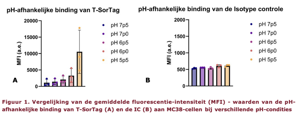
Dit wijst erop dat T-SorTag onder zure omstandigheden sterker met het celmembraan interageert. Daarmee werd een volgende stap gezet richting het gebruik van T-SorTag als mogelijke tracer voor kankerbeeldvorming.
Van eiwit naar radioactieve speurstof
Om een tumor op een medische scan zichtbaar te maken, moet het eiwit ook van buiten het lichaam kunnen worden gedetecteerd. Dat doen we door T-SorTag te voorzien van een radioactief label. Zo zou het in de toekomst kunnen dienen als tracer: een speurstof waarvan de verspreiding en ophoping in het lichaam met medische beeldvorming gevolgd kan worden.
Het eiwit werd gelabeled met gallium-68, een radioactieve isotoop die geschikt is voor PET-scans (positronemissietomografie). Om gallium-68 aan T-SorTag te koppelen, werd een soort hulpstof, een chelator, gebruikt die de radio-isotoop kan vasthouden.
De belangrijkste vraag was of het eiwit T-SorTag na deze radioactieve labeling nog steeds op een zure omgeving zou kunnen reageren…
Dat bleek uit de celbindingsstudie met radioactief gelabeld T-SorTag. Hoe zuurder de omgeving werd, hoe meer radioactiviteit aan de cellen gebonden bleef. Bij pH 5,5 was die celgebonden activiteit ongeveer 260% hoger dan bij pH 7,5. Figuur 2 toont hoe die stijging geleidelijk zichtbaar wordt naarmate de pH daalt.
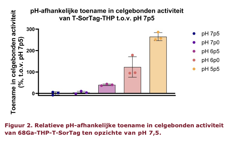
Hoewel de koppeling aan de chelator alsook de radioactieve labeling tijdens het onderzoek meerdere pogingen en optimalisaties vergde, bleef het pH-afhankelijke bindingsgedrag van T-SorTag uiteindelijk zichtbaar. Dat is belangrijk, omdat het eiwit voor een mogelijke toepassing bij PET-beeldvorming radioactief gemerkt moet kunnen worden zonder zijn zuurgevoelige eigenschappen te verliezen.
De grootste uitdaging moet nog komen
T-SorTag bevat een aantal veelbelovende eigenschappen: het reageert op een lagere pH, interageert onder zure omstandigheden sterker met de onderzochte kankercellen en behoudt dat gedrag ook na radioactieve labeling. Daarmee is het een interessante kandidaat voor verder onderzoek naar kankerbeeldvorming.
Toch betekent dit niet dat T-SorTag al gebruikt kan worden om kanker bij patiënten op te sporen. De experimenten vonden plaats in het laboratorium en verdere optimalisatie richting PET-beeldvorming is nodig, vooraleer er in vivo kan getest worden.
Bovendien werd de sterkste reactie gemeten bij pH 5,5, een zuurtegraad die lager ligt dan die van veel tumoromgevingen. Verder onderzoek moet uitwijzen of T-SorTag zich ook in een levend organisme voldoende gericht in tumorweefsel kan ophopen om tumoren zichtbaar te maken op een PET-scan.
Bibliografie
Andreev, O. A., Dupuy, A. D., Segala, M., Sandugu, S., Serra, D. A., Chichester, C. O., Engelman, D. M., & Reshetnyak, Y. K. (2007). Mechanism and uses of a membrane peptide that targets tumors and other acidic tissues in vivo. Proceedings of the National Academy of Sciences of the United States of America, 104(19), 7893–7898. https://doi.org/10.1073/pnas.0702439104
Andreev, O. A., Engelman, D. M., & Reshetnyak, Y. K. (2010). pH-sensitive membrane peptides (pHLIPs) as a novel class of delivery agents. Molecular Membrane Biology, 27(7), 341. https://doi.org/10.3109/09687688.2010.509285
Anemone, A., Consolino, L., Arena, F., Capozza, M., & Longo, D. L. (2019a). Imaging tumor acidosis: a survey of the available techniques for mapping in vivo tumor pH. Cancer Metastasis Reviews, 38(1–2), 25–49. https://doi.org/10.1007/s10555-019-09782-9
Anemone, A., Consolino, L., Arena, F., Capozza, M., & Longo, D. L. (2019b). Imaging tumor acidosis: a survey of the available techniques for mapping in vivo tumor pH. Cancer Metastasis Reviews, 38(1–2), 25–49. https://doi.org/10.1007/s10555-019-09782-9
Baudhuin, H., Cousaert, J., Vanwolleghem, P., Raes, G., Caveliers, V., Keyaerts, M., Lahoutte, T., & Xavier, C. (2021). 68Ga-labeling: Laying the foundation for an anti-radiolytic formulation for nota-sdab pet tracers. Pharmaceuticals, 14(5), 448. https://doi.org/10.3390/PH14050448/S1
Bauer, D., Visca, H., Weerakkody, A., Carter, L. M., Samuels, Z., Kaminsky, S., Andreev, O. A., Reshetnyak, Y. K., & Lewis, J. S. (2022). PET Imaging of Acidic Tumor Environment With 89Zr-labeled pHLIP Probes. Frontiers in Oncology, 12, 882541. https://doi.org/10.3389/fonc.2022.882541
Boyken, S. E., Benhaim, M. A., Busch, F., Jia, M., Bick, M. J., Choi, H., Klima, J. C., Chen, Z., Walkey, C., Mileant, A., Sahasrabuddhe, A., Wei, K. Y., Hodge, E. A., Byron, S., Quijano-Rubio, A., Sankaran, B., King, N. P., Lippincott-Schwartz, J., Wysocki, V. H., … Baker, D. (2019). De novo design of tunable, pH-driven conformational changes. Science, 364(6442), 658–664. https://doi.org/10.1126/SCIENCE.AAV7897;PAGE:STRING:ARTICLE/CHAPTER
Brown, J. S., Amend, S. R., Austin, R. H., Gatenby, R. A., Hammarlund, E. U., & Pienta, K. J. (2023). Updating the Definition of Cancer. Molecular Cancer Research, 21(11), 1142–1147. https://doi.org/10.1158/1541-7786.MCR-23-0411/727658/AM/UPDATING-THE-DEFIN ITION-OF-CANCERUPDATING -THE
Choe, S., Bennett, M. J., Fujii, G., Curmi, P. M. G., Kantardjieff, K. A., Collier, R. J., & Eisenberg, D. (1992). The crystal structure of diphtheria toxin. Nature, 357(6375), 216–222. https://doi.org/10.1038/357216a0
Chou, D. K., Krishnamurthy, R., Randolph, T. W., Carpenter, J. F., & Manning, M. C. (2005). Effects of Tween 20® and Tween 80® on the stability of Albutropin during agitation. Journal of Pharmaceutical Sciences, 94(6), 1368–1381. https://doi.org/10.1002/jps.20365
Demoin, D. W., Wyatt, L. C., Edwards, K. J., Abdel-Atti, D., Sarparanta, M., Pourat, J., Longo, V. A., Carlin, S. D., Engelman, D. M., Andreev, O. A., Reshetnyak, Y. K., Viola-Villegas, N., & Lewis, J. S. (2016). PET Imaging of Extracellular pH in Tumors with (64)Cu- and (18)F-Labeled pHLIP Peptides: A Structure-Activity Optimization Study. Bioconjugate Chemistry, 27(9), 2014–2023. https://doi.org/10.1021/acs.bioconjchem.6b00306
drs. T.W.H. Meijer, dr. P.N. Span, prof. dr. L-F de Geus-Oei, & prof. dr. J.H.A.M. Kaanders. (n.d.). Tumormetabolisme en het Warburg-effect: translatie naar de klinische praktijk - NTVO. Retrieved March 1, 2026, from https://www.ntvo.nl/journal-article/tumormetabolisme-e n-het-warburg-ef fect-translatie-naar-de-klinische-praktijk/
Falnes, P., & Sandvig, K. (2000). Penetration of protein toxins into cells. Current Opinion in Cell Biology, 12(4), 407–413. https://doi.org/10.1016/S0955-0674(00)00109-5
Gillies, R. J., Raghunand, N., Karczmar, G. S., & Bhujwalla, Z. M. (2002). MRI of the tumor microenvironment. Journal of Magnetic Resonance Imaging : JMRI , 16(4), 430–450. https://doi.org/10.1002/jmri.10181
Goedaardige of kwaadaardige tumor? - UMCG Protonentherapiecentrum. (n.d.). Retrieved March 1, 2026, from https://umcgprotonentherapiecentrum.nl/goedaardige-of-kwaadaardige-tumor/
Gorasso, V., Vandevijvere, S., Van der Heyden, J., Pelgrims, I., Hilderink, H., Nusselder, W., Demoury, C., Schmidt, M., Vansteelandt, S., De Smedt, D., & Devleesschauwer, B. (2024). The incremental healthcare cost associated with cancer in Belgium: A registry-based data analysis. Cancer Medicine, 13(3), e6659. https://doi.org/10.1002/CAM4.6659;WGROUP:STRING:PUBLICATION
Gourni, E., Del Pozzo, L., Bartholomä, M., Kief er, Y., T. Meyer, P., Maecke, H. R., & Holland, J. P. (2017). Radiochemistry and Preclinical PET Imaging of 68Ga-Desferrioxamine Radiotracers Targeting Prostate-Specific Membrane Antigen. Molecular Imaging, 16. https://doi.org/10.1177/1536012117737010
Greenlee, J. D., & King, M. R. (2022). A syngeneic MC38 orthotopic mouse model of colorectal cancer metastasis. Biology Methods and Protocols, 7(1). https://doi.org/10.1093/biomethods/bpac024
Guckeisen, T., Hosseinpour, S., & Peukert, W. (2021). Effect of pH and urea on the proteins secondary structure at the water/air interface and in solution. Journal of Colloid and Interface Science, 590, 38–49. https://doi.org/10.1016/j.jcis.2021.01.015
Harsha. (2024, March). De verschillen begrijpen: SPECT-scan versus PET-scan. https://kiranpetct.com/understanding-the-differences-spect-scan-vs-pet-scan/
Hashim, A. I., Zhang, X., Wojtkowiak, J. W., Martinez, G. V., & Gillies, R. J. (2011). Imaging pH and Metastasis. NMR in Biomedicine, 24(6), 582. https://doi.org/10.1002/nbm.1644
Heiden, M. G. V., Cantley, L. C., & Thompson, C. B. (2009). Understanding the Warburg Effect: The Metabolic Requirements of Cell Proliferation. Science (New York, N.Y.), 324(5930), 1029. https://doi.org/10.1126/science.1160809
Jardim-Perassi, B. V., Irrera, P., Lau, J. Y. C., Budzevich, M., Whelan, C. J., Abrahams, D., Ruiz, E., Ibrahim-Hashim, A., Damgaci Erturk, S., Longo, D. L., Pilon-Thomas, S. A., & Gillies, R. J. (2023). Intraperitoneal Delivery of Iopamidol to Assess Extracellular pH of Orthotopic Pancreatic Tumor Model by CEST-MRI. Contrast Media & Molecular Imaging, 2023, 1944970. https://doi.org/10.1155/2023/1944970
Juweid, M. E., Al-Qasem, S. F., Khuri, F. R., Gallamini, A., Lohmann, P., Ziellenbach, H., & Mottaghy, F. M. (2025). Beyond f luorodeoxyglucose: Molecular imaging of cancer in precision medicine. CA: A Cancer Journal for Clinicians, 75(3), 226–242. https://doi.org/10.3322/CAAC.70007;REQUESTEDJOURNAL:JOURNAL:1542486 3;WGROUP:STRING:PUBLICATION
Kanker - Naar een gezond België. (n.d.). Retrieved March 1, 2026, from https://www.gezondbelgie.be/nl/gezondheidstoestand/niet-overdraagbare-aandoeningen/kanker
Kanker.nl. (n.d.). Goedaardige en kwaadaardige tumoren. Retrieved March 1, 2026, from https://www.kanker.nl/algemene-onderwerpen/wat-is-kanker/algemeen/goedaardige-en-kwaadaardige-tumoren
Kelley Murfin. (n.d.). What is the tumor microenvironment? | MD Anderson Cancer Center. Retrieved March 1, 2026, from https://www.mdanderson.org/cancerwise/what-is-the-tum or-microenvironment-3-things-to-k now.h00-159460056. html
Lauren Rodriguez, & Larry Joe. (n.d.). Protein structure and function (article) | Khan Academy. Retrieved March 1, 2026, from https://www.khanacademy.org/science/hs-bio/x230b3ff 252126bb6:gene-expression-and-regulation/x230b3ff 252126bb6:untitled-348/a/protein-structure-and-function
Longo, D. L., Bartoli, A., Consolino, L., Bardini, P., Arena, F., Schwaiger, M., & Aime, S. (2016). In Vivo Imaging of Tumor Metabolism and Acidosis by Combining PET and MRI-CEST pH Imaging. Cancer Research, 76(22), 6463–6470. https://doi.org/10.1158/0008-5472.CAN-16-0825
Low, H. Y., Yang, C. T., Xia, B., He, T., Lam, W. W. C., & Ng, D. C. E. (2023). Radiolabeled Liposomes for Nuclear Imaging Probes. Molecules 2023, Vol. 28, 28(9). https://doi.org/10.3390/molecules28093798
M E Phelps. (2000). PET: the merging of biology and imaging into molecular imaging - PubMed. Journal of Nuclear Medicine. https://pubmed.ncbi.nlm.nih.gov/10768568/
Martins Pinto, M., Paumard, P., Bouchez, C., Ransac, S., Duvezin-Caubet, S., Mazat, J. P., Rigoulet, M., & Devin, A. (2023). The Warburg effect and mitochondrial oxidative phosphorylation: Friends or foes? Biochimica et Biophysica Acta - Bioenergetics, 1864(1). https://doi.org/10.1016/j.bbabio.2022.148931
McDonald, P. C., Chafe, S. C., Brown, W. S., Saberi, S., Swayampakula, M., Venkateswaran, G., Nemirovsky, O., Gillespie, J. A., Karasinska, J. M., Kalloger, S. E., Supuran, C. T., Schaeffer, D. F., Bashashati, A., Shah, S. P., Topham, J. T., Yapp, D. T., Li, J., Renouf, D. J., Stanger, B. Z., & Dedhar, S. (2019). Regulation of pH by Carbonic Anhydrase 9 Mediates Survival of Pancreatic Cancer Cells With Activated KRAS in Response to Hypoxia. Gastroenterology, 157(3), 823–837. https://doi.org/10.1053/j.gastro.2019.05.004
Melanie M. Cooper, & Michael W. Klymkowsky. (n.d.). 9.3: Amino Acids, Proteins, and pH - Chemistry LibreTexts. Retrieved May 24, 2026, from https://chem.libretexts.org/Bookshelves/General_Chemistry/CLUE%3A_Chemis try_Life_the_Universe_and_Everything/09%3A_Reaction_Systems/9.3%3A_Am ino_Acids_Proteins_and_pH
Nakhjavani, M., Giles, B., Strom, M., Vi, C., Attenborough, S., & Shigdar, S. (2022). A Flow Cytometry-Based Cell Surface Protein Binding Assay for Assessing Selectivity and Specificity of an Anticancer Aptamer. Journal of Visualized Experiments (JoVE), 2022(187), e64304. https://doi.org/10.3791/64304
O’Brien, E. P., Brooks, B. R., & Thirumalai, D. (2011). Effects of pH on proteins: Predictions for ensemble and single molecule pulling experiments. Journal of the American Chemical Society, 134(2), 979. https://doi.org/10.1021/ja206557y
OECD, & Commission, E. (2025). Landenprofiel voor Kanker: België 2025. EU Country Cancer Profiles, EU Country Cancer Profiles . https://doi.org/10.1787/5c4dfa17-nl
Pometti, M. A., Di Natale, G., Geremia, G., Gauswami, N., Garufi, G., Ricciardi, G., Sciortino, M., Scopelliti, F., Russo, G., & Ippolito, M. (2024). A Kinetically Controlled Bioconjugation Method for the Synthesis of Radioimmunoconjugates and the Development of a Domain Mapping MS-Workflow for Its Characterization. Bioconjugate Chemistry, 35(3), 324–332. https://doi.org/10.1021/ACS.BIOCONJCHEM.3C00519
Price, E. W., & Orvig, C. (2013). Matching chelators to radiometals for radiopharmaceuticals. Chemical Society Reviews, 43(1), 260–290. https://doi.org/10.1039/c3cs60304k
Rahman, A., Janic, B., Rahman, T., Singh, H., Ali, H., Rattan, R., Kazi, M., & Ali, M. M. (2023). Immunotherapy Enhancement by Targeting Extracellular Tumor pH in Triple-Negative Breast Cancer Mouse Model. Cancers, 15(20), 4931. https://doi.org/10.3390/CANCERS15204931/S1
Rahman, M. A., Yadab, M. K., & Ali, M. M. (2024a). Emerging Role of Extracellular pH in Tumor Microenvironment as a Therapeutic Target for Cancer Immunotherapy. Cells 2024, Vol. 13, 13(22). https://doi.org/10.3390/cells13221924
Rahman, M. A., Yadab, M. K., & Ali, M. M. (2024b). Emerging Role of Extracellular pH in Tumor Microenvironment as a Therapeutic Target for Cancer Immunotherapy. Cells 2024, Vol. 13, 13(22). https://doi.org/10.3390/cells13221924
Reshetnyak, Y. K., Andreev, O. A., & Engelman, D. M. (2024). Aiming the magic bullet: targeted delivery of imaging and therapeutic agents to solid tumors by pHLIP peptides. Frontiers in Pharmacology, 15. https://doi.org/10.3389/fphar.2024. 1355893
Rowe, S. P., & Pomper, M. G. (2021). Molecular Imaging in Oncology: Current Impact and Future Directions. CA: A Cancer Journal for Clinicians, 72(4), 333. https://doi.org/10.3322/caac.21713
Saeed, E., Bibi, K., Mushtaq, S., Bibi, I., & Park, J. A. (2025). Advancing Photodynamic Therapy: The Role of PET and SPECT in Imaging-Guided Theranostics. Molecular Pharmaceutics, 22(7), 3637–3653. https://doi.org/10.1021/ACS.MOLPHARMACEUT.5C00551
’Sharma, N. C., ’Efstratiou, A., ’Mokrousov, I., ’Mutreja, A., ’Das, B., & ’Ramamurthy, T. (2019). Diphtheria. Suzuki, A., Maeda, T., Baba, Y., Shimamura, K., & Kato, Y. (2014). Acidic extracellular pH promotes epithelial mesenchymal transition in Lewis lung carcinoma model. Cancer Cell International 2014 14:1, 14(1), 129-. https://doi.org/10.1186/S12935-014-0129-1
Taniura, T., Iida, Y., Kotani, H., Ishitobi, K., Tajima, Y., & Harada, M. (2020). Immunogenic chemotherapy in two mouse colon cancer models. Cancer Science, 111(10), 3527. https://doi.org/10.1111/cas.14624
Visca, H., DuPont, M., Moshnikova, A., Crawford, T., Engelman, D. M., Andreev, O. A., & Reshetnyak, Y. K. (2022). pHLIP Peptides Target Acidity in Activated Macrophages. Molecular Imaging and Biology, 24(6), 874–885. https://doi.org/10.1007/s11307-022-01737-x
Wyatt, L. C., Lewis, J. S., Andreev, O. A., Reshetnyak, Y. K., & Engelman, D. M. (2017). Applications of pHLIP Technology for Cancer Imaging and Therapy. Trends in Biotechnology, 35(7), 653–664. https://doi.org/10.1016/j.tibtech.2017.03.014
Zhang, X., Lin, Y., & Gillies, R. J. (2010). Tumor pH and its measurement. Journal of Nuclear Medicine : Official Publication, Society of Nuclear Medicine , 51(8), 1167–1170. https://doi.org/10.2967/jnumed.109.068981