POEMBAKterie: het verborgen leven in onze keukengootsteen
POEMBAKterie: het verborgen leven in onze keukengootsteen
Elke dag komen we in contact met duizenden micro-organismen zowel op onze werkplek als in publieke ruimtes, maar natuurlijk ook thuis. De keukengootsteen is een van de plekken die we dagelijks gebruiken. We wassen er onze handen, doen er de afwas en gebruiken het voor allerlei andere dagelijkse taken. Daarna draaien we de kraan dicht en gaan we verder met onze dag. Wat er in de gootsteen achterblijft, daar staan we meestal niet lang bij stil.
Toch vormt de keukengootsteen een opmerkelijke leefomgeving voor bacteriën. Door de voortdurende aanvoer van vocht, organisch materiaal en micro-organismen ontstaat er een omgeving waarin verschillende bacteriesoorten kunnen groeien en samenleven.
Onderzoek naar het microbioom van gootstenen heeft zich tot nu toe voornamelijk gericht op ziekenhuizen. Daarbij werd aangetoond dat gootstenen onder meer opportunistische ziekteverwekkers en antibioticaresistente bacteriën kunnen bevatten. Over het microbioom van keukengootstenen in huishoudens is daarentegen nog opvallend weinig bekend.
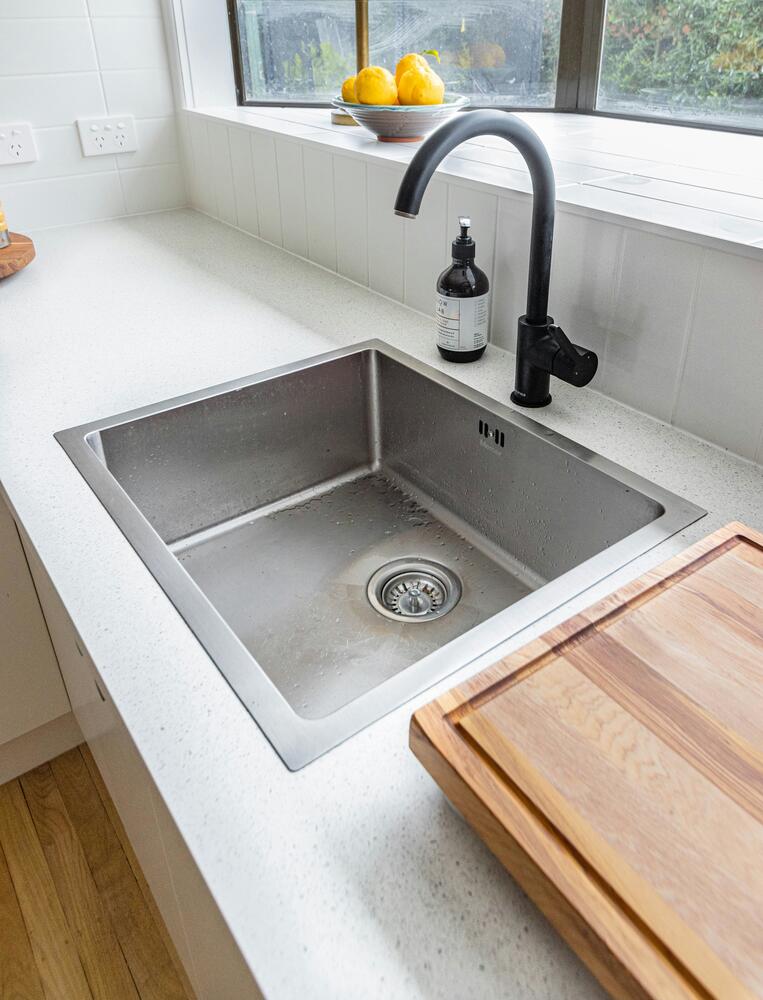 Om die kenniskloof te helpen dichten, werd het POEMBAKterie-onderzoek opgestart: een burgerwetenschappelijk onderzoek naar het microbioom van Antwerpse keukengootstenen. POEMBAKterie is een woordspeling op het Antwerpse dialectwoord poembak, een benaming voor een gootsteen, en het woord bacterie. De naam verwijst zo zowel naar de Antwerpse keukenpoembak als naar haar bacteriële bewoners.
Bij burgerwetenschap worden vrijwilligers actief betrokken bij wetenschappelijk onderzoek. In dit project gingen Antwerpenaren zelf aan de slag in hun keuken en verzamelden ze stalen uit hun gootsteen en kraanwater. Daarnaast deelden ze informatie over hun gootsteen, het gebruik ervan en hun huishouden.
Voor mijn thesis onderzocht ik de eerste resultaten van een pilootstudie bij twaalf huishoudens. De deelnemers verzamelden stalen van het gootsteenoppervlak, de afvoer en het kraanwater. We gebruikten twee complementaire technieken. DNA-onderzoek gaf ons een overzicht van de bacteriegemeenschappen in de gootsteen, terwijl kweekexperimenten aantoonden of levende E. coli aanwezig was.
Naast het analyseren van de stalen hielp ik mee aan de opstart van het grootschalige project. We maakten meer dan 850 staalnamekits en brachten ze naar deelnemers verspreid over heel Antwerpen. Fietsend door de stad met zakken vol kits werd de schaal van het project tastbaar: een onderzoek naar de Antwerpse poembak dat inwoners uit alle hoeken van de stad samenbracht.
Een ecosysteem onder de kraan
Achter de alledaagse keukengootsteen gaat een verrassend divers microbieel ecosysteem schuil. In de onderzochte stalen vonden we een grote diversiteit aan bacteriën: 28 bacteriële hoofdgroepen en 905 verschillende bacteriegeslachten. Vooral Pseudomonadota, Actinomycetota, Bacillota en Bacteroidota kwamen veel voor. Tot de meest voorkomende geslachten behoorden onder andere Microbacterium, Acinetobacter, Paracoccus, Pseudomonas en Moraxella.
Die namen zeggen de meeste mensen weinig, maar ze vertellen wel iets over waar die bacteriën vandaan komen. Sommige worden teruggevonden in water of vochtige omgevingen, andere zijn gelinkt aan voedsel, de bodem of aan de mens. Dit bewijst dat de keukengootsteen een ontmoetingsplaats vormt waar bacteriën uit verschillende bronnen samenkomen.
De bacteriegemeenschappen in de afvoer verschilden van die op het gootsteenoppervlak. Dat is niet zo verrassend, het oppervlak droogt regelmatig op, komt in direct contact met handen en voedsel, en wordt vaker schoongemaakt. De afvoer daarentegen blijft langer vochtig en biedt andere omstandigheden voor bacteriën. Op basis van deze pilootstudie kunnen we nog niet bepalen welke factoren precies verantwoordelijk zijn voor de verschillen, maar de resultaten tonen wel aan dat zelfs binnen één gootsteen verschillende microbiële leefomgevingen bestaan.
Een paar opvallende bewoners
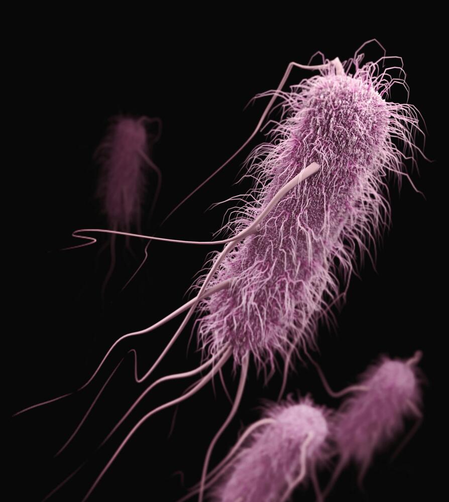 Naast de algemene bacteriële samenstelling gingen we ook op zoek naar enkele bacteriesoorten die relevant kunnen zijn voor voedselveiligheid. In de DNA-analyses werden tien van die soorten teruggevonden, waaronder Bacillus cereus, Salmonella enterica en Escherichia coli, beter bekend als E. coli.
Enkele hiervan werden in meerdere stalen gedetecteerd. Dat betekent echter niet dat deze bacteriën ziekmakend waren. Binnen de soort bestaan zowel onschuldige als ziekteverwekkende varianten, en met onze methode konden we dat onderscheid niet maken. Bovendien toont DNA-onderzoek alleen aan dat sporen van een bacterie aanwezig zijn, niet of die bacterie nog leeft. Daarom was de kweekmethode een belangrijke aanvulling. Met deze methode konden we in acht van de twaalf afvoerstalen levende E. coli isoleren.
Opvallend was dat de twee onderzoeksmethoden niet altijd dezelfde bacteriën opleverden. Dat is niet noodzakelijk tegenstrijdig. DNA kan afkomstig zijn van bacteriën die intussen niet meer leven, terwijl niet elke levende bacterie onder de gekozen laboratoriumomstandigheden zal groeien. De twee methodes vormen dus elk een ander stukje van de puzzel.
Belangrijk is ook dat de soorten waarnaar we gericht zochten slechts een klein deel van de totale bacteriegemeenschap vormden. Hun aanwezigheid betekent daarom niet automatisch dat een gootsteen een gezondheidsrisico vormt. Daarvoor is verder onderzoek nodig.
Een eerste antwoord, nog vele vragen
Mijn thesis vormt een eerste stap in het onderzoek naar het microbioom van huishoudelijke keukengootstenen. Omdat het om een pilootstudie met twaalf huishoudens ging, moeten de resultaten voorzichtig worden geïnterpreteerd. We konden verschillen vaststellen tussen het gootsteenoppervlak en de afvoer, maar nog niet achterhalen welke factoren daarvoor precies verantwoordelijk zijn.
Daar ligt meteen de meerwaarde van de grotere POEMBAKterie-studie. In totaal werden 862 staalnamekits uitgezonden, waarvan 758 hun stalen terugstuurden tegen begin juni. Met honderden extra stalen wordt het mogelijk om verder te kijken dan enkel welke bacteriën aanwezig zijn. De verzamelde informatie over huishoudens, schoonmaakroutines en gebruiksgewoonten kan helpen om te achterhalen waarom het microbioom van de ene gootsteen verschilt van dat van de andere.
Er blijven nog veel vragen onbeantwoord. Veranderen de bacteriën doorheen de tijd? Welke soorten verdwijnen snel en welke blijven aanwezig? Welke rol spelen schoonmaken, voedselresten en vocht? En wanneer wordt de aanwezigheid van een bepaalde bacterie daadwerkelijk relevant? Ook kan verder onderzoek nagaan of dezelfde bacteriestammen op meerdere plekken in de stad voorkomen.
Mijn thesis biedt een eerste blik op het verborgen microbiële leven in een omgeving die we dagelijks gebruiken. Tegelijk laat ze zien hoeveel er nog te ontdekken valt. De keukengootsteen is geen steriele omgeving, maar dat is op zich geen reden tot ongerustheid. Wie wil begrijpen welke micro-organismen deel uitmaken van onze dagelijkse leefomgeving, vindt in de poembak alvast een veelbelovend vertrekpunt.
Bibliografie
1. United Nations Department of Economic and Social Affairs. World Urbanization Prospects 2025: Summary of Results [Internet]. United Nations; 2025 [cited 2026 Jan 14]. (Statistical Papers - United Nations (Ser. A), Population and Vital Statistics Report). Available from: https://www.un-library.org/content/books/9789211576788 doi:10.18356/9789211576788
2. Gilbert JA, Stephens B. Microbiology of the built environment. Nat Rev Microbiol. 2018 Nov;16(11):661–70. doi:10.1038/s41579-018-0065-5
3. Bruno A, Fumagalli S, Ghisleni G, Labra M, Bruno A, Fumagalli S, et al. The Microbiome of the Built Environment: The Nexus for Urban Regeneration for the Cities of Tomorrow. Microorganisms. 2022 Nov 22;10(12). doi:10.3390/microorganisms10122311
4. Hsu T, Joice R, Vallarino J, Abu-Ali G, Hartmann EM, Shafquat A, et al. Urban Transit System Microbial Communities Differ by Surface Type and Interaction with Humans and the Environment. mSystems. 2016 Jun 28;1(3):e00018-16. doi:10.1128/mSystems.00018-16 PubMed PMID: 27822528; PubMed Central PMCID: PMC5069760.
5. Danko D, Bezdan D, Afshin EE, Ahsanuddin S, Bhattacharya C, Butler DJ, et al. A global metagenomic map of urban microbiomes and antimicrobial resistance. Cell. 2021 Jun;184(13):3376-3393.e17. doi:10.1016/j.cell.2021.05.002
6. Wu J, Danko D, Afshinnekoo E, Bezdan D, Bhattacharyya M, Castro-Nallar E, et al. Annotating unknown species of urban microorganisms on a global scale unveils novel functional diversity and local environment association. Environmental Research. 2022 May;207:112183. doi:10.1016/j.envres.2021.112183
7. Guevarra RB, Hwang J, Lee H, Kim HJ, Lee Y, Danko D, et al. Metagenomic characterization of bacterial community and antibiotic resistance genes found in the mass transit system in Seoul, South Korea. Ecotoxicology and Environmental Safety. 2022 Nov;246:114176. doi:10.1016/j.ecoenv.2022.114176
8. Dunn RR, Fierer N, Henley JB, Leff JW, Menninger HL. Home Life: Factors Structuring the Bacterial Diversity Found within and between Homes. PLoS One. 2013 May 22;8(5):e64133. doi:10.1371/journal.pone.0064133 PubMed PMID: 23717552; PubMed Central PMCID: PMC3661444.
9. Adams RI, Bateman AC, Bik HM, Meadow JF. Microbiota of the indoor environment: a meta-analysis. Microbiome. 2015 Oct 13;3:49. doi:10.1186/s40168-015-0108-3 PubMed PMID: 26459172; PubMed Central PMCID: PMC4604073.
10. Kelley ST, Gilbert JA. Studying the microbiology of the indoor environment. Genome Biol. 2013;14(2):202. doi:10.1186/gb-2013-14-2-202 PubMed PMID: 23514020; PubMed Central PMCID: PMC3663111.
11. Lax S, Smith DP, Hampton-Marcell J, Owens SM, Handley KM, Scott NM, et al. Longitudinal analysis of microbial interaction between humans and the indoor environment. Science. 2014 Aug 29;345(6200):1048–52. doi:10.1126/science.1254529
12. Cao H, Ye H, Tian Y, Zhang J, Xie Y, Chen Y, et al. Functional areas shape indoor microbial structure and potential risks in university dormitories. Front Microbiol. 2025 Jul 15;16. doi:10.3389/fmicb.2025.1604064
13. Hewitt KM, Gerba CP, Maxwell SL, Kelley ST. Office Space Bacterial Abundance and Diversity in Three Metropolitan Areas. PLoS One. 2012 May 30;7(5):e37849. doi:10.1371/journal.pone.0037849 PubMed PMID: 22666400; PubMed Central PMCID: PMC3364274.
14. Adams RI, Bhangar S, Dannemiller KC, Eisen JA, Fierer N, Gilbert JA, et al. Ten questions concerning the microbiomes of buildings. Building and Environment. 2016 Nov;109:224–34. doi:10.1016/j.buildenv.2016.09.001
15. Flores GE, Bates ST, Knights D, Lauber CL, Stombaugh J, Knight R, et al. Microbial Biogeography of Public Restroom Surfaces. PLoS One. 2011 Nov 23;6(11):e28132. doi:10.1371/journal.pone.0028132 PubMed PMID: 22132229; PubMed Central PMCID: PMC3223236.
16. Thompson TP, Rice CJ, Athanasakis E, Mawhinney J, Gilmore BF, Fitzgerald P, et al. The effect of sample type and location on industrial workplace sink and hand dryer microbiomes. BMC Microbiol. 2025 May 26;25(1):325. doi:10.1186/s12866-025-04054-9
17. Meadow JF, Altrichter AE, Kembel SW, Kline J, Mhuireach G, Moriyama M, et al. Indoor airborne bacterial communities are influenced by ventilation, occupancy, and outdoor air source. Indoor Air. 2014 Feb;24(1):41–8. doi:10.1111/ina.12047 PubMed PMID: 23621155; PubMed Central PMCID: PMC4285785.
18. Flores GE, Bates ST, Caporaso JG, Lauber CL, Leff JW, Knight R, et al. Diversity, distribution and sources of bacteria in residential kitchens. Environmental Microbiology. 2013 Feb;15(2):588–96. doi:10.1111/1462-2920.12036
19. Liu S, Gunawan C, Barraud N, Rice SA, Harry EJ, Amal R. Understanding, Monitoring, and Controlling Biofilm Growth in Drinking Water Distribution Systems. Environ Sci Technol. 2016 Sep 6;50(17):8954–76. doi:10.1021/acs.est.6b00835
20. Novak Babič M, Gostinčar C, Gunde-Cimerman N. Microorganisms populating the water-related indoor biome. Appl Microbiol Biotechnol. 2020 Aug;104(15):6443–62. doi:10.1007/s00253-020-10719-4
21. Withey Z, Goodall T, MacIntyre S, Gweon HS. Characterization of communal sink drain communities of a university campus. Environmental DNA. 2021;3(5):901–11. doi:10.1002/edn3.196
22. De Geyter D, Blommaert L, Verbraeken N, Sevenois M, Huyghens L, Martini H, et al. The sink as a potential source of transmission of carbapenemase-producing Enterobacteriaceae in the intensive care unit. Antimicrob Resist Infect Control. 2017 Feb 16;6(1):24. doi:10.1186/s13756-017-0182-3
23. Franco LC, Tanner W, Ganim C, Davy T, Edwards J, Donlan R. A microbiological survey of handwashing sinks in the hospital built environment reveals differences in patient room and healthcare personnel sinks. Sci Rep. 2020 May 19;10(1):8234. doi:10.1038/s41598-020-65052-7
24. Healy HG, Pawluk E, Dieter L, Roberts SC, Tanner W, Mathew T, et al. Bacterial recolonization of hospital sink biofilms. Journal of Hospital Infection. 2025 Aug;162:95–105. doi:10.1016/j.jhin.2025.05.013
25. Aranega-Bou P, Ellaby N, Ellington MJ, Moore G. Migration of Escherichia coli and Klebsiella pneumoniae Carbapenemase (KPC)-Producing Enterobacter cloacae through Wastewater Pipework and Establishment in Hospital Sink Waste Traps in a Laboratory Model System. Microorganisms. 2021 Sep 3;9(9):1868. doi:10.3390/microorganisms9091868 PubMed PMID: 34576763; PubMed Central PMCID: PMC8468231.
26. Kotay S, Chai W, Guilford W, Barry K, Mathers AJ. Spread from the Sink to the Patient: In Situ Study Using Green Fluorescent Protein (GFP)-Expressing Escherichia coli To Model Bacterial Dispersion from Hand-Washing Sink-Trap Reservoirs. Appl Environ Microbiol. 2017 Mar 31;83(8):e03327-16. doi:10.1128/AEM.03327-16 PubMed PMID: 28235877; PubMed Central PMCID: PMC5377511.
27. Kotay SM, Donlan RM, Ganim C, Barry K, Christensen BE, Mathers AJ. Droplet- Rather than Aerosol-Mediated Dispersion Is the Primary Mechanism of Bacterial Transmission from Contaminated Hand-Washing Sink Traps. Appl Environ Microbiol. 2019 Jan 9;85(2):e01997-18. doi:10.1128/AEM.01997-18 PubMed PMID: 30367005; PubMed Central PMCID: PMC6328769.
28. Mahnert A, Moissl-Eichinger C, Zojer M, Bogumil D, Mizrahi I, Rattei T, et al. Man-made microbial resistances in built environments. Nat Commun. 2019 Feb 27;10:968. doi:10.1038/s41467-019-08864-0 PubMed PMID: 30814504; PubMed Central PMCID: PMC6393488.
29. Bourdin T, Benoit MÈ, Prévost M, Charron D, Quach C, Déziel E, et al. Disinfection of sink drains to reduce a source of three opportunistic pathogens, during Serratia marcescens clusters in a neonatal intensive care unit. PLoS One. 2024 Jun 12;19(6):e0304378. doi:10.1371/journal.pone.0304378 PubMed PMID: 38865328; PubMed Central PMCID: PMC11168660.
30. Hennebique A, Monge-Ruiz J, Roger-Margueritat M, Morand P, Terreaux-Masson C, Maurin M, et al. The hospital sink drain biofilm resistome is independent of the corresponding microbiota, the environment and disinfection measures [Internet]. Public and Global Health; 2024 [cited 2026 Jan 8]. Available from: http://medrxiv.org/lookup/doi/10.1101/2024.12.07.24318639 doi:10.1101/2024.12.07.24318639
31. Bourdin T, Benoit MÈ, Monnier A, Bédard E, Prévost M, Charron D, et al. Serratia marcescens Colonization in a Neonatal Intensive Care Unit Has Multiple Sources, with Sink Drains as a Major Reservoir. Appl Environ Microbiol. 89(5):e00105-23. doi:10.1128/aem.00105-23 PubMed PMID: 37067412; PubMed Central PMCID: PMC10231179.
32. Weingarten RA, Johnson RC, Conlan S, Ramsburg AM, Dekker JP, Lau AF, et al. Genomic Analysis of Hospital Plumbing Reveals Diverse Reservoir of Bacterial Plasmids Conferring Carbapenem Resistance. mBio. 2018 Feb 6;9(1):e02011-17. doi:10.1128/mBio.02011-17 PubMed PMID: 29437920; PubMed Central PMCID: PMC5801463.
33. Nieto-Rosado M, Sands K, Portal EAR, Thomson KM, Carvalho MJ, Mathias J, et al. Colonisation of hospital surfaces from low- and middle-income countries by extended spectrum β-lactamase- and carbapenemase-producing bacteria. Nat Commun. 2024 Mar 29;15(1):2758. doi:10.1038/s41467-024-46684-z
34. Pirzadian J, Souhoka T, Herweijer M, Van Heel L, Van Wamel WJB, Goossens RHM, et al. Impact of sink design on bacterial transmission from hospital sink drains to the surrounding sink environment tested using a fluorescent marker. Journal of Hospital Infection. 2022 Sep;127:39–43. doi:10.1016/j.jhin.2022.04.017
35. Snell LB, Prossomariti D, Alcolea-Medina A, Sasson M, Dibbens M, Al-Yaakoubi N, et al. The drainome: longitudinal metagenomic characterization of wastewater from hospital ward sinks to characterize the microbiome and resistome and to assess the effects of decontamination interventions. Journal of Hospital Infection. 2024 Nov;153:55–62. doi:10.1016/j.jhin.2024.06.005
36. Diorio-Toth L, Wallace MA, Farnsworth CW, Wang B, Gul D, Kwon JH, et al. Intensive care unit sinks are persistently colonized with multidrug resistant bacteria and mobilizable, resistance-conferring plasmids. mSystems. 8(4):e00206-23. doi:10.1128/msystems.00206-23 PubMed PMID: 37439570; PubMed Central PMCID: PMC10469867.
37. Moen B, Langsrud S, Berget I, Maugesten T, Møretrø T. Mapping the Kitchen Microbiota in Five European Countries Reveals a Set of Core Bacteria across Countries, Kitchen Surfaces, and Cleaning Utensils. Elkins CA, editor. Appl Environ Microbiol. 2023 Jun 28;89(6):e00267-23. doi:10.1128/aem.00267-23
38. Carstens CK, Salazar JK, Sharma SV, Chan W, Darkoh C. Evaluation of the kitchen microbiome and food safety behaviors of predominantly low-income families. Front Microbiol. 2022 Sep 29;13:987925. doi:10.3389/fmicb.2022.987925
39. Rutala WA, Weber DJ, Barbee SL, Gergen MF, Sobsey MD, Samsa GP, et al. Evaluation of antibiotic-resistant bacteria in home kitchens and bathrooms: Is there a link between home disinfectant use and antibiotic resistance? American Journal of Infection Control. 2023 Nov;51(11):A158–63. doi:10.1016/j.ajic.2023.04.005
40. Riley LW. Distinguishing Pathovars from Nonpathovars: Escherichia coli. Microbiol Spectr. 8(4):10.1128/microbiolspec.ame-0014–2020. doi:10.1128/microbiolspec.ame-0014-2020 PubMed PMID: 33385193; PubMed Central PMCID: PMC10773148.
41. Geurtsen J, de Been M, Weerdenburg E, Zomer A, McNally A, Poolman J. Genomics and pathotypes of the many faces of Escherichia coli. FEMS Microbiol Rev. 2022 Jun 24;46(6):fuac031. doi:10.1093/femsre/fuac031 PubMed PMID: 35749579; PubMed Central PMCID: PMC9629502.
42. Antimicrobial resistance in the EU/EEA (EARS-Net) - Annual epidemiological report for 2024. 2024.
43. Global burden of bacterial antimicrobial resistance in 2019: a systematic analysis. Lancet. 2022;399(10325):629–55. doi:10.1016/S0140-6736(21)02724-0 PubMed PMID: 35065702; PubMed Central PMCID: PMC8841637.
44. Constantinides B, Chau KK, Quan TP, Rodger G, Andersson MI, Jeffery K, et al. Genomic surveillance of Escherichia coli and Klebsiella spp. in hospital sink drains and patients. Microb Genom. 2020 Jun 18;6(7):mgen000391. doi:10.1099/mgen.0.000391 PubMed PMID: 32553019; PubMed Central PMCID: PMC7478627.
45. Decraene V, Phan HTT, George R, Wyllie DH, Akinremi O, Aiken Z, et al. A Large, Refractory Nosocomial Outbreak of Klebsiella pneumoniae Carbapenemase-Producing Escherichia coli Demonstrates Carbapenemase Gene Outbreaks Involving Sink Sites Require Novel Approaches to Infection Control. Antimicrob Agents Chemother. 2018 Nov 26;62(12):e01689-18. doi:10.1128/AAC.01689-18 PubMed PMID: 30249685; PubMed Central PMCID: PMC6256751.
46. Emerson JB, Adams RI, Román CMB, Brooks B, Coil DA, Dahlhausen K, et al. Schrödinger’s microbes: Tools for distinguishing the living from the dead in microbial ecosystems. Microbiome. 2017 Aug 16;5:86. doi:10.1186/s40168-017-0285-3 PubMed PMID: 28810907; PubMed Central PMCID: PMC5558654.
47. Johnson JS, Spakowicz DJ, Hong BY, Petersen LM, Demkowicz P, Chen L, et al. Evaluation of 16S rRNA gene sequencing for species and strain-level microbiome analysis. Nat Commun. 2019 Nov 6;10:5029. doi:10.1038/s41467-019-13036-1 PubMed PMID: 31695033; PubMed Central PMCID: PMC6834636.
48. Szoboszlay M, Schramm L, Pinzauti D, Scerri J, Sandionigi A, Biazzo M. Nanopore Is Preferable over Illumina for 16S Amplicon Sequencing of the Gut Microbiota When Species-Level Taxonomic Classification, Accurate Estimation of Richness, or Focus on Rare Taxa Is Required. Microorganisms. 2023 Mar 21;11(3):804. doi:10.3390/microorganisms11030804 PubMed PMID: 36985377; PubMed Central PMCID: PMC10059749.
49. Veselovsky V, Romanov M, Zoruk P, Larin A, Babenko V, Morozov M, et al. Comparative evaluation of sequencing platforms: Pacific Biosciences, Oxford Nanopore Technologies, and Illumina for 16S rRNA-based soil microbiome profiling. Front Microbiol. 2025 Aug 6;16:1633360. doi:10.3389/fmicb.2025.1633360 PubMed PMID: 40842827; PubMed Central PMCID: PMC12365774.
50. Benítez-Páez A, Sanz Y. Multi-locus and long amplicon sequencing approach to study microbial diversity at species level using the MinIONTM portable nanopore sequencer. GigaScience. 2017 Jul 1;6(7):gix043. doi:10.1093/gigascience/gix043
51. Child HT, Wierzbicki L, Joslin GR, Tennant RK. Comparative evaluation of soil DNA extraction kits for long read metagenomic sequencing. Access Microbiol. 2024 Sep 27;6(9):000868.v3. doi:10.1099/acmi.0.000868.v3 PubMed PMID: 39346682; PubMed Central PMCID: PMC11432601.
52. Lu H, Giordano F, Ning Z. Oxford Nanopore MinION Sequencing and Genome Assembly. Genomics, Proteomics & Bioinformatics. 2016 Oct 1;14(5):265–79. doi:10.1016/j.gpb.2016.05.004
53. Lebeer S, Ahannach S, Gehrmann T, Wittouck S, Eilers T, Oerlemans E, et al. A citizen-science-enabled catalogue of the vaginal microbiome and associated factors. Nat Microbiol. 2023 Nov;8(11):2183–95. doi:10.1038/s41564-023-01500-0
54. Citizen Science | Science for everyone | University of Antwerp [Internet]. [cited 2026 Jan 14]. Available from: https://www.uantwerpen.be/en/research/science-for-everyone/citizen-scie…
55. Rozas M, Brillet F, Callewaert C, Paetzold B. MinIONTM Nanopore Sequencing of Skin Microbiome 16S and 16S-23S rRNA Gene Amplicons. Front Cell Infect Microbiol. 2022 Jan 5;11:806476. doi:10.3389/fcimb.2021.806476 PubMed PMID: 35071053; PubMed Central PMCID: PMC8766866.
56. Volksgezondheid FOD. Voedselinfecties en -vergiftigingen | FOD Volksgezondheid [Internet]. 2026 [cited 2026 May 28]. Available from: https://www.health.belgium.be/nl/themas/voeding/voedselveiligheid/micro…
57. Cardinale M, Kaiser D, Lueders T, Schnell S, Egert M. Microbiome analysis and confocal microscopy of used kitchen sponges reveal massive colonization by Acinetobacter, Moraxella and Chryseobacterium species. Sci Rep. 2017 Jul 19;7(1):5791. doi:10.1038/s41598-017-06055-9
58. Hayward C, Ross KE, Brown MH, Bentham R, Whiley H. The Presence of Opportunistic Premise Plumbing Pathogens in Residential Buildings: A Literature Review. Water. 2022 Jan;14(7):1129. doi:10.3390/w14071129
59. Milligan EG, Calarco J, Davis BC, Keenum IM, Liguori K, Pruden A, et al. A Systematic Review of Culture-Based Methods for Monitoring Antibiotic-Resistant Acinetobacter, Aeromonas, and Pseudomonas as Environmentally Relevant Pathogens in Wastewater and Surface Water. Curr Envir Health Rpt. 2023 Jun 1;10(2):154–71. doi:10.1007/s40572-023-00393-9
60. Barcenilla C, Cobo-Díaz JF, Puente A, Valentino V, De Filippis F, Ercolini D, et al. In-depth characterization of food and environmental microbiomes across different meat processing plants. Microbiome. 2024 Oct 15;12(1):199. doi:10.1186/s40168-024-01856-3
61. Maes S, Heyndrickx M, Vackier T, Steenackers H, Verplaetse A, Reu KD. Identification and Spoilage Potential of the Remaining Dominant Microbiota on Food Contact Surfaces after Cleaning and Disinfection in Different Food Industries. Journal of Food Protection. 2019 Feb 1;82(2):262–75. doi:10.4315/0362-028X.JFP-18-226
62. Xu ZS, Ju T, Yang X, Gänzle M. A Meta-Analysis of Bacterial Communities in Food Processing Facilities: Driving Forces for Assembly of Core and Accessory Microbiomes across Different Food Commodities. Microorganisms. 2023 Jun;11(6):1575. doi:10.3390/microorganisms11061575
63. van den Munckhof EHA, Hafkamp HC, de Kluijver J, Kuijper EJ, de Koning MNC, Quint WGV, et al. Nasal microbiota dominated by Moraxella spp. is associated with respiratory health in the elderly population: a case control study. Respir Res. 2020 Jul 14;21(1):181. doi:10.1186/s12931-020-01443-8
64. Yi H, Yong D, Lee K, Cho YJ, Chun J. Profiling bacterial community in upper respiratory tracts. BMC Infect Dis. 2014 Nov 13;14(1):583. doi:10.1186/s12879-014-0583-3
65. McCauley KE, DeMuri G, Lynch K, Fadrosh DW, Santee C, Nagalingam NN, et al. Moraxella-dominated pediatric nasopharyngeal microbiota associate with upper respiratory infection and sinusitis. PLOS ONE. 2021 Dec 28;16(12):e0261179. doi:10.1371/journal.pone.0261179
66. Ziogou A, Giannakodimos I, Giannakodimos A, Baliou S, Ioannou P. Kocuria Species Infections in Humans—A Narrative Review. Microorganisms. 2023 Sep 21;11(9):2362. doi:10.3390/microorganisms11092362 PubMed PMID: 37764205; PubMed Central PMCID: PMC10535236.
67. Wu Y, Zaiden N, Cao B. The Core- and Pan-Genomic Analyses of the Genus Comamonas: From Environmental Adaptation to Potential Virulence. Front Microbiol. 2018 Dec 12;9:3096. doi:10.3389/fmicb.2018.03096 PubMed PMID: 30619175; PubMed Central PMCID: PMC6299040.
68. Yadav AN, Kour D, Kaur T, Devi R, Yadav A, Dikilitas M, et al. Biodiversity, and biotechnological contribution of beneficial soil microbiomes for nutrient cycling, plant growth improvement and nutrient uptake. Biocatalysis and Agricultural Biotechnology. 2021 May 1;33:102009. doi:10.1016/j.bcab.2021.102009
69. Chen P, Sun Z, Wang J, Liu X, Bai Y, Chen J, et al. Portable nanopore-sequencing technology: Trends in development and applications. Front Microbiol. 2023 Feb 1;14. doi:10.3389/fmicb.2023.1043967
70. Lagier JC, Hugon P, Khelaifia S, Fournier PE, La Scola B, Raoult D. The Rebirth of Culture in Microbiology through the Example of Culturomics To Study Human Gut Microbiota. Clinical Microbiology Reviews. 2015 Jan;28(1):237–64. doi:10.1128/cmr.00014-14
71. Lagier JC, Edouard S, Pagnier I, Mediannikov O, Drancourt M, Raoult D. Current and Past Strategies for Bacterial Culture in Clinical Microbiology. Clinical Microbiology Reviews. 2015 Jan;28(1):208–36. doi:10.1128/cmr.00110-14
72. Stewart EJ. Growing Unculturable Bacteria. J Bacteriol. 2012 Aug;194(16):4151–60. doi:10.1128/JB.00345-12 PubMed PMID: 22661685; PubMed Central PMCID: PMC3416243.
73. Oliver JD. Recent findings on the viable but nonculturable state in pathogenic bacteria. FEMS Microbiol Rev. 2010 Jul;34(4):415–25. doi:10.1111/j.1574-6976.2009.00200.x
74. Salter SJ, Cox MJ, Turek EM, Calus ST, Cookson WO, Moffatt MF, et al. Reagent and laboratory contamination can critically impact sequence-based microbiome analyses. BMC Biol. 2014 Nov 12;12:87. doi:10.1186/s12915-014-0087-z PubMed PMID: 25387460; PubMed Central PMCID: PMC4228153.
75. Kim DW, Cha CJ. Antibiotic resistome from the One-Health perspective: understanding and controlling antimicrobial resistance transmission. Exp Mol Med. 2021 Mar 1;53(3):301–9. doi:10.1038/s12276-021-00569-z PubMed PMID: 33642573; PubMed Central PMCID: PMC8080597.
76. Gillings MR. Evolutionary consequences of antibiotic use for the resistome, mobilome and microbial pangenome. Front Microbiol. 2013 Jan 22;4. doi:10.3389/fmicb.2013.00004