Een kaart van je hart: hoe AI elektrische signalen kan gebruiken om hartritmestoornissen beter te begrijpen
Volgens de European Society of Cardiology zal 1 op de 3 mensen ooit een mogelijk dodelijke hartritmestoornis krijgen. Zo'n stoornis ontstaat wanneer de elektrische signalen in het hart zich niet meer normaal verspreiden, waardoor het hart niet meer efficiënt kan pompen. Dit wordt vaak getriggerd door bijvoorbeeld alcoholgebruik of stress.
Bij hartritmestoornissen wordt de elektrische activiteit in het hart vertraagd of volledig geblokkeerd. Een volledige blokkade en een vertraging vereisen een andere behandeling, maar de kaarten die cardiologen vandaag gebruiken, kunnen dit onderscheid moeilijk maken. In mijn masterthesis onderzocht ik via simulaties of de elektrische signalen in het hart voldoende informatie bevatten om dit onderscheid wél te maken.
Wat gebeurt er bij een hartritmestoornis?
Elektrische signalen zorgen ervoor dat de hartspier op het juiste moment samentrekt. Wanneer deze signalen vertraagd worden of vastlopen, kan het hartritme verstoord raken. Zo kan een elektrisch signaal bijvoorbeeld rond een zone van aangetast weefsel blijven ronddraaien, waardoor het hart niet meer normaal samentrekt. Dit aangetast weefsel kan ontstaan door bijvoorbeeld een eerder hartinfarct, hoge bloeddruk of veroudering. Daarbij kan de elektrische geleiding sterk vertragen of zelfs worden geblokkeerd. Het is daarom belangrijk om niet alleen te weten waar het aangetaste weefsel zich bevindt, maar ook hoe sterk de elektrische geleiding er verstoord is.
Een blinde vlek in huidige kaarten
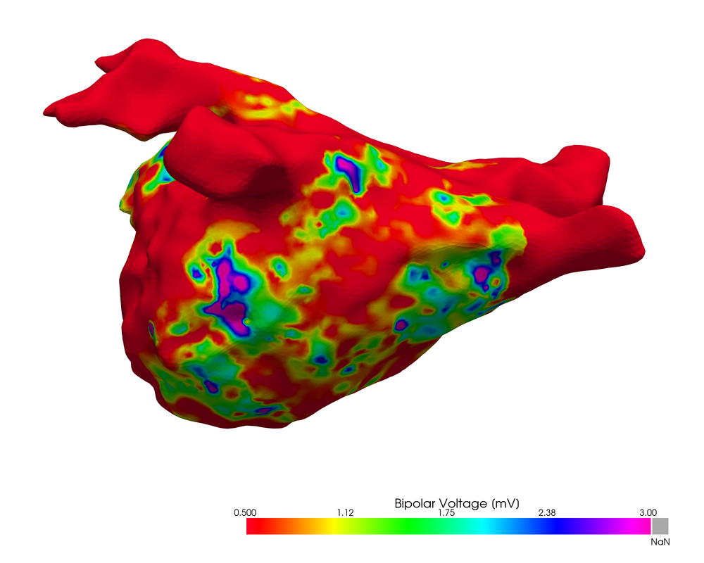
Om hartweefsel in kaart te brengen, gebruiken cardiologen vandaag voltagekaarten die tonen hoe sterk de spanning op elke plaats in het hart is. Zowel trage geleiding als littekenweefsel verzwakken de spanning echter op een vergelijkbare manier, waardoor een arts met een voltagekaart alleen moeilijk kan zien of weefsel nog te behandelen of al afgestorven is.
Mijn hypothese is dat je dit onderscheid wél kan maken door niet enkel naar de spanning te kijken, maar ook andere signaalkenmerken in rekening te brengen. Als deze kenmerken voldoende informatie bevatten om de lokale snelheid van het elektrische signaal te berekenen, kan je in plaats van een voltagekaart een 'snelheidskaart' maken die de geleidingssnelheid op elke plaats in het hart weergeeft. Dit zou een nieuwe kaart van het hart zijn die de behandeling van hartritmestoornissen bevordert.
Experimenteren met een virtueel hart
Omdat het hart zich midden in de borstkas bevindt, is het niet vanzelfsprekend om de elektrische signalen ervan rechtstreeks in beeld te brengen. Een digitaal gesimuleerd hart biedt hiervoor een oplossing: de eigenschappen van het hartweefsel zijn exact gekend en volledig aanpasbaar, waardoor je precies kunt nagaan wat het effect is van bijvoorbeeld littekenweefsel op de gemeten signalen. Klinisch opgemeten signalen kunnen vervolgens vergeleken worden met deze gesimuleerde signalen, wat kan leiden tot een betere diagnose.
Hoe breng je een hartritmestoornis in kaart?
Zoals veel mensen weten, is de eerste stap bij hartziekten een elektrocardiogram (ECG), waarbij elektroden op de huid de elektrische signalen van het hart opmeten. Het nadeel is dat deze signalen eerst door ander weefsel moeten reizen voordat ze de elektrode bereiken en dat de exacte locatie op het hart waar het signaal vandaan komt moeilijk te bepalen is.
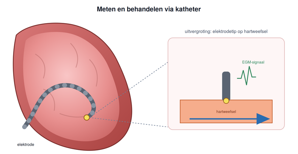
Bij het behandelen van hartritmestoornissen wordt hartweefsel waar de elektrische signalen vastlopen weggebrand, zodat de signalen weer het juiste pad volgen. Het is dus belangrijk om te achterhalen waar in het hart de signalen verstoord worden. Daarom gebruiken cardiologen vaak een elektrogram (EGM) om deze procedure te plannen. Hierbij wordt de elektrode via een bloedvat in de lies ingebracht om vervolgens in het hart uit te komen. Zo kan men metingen rechtstreeks op het hart verrichten.
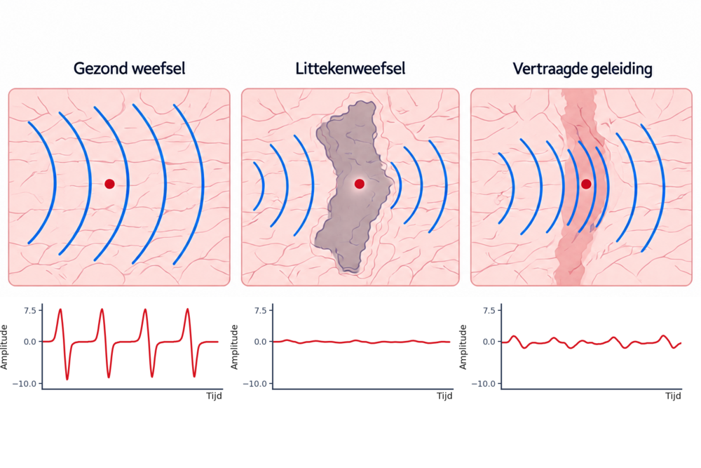
Door op verschillende plaatsen EGM-metingen uit te voeren en te zoeken naar afwijkingen in de signalen, kunnen plaatsen met aangetast weefsel worden opgespoord. Een signaal uit aangetast weefsel zal over het algemeen langer duren, een lagere amplitude hebben, meer asymmetrie vertonen en vaak meerdere pieken hebben. In klinische situaties is er vaak veel ruis op deze signalen omdat de patiënt ademt en het hart klopt, wat betekent dat de elektrode niet altijd even hard op het hartweefsel duwt. Het is dus moeilijk om het weefsel correct onder te verdelen door de signalen visueel te interpreteren.
Artificiële hulp inschakelen
Men kan ook signaalkenmerken, zoals bijvoorbeeld de maximale of minimale amplitude, van het signaal berekenen. Dit resulteert dan in een set van getallen die het signaal kwantitatief beschrijven. Dergelijke kenmerken worden al vaak gebruikt om EGM-signalen te analyseren, maar meestal is deze set erg beperkt. Bovendien onderzoekt men zelden hoeveel informatie elk kenmerk afzonderlijk bevat en of verschillende kenmerken elkaar deels overlappen. Zo beschrijft bijvoorbeeld het bereik van het signaal, gedefinieerd als het verschil tussen maximum en minimum, uiteraard gelijkaardige informatie als het maximum en minimum zelf.
Om dit probleem aan te pakken berekende ik 11 signaalkenmerken voor elk gesimuleerd signaal. Dat zijn een stuk meer kenmerken dan men in de meeste studies gebruikt en 2 van deze kenmerken zijn nog niet eerder op EGM-signalen toegepast. Deze kenmerken kunnen het opgemeten signaal in 11 cijfers kwantificeren. Ik verzamelde signalen voor 5 verschillende scenario's, waarbij in elk scenario de parameters, zoals de breedte en de geleidingssnelheid, gevarieerd werden. Zo ontstond een dataset van 1,7 miljoen EGM-signalen, elk samengevat in elf getallen.
Vervolgens onderzocht ik wat de overlap tussen deze verschillende kenmerken was en of ze gecombineerd konden worden tot een kleinere set getallen. Door de elf kenmerken op een slimme manier te combineren, konden vier nieuwe getallen berekend worden die volstonden om 95% van de informatie te behouden. Met deze vier getallen voorspelde het machine learning-model de lokale geleidingssnelheid met een gemiddelde fout van slechts 1,2%.
Deze resultaten komen uit relatief eenvoudige simulaties zonder ruis. De volgende stap is daarom om te onderzoeken of dezelfde aanpak werkt op complexere simulaties en uiteindelijk op EGM-signalen van echte patiënten. Als dat lukt, kan een kaart van de geleidingssnelheid artsen en onderzoekers helpen om verschillende vormen van aangetast hartweefsel beter van elkaar te onderscheiden. Aangezien hartritmestoornissen vaak voorkomen, kan dit jaarlijks miljoenen mensen helpen.
Bibliografie
1] J. W. Calvert and D. J. Lefer, “Overview of cardiac muscle physiology,” in Cardiovascular Physiology Concepts, 2nd ed., R. E. Klabunde, Ed. Lippincott Williams & Wilkins, 2012, ch. 6. [2] J. Rice, Medical Terminology for Health Care Professionals, 9th ed. USA: Pearson, 2017, [Online]. [Online]. Available: https://www.pearsonhighered.com/assets/samplechapter/0 /1/3/4/0134746279.pdf [3] J. E. Hall and M. E. Hall, Guyton and Hall Textbook of Medical Physiology, 14th ed. Philadelphia, PA: Elsevier, 2021. [4] A. Sirajuddin, M. Y. Chen, C. S. White, and A. E. Arai, “Coronary venous anatomy and anomalies,” Journal of Cardiovascular Computed Tomography, vol. 14, no. 1, pp. 80–90, 2020. [5] H. E. Durham Jr., Cardiac Anatomy. John Wiley Sons, Ltd, 2017, ch. 1, pp. 5–21. [Online]. Available: https://onlinelibrary.wiley.com/doi/abs/10.1002/9781119357407.ch1 [6] T. Mikawa and T. Brand, “Chapter 5.1 - epicardial lineage: Origins and fates,” in Heart Development and Regeneration, N. Rosenthal and R. P. Harvey, Eds. Boston: Academic Press, 2010, pp. 325–344. [Online]. Available: https: //www.sciencedirect.com/science/article/pii/B9780123813329000165 [7] Wikiversity, “Wikijournal of medicine/medical gallery of blausen medical 2014 — wikiversity,,” 2026, [Online; accessed 28-May-2026]. [Online]. Available: https: //en.wikiversity.org/w/index.php?title=WikiJournal of Medicine/Medical gallery of B lausen Medical 2014&oldid=2785552 [8] R. L. Maynard and N. Downes, “Chapter 8 - histology of the vascular system,” in Anatomy and Histology of the Laboratory Rat in Toxicology and Biomedical Research. Academic Press, 2019, pp. 91–95. [Online]. Available: https://www.sciencedirect.com/sc ience/article/pii/B9780128118375000083 [9] P. Kohl and M. Helmes, “Cardiovascular system – the heart,” in Lecture Notes: Human Physiology, 5th ed., O. H. Petersen, Ed. Blackwell Publishing Ltd, 2006, vol. 5, pp. 335–371. [10] K. Nagao, J. Toyama, I. Kodama, and K. Yamada, “Role of the conduction system in the endocardial excitation spread in the right ventricle,” The American Journal of Cardiology, vol. 48, no. 5, pp. 864–870, 1981. [Online]. Available: https://www.sciencedirect.com/science/article/pii/0002914981903519 [11] Z. F. Issa, J. M. Miller, and D. P. Zipes, “Chapter 27 - epicardial ventricular tachycardia,” in Clinical Arrhythmology and Electrophysiology: A Companion to Braunwald’s Heart Disease (Second Edition), second edition ed. Philadelphia: W.B. Saunders, 2012, pp. 608–617. [Online]. Available: https://www.sciencedirect.com/science/article/pii/B97814 55712748000270 [12] S. B. Yeon and N. Oyama, “Chapter 36 - the pericardium: Normal anatomy and spectrum of disease,” in Cardiovascular Magnetic Resonance (Second Edition), second edition ed., W. J. Manning and D. J. Pennell, Eds. Philadelphia: Churchill Livingstone, 2010, pp. 488–497. [Online]. Available: https://www.sciencedirect.com/science/article/pi i/B9780443066863000369 [13] J. Pinnell, S. Turner, and S. Howell, “Cardiac muscle physiology,” Continuing Education in Anaesthesia Critical Care Pain, vol. 7, no. 3, pp. 85–88, 2007. [Online]. Available: https://www.sciencedirect.com/science/article/pii/S1743181617304900 [14] C. Antzelevitch and A. Burashnikov, “Overview of basic mechanisms of cardiac arrhythmia,” Cardiac Electrophysiology Clinics, vol. 3, no. 1, pp. 23–45, 2011. [15] B. F. Fremgen and S. S. Frucht, Medical Terminology: A Living Language, 6th ed. Pearson, 2018. [16] Y. Wang, D. DeMazumder, and J. A. Hill, “Chapter 7 - ionic fluxes and genesis of the cardiac action potential,” in Muscle, J. A. Hill and E. N. Olson, Eds. Boston/Waltham: Academic Press, 2012, pp. 67–85. [Online]. Available: https://www.sciencedirect.com/science/article/pii/B9780123815101000077 [17] M. J. Ackerman and D. E. Clapham, “Ion channels — basic science and clinical disease,” New England Journal of Medicine, vol. 336, no. 22, pp. 1575–1586, 1997. [Online]. Available: https://www.nejm.org/doi/full/10.1056/NEJM199705293362207 [18] I. Kotadia, J. Whitaker, C. Roney, S. Niederer, M. O’Neill, M. Bishop, and M. Wright, “Anisotropic cardiac conduction,” Arrhythmia & Electrophysiology Review, vol. 9, no. 4, p. 202, 2020. [19] S. A. Bernstein and G. E. Morley, “Gap junctions and propagation,” Cardiovascular Gap Junctions, vol. 42, pp. 71–85, 2006. [20] X. Wei, S. Yohannan, and J. R. Richards, “Physiology, cardiac repolarization dispersion and reserve,” in StatPearls [Internet]. StatPearls Publishing, 2023. [21] E. L. Kessler, M. Boulaksil, H. V. van Rijen, M. A. Vos, and T. A. van Veen, “Passive ventricular remodeling in cardiac disease: focus on heterogeneity,” Frontiers in physiology, vol. 5, p. 482, 2014. [22] A. Yartsev, “Normal processes of cardiac excitation and electrical activity,” https://dera ngedphysiology.com/main/cicm-primary-exam/cardiovascular-system/Chapter-010/nor mal-processes-cardiac-excitation-and-electrical-activity, 2020, deranged Physiology. Last updated: 2025-01-09. Accessed: 2025-05-05. [23] P. A. Iaizzo, Handbook of cardiac anatomy, physiology, and devices. Springer Science & Business Media, 2010. [24] E. Prystowsky, “Tachycardia-induced-tachycardia: a mechanism of initiation of atrial fibrillation,” Atrial Arrhythmias: State of the Art. Armonk, NY: Futura, pp. 81–95, 1995. [25] Z. Chu, D. Yang, and X. Huang, “Conditions for the genesis of early afterdepolarization in a model of a ventricular myocyte,” Chaos: An Interdisciplinary Journal of Nonlinear Science, vol. 30, no. 4, 2020.
26] A. L. Wit and P. A. Boyden, “Triggered activity and atrial fibrillation,” Heart rhythm, vol. 4, no. 3, pp. S17–S23, 2007. [27] G. Larraitz, M. F. E., and B. B. P., “Mechanisms of cardiac arrhythmias,” Revista Espa˜nola de Cardiolog´ıa (English Edition), vol. 65, no. 2, pp. 113–201, 2012. [28] N. Vandersickel, S. Hendrickx, R. Van den Abeele, and B. Verstraeten, “Impact of topology on the number of loops during macro-re-entrant atrial tachycardia,” 2024. [29] N. Vandersickel, “Revelation after three decades of ablation therapy: two loops instead of a single re-entry loop drive atrial tachycardia,” Project Repository Journal, vol. 18, pp. 20–23, 2023, accessed: 30 May 2026. [Online]. Available: https://www.europeandissemination.eu/article/revelation-after-three-dec… n-therapy-two-loops-instead-of-a-single-re-entry-loop-drive-atrial-tachycardia/20794 [30] H. Lin, R. Liu, and Z. Liu, “Electrocardiogram signal denoising,” Encyclopedia, accessed: 30 May 2026. [Online]. Available: https://encyclopedia.pub/entry/52174 [31] A. K. Gupta, A. Maheshwari, R. Thakur, and Y. Y. Lokhandwala, “Cardiac mapping: utility or futility?” Indian Pacing and Electrophysiology Journal, vol. 2, no. 1, p. 20, 2002. [32] J. S´anchez and A. Loewe, “A review of healthy and fibrotic myocardium microstructure modeling and corresponding intracardiac electrograms,” Frontiers in Physiology, vol. 13, p. 908069, 2022. [33] S. Saha, D. Linz, D. Saha, A. McEwan, and M. Baumert, “Overcoming uncertainties in electrogram-based atrial fibrillation mapping: A review,” Cardiovascular Engineering and Technology, vol. 15, pp. 52–64, 2023. [34] N. M. S. de Groot, D. Shah, P. M. Boyle, E. Anter, G. D. Clifford, I. Deisenhofer, T. Deneke, P. van Dessel, O. Doessel, P. Dilaveris et al., “Critical appraisal of technologies to assess electrical activity during atrial fibrillation: a position paper from the european heart rhythm association and european society of cardiology working group on ecardiology in collaboration with the heart rhythm society, asia pacific heart rhythm society, latin american heart rhythm society and computing in cardiology,” EP Europace, vol. 24, no. 2, pp. 313–330, 2022. [Online]. Available: https://doi.org/10.1093/europace/euab254 [35] S. Saha, D. Linz, P. Sanders, and M. Baumert, “Beamforming-inspired spatial filtering technique for intracardiac electrograms,” in 2019 41st Annual International Conference of the IEEE Engineering in Medicine and Biology Society (EMBC), 2019, pp. 4254–4257. [36] B. Abdi, M. S. van Schie, N. M. de Groot, and R. C. Hendriks, “Analyzing the effect of electrode size on electrogram and activation map properties,” Computers in Biology and Medicine, vol. 134, p. 104467, 2021. [Online]. Available: https://www.sciencedirect.com/science/article/pii/S0010482521002614 [37] E. Anter and M. E. Josephson, “Bipolar voltage amplitude: What does it really mean?” Heart Rhythm, vol. 13, no. 1, pp. 326–327, 2016. [Online]. Available: https://doi.org/10.1016/j.hrthm.2015.09.033 [38] S. Gaeta, T. D. Bahnson, and C. Henriquez, “Mechanism and magnitude of bipolar electrogram directional sensitivity: Characterizing underlying determinants of bipolar amplitude,” Heart Rhythm, vol. 17, no. 5, Part A, pp. 777–785, 2020. [Online]. Available: https://www.sciencedirect.com/science/article/pii/S1547527119311026
[39] J. M. Stinnett-Donnelly, N. Thompson, N. Habel, V. Petrov-Kondratov, D. D. C. de Sa, J. H. Bates, and P. S. Spector, “Effects of electrode size and spacing on the resolution of intracardiac electrograms,” Coronary Artery Disease, vol. 23, no. 2, pp. 126–132, March 2012. [Online]. Available: https://doi.org/10.1097/MCA.0b013e3283507a9b [40] D. Nairn, H. Lehrmann, B. M¨uller-Edenborn, S. Schuler, T. Arentz, O. D¨ossel, A. Jadidi, and A. Loewe, “Comparison of unipolar and bipolar voltage mapping for localization of left atrial arrhythmogenic substrate in patients with atrial fibrillation,” Frontiers in Physiology, vol. 11, p. 575846, 2020. [41] L. J. van der Does, P. Knops, C. P. Teuwen, C. Serban, R. Starreveld, E. A. Lanters, E. M. Mouws, C. Kik, A. J. Bogers, and N. M. de Groot, “Unipolar atrial electrogram morphology from an epicardial and endocardial perspective,” Heart Rhythm, vol. 15, no. 6, pp. 879–887, Jun. 2018. [42] Z. Ye, M. S. van Schie, L. Pool, A. Heida, P. Knops, Y. J. H. J. Taverne, B. J. J. M. Brundel, and N. M. S. de Groot, “Characterization of unipolar electrogram morphology: a novel tool for quantifying conduction inhomogeneity,” Europace, vol. 25, no. 11, p. euad324, 2023. [43] V. Schlageter, A. Luca, P. Badertscher, P. Krisai, T. Kueffer, D. Spreen, J. Katic, S. Osswald, B. Schaer, C. Sticherling, M. K¨uhne, and S. Knecht, “Effect of electrode size and distance to tissue on unipolar and bipolar voltage electrograms and their implications for a near-field cutoff,” Scientific Reports, vol. 14, p. 27184, 2024. [Online]. Available: https://www.nature.com/articles/s41598-024-78627-5 [44] M. Takigawa, T. Kitamura, S. Basu, M. Bartal, C. A. Martin, R. Martin, G. Cheniti, K. Vlachos, X. Pillois, A. Frontera, G. Massoulli´e, N. Thompson, F. Bourier, A. Lam, J. Duchateau, T. Pambrun, A. Denis, N. Derval, H. Cochet, M. Ha¨ıssaguerre, F. Sacher, M. Hocini, and P. Ja¨ıs, “Effect of electrode size and spacing on electrograms: Optimized electrode configuration for near-field electrogram characterization,” Heart Rhythm, vol. 19, no. 1, pp. 102–112, 2022. [Online]. Available: https://www.sciencedirect.com/science/article/pii/S1547527121021147 [45] M. Hwang, J. Kim, B. Lim, J.-S. Song, B. Joung, E. B. Shim, and H.-N. Pak, “Multiple factors influence the morphology of the bipolar electrogram: An in silico modeling study,” PLoS Computational Biology, vol. 15, no. 4, p. e1006765, 2019. [46] P. Mankad and G. Kalahasty, “Antiarrhythmic drugs: risks and benefits,” Medical Clinics of North America, vol. 103, no. 5, pp. 821–834, 2019. [47] D. E. Krummen, G. Ho, C. T. Villongco, J. Hayase, and A. A. Schricker, “Ventricular fibrillation: triggers, mechanisms and therapies,” Future cardiology, vol. 12, no. 3, pp. 373–390, 2016. [48] R. Parameswaran, A. M. Al-Kaisey, and J. M. Kalman, “Catheter ablation for atrial fibrillation: current indications and evolving technologies,” Nature Reviews Cardiology, vol. 18, no. 3, pp. 210–225, 2021. [49] A. L. Hodgkin and A. F. Huxley, “A quantitative description of membrane current and its application to conduction and excitation in nerve,” The Journal of Physiology, vol. 117, no. 4, pp. 500–544, 1952. [50] O. H. Schmitt, Biological Information Processing Using the Concept of Interpenetrating Domains. Berlin, Heidelberg: Springer Berlin Heidelberg, 1969, pp. 325–331. [Online]. Available: https://doi.org/10.1007/978-3-642-87086-6 18
51] L. Tung, “A bi-domain model for describing ischemic myocardial d-c potentials,” PhD thesis, Massachusetts Institute of Technology, Cambridge, MA, 1978. [Online]. Available: https://dspace.mit.edu/entities/publication/375f998c-5980-4274-83de-b22… [52] R. H. Clayton, O. Bernus, E. M. Cherry, H. Dierckx, F. H. Fenton, L. Mirabella, A. V. Panfilov, F. B. Sachse, G. Seemann, and H. Zhang, “Models of cardiac tissue electrophysiology: Progress, challenges and open questions,” Progress in Biophysics and Molecular Biology, vol. 104, no. 1–3, pp. 22–48, 2011. [53] M. Potse, B. Dub´e, A. Vinet, and R. Cardinal, “A comparison of monodomain and bidomain propagation models for the human heart,” in 2006 International Conference of the IEEE Engineering in Medicine and Biology Society. IEEE, 2006, pp. 3895–3898. [54] M. J. Bishop and G. Plank, “Representing cardiac bidomain bath-loading effects by an augmented monodomain approach: Application to complex ventricular models,” IEEE Transactions on Biomedical Engineering, vol. 58, no. 4, pp. 1066–1075, Apr. 2011. [55] S. Rossi and B. E. Griffith, “Incorporating inductances in tissue-scale models of cardiac electrophysiology,” Chaos: An Interdisciplinary Journal of Nonlinear Science, vol. 27, no. 9, p. 093926, September 2017. [Online]. Available: https://doi.org/10.1063/1.5000706 [56] J. S´anchez, J. F. Gomez, L. Martinez-Mateu, L. Romero, J. Saiz, and B. Trenor, “Heterogeneous effects of fibroblast-myocyte coupling in different regions of the human atria under conditions of atrial fibrillation,” Frontiers in Physiology, vol. 10, p. 847, Jul. 2019. [57] E. J. Vigmond, M. Hughes, G. Plank, and L. J. Leon, “Computational tools for modeling electrical activity in cardiac tissue,” Journal of Electrocardiology, vol. 36, no. Supplement 1, pp. 69–74, Dec. 2003. [58] G. Plank, A. Loewe, A. Neic, C. Augustin, Y.-L. Huang, M. A. Gsell, E. Karabelas, M. Nothstein, A. J. Prassl, J. S´anchez, G. Seemann, and E. J. Vigmond, “The opencarp simulation environment for cardiac electrophysiology,” Computer Methods and Programs in Biomedicine, vol. 208, p. 106223, Sep. 2021. [59] M. Courtemanche, R. J. Ramirez, and S. Nattel, “Ionic mechanisms underlying human atrial action potential properties: insights from a mathematical model,” American Journal of Physiology-Heart and Circulatory Physiology, vol. 275, no. 1, pp. H301–H321, 1998. [60] C. Corrado, J. Whitaker, H. Chubb, S. Williams, M. Wright, J. Gill, M. O’Neill, and S. A. Niederer, “Personalized models of human atrial electrophysiology derived from endocardial electrograms,” IEEE Transactions on Biomedical Engineering, vol. 64, no. 4, pp. 735–742, 2017. [61] M. Wilhelms, H. Hettmann, M. M. Maleckar, J. T. Koivum¨aki, O. D¨ossel, and G. Seemann, “Benchmarking electrophysiological models of human atrial myocytes,” Frontiers in Physiology, vol. 3, p. 487, 2013. [62] N. F. Otani and R. F. J. Gilmour, “Memory models for the electrical properties of local cardiac systems,” Journal of Theoretical Biology, vol. 187, no. 3, pp. 409–436, 1997. [63] A. Baher, Z. Qu, A. Hayatdavoudi, S. T. Lamp, M.-J. Yang, F. Xie, S. Turner, A. Garfinkel, and J. N. Weiss, “Short-term cardiac memory and mother rotor fibrillation,” American Journal of Physiology-Heart and Circulatory Physiology, vol. 292, no. 1, pp. H180–H189, 2007.
64] E. M. Cherry, H. M. Hastings, and S. J. Evans, “Dynamics of human atrial cell models: Restitution, memory, and intracellular calcium dynamics in single cells,” Progress in Biophysics and Molecular Biology, vol. 98, no. 1, pp. 24–37, 2008. [65] T. Nezlobinsky and A. Okenov, “Finitewave,” n.d., mIT License, accessed 2026-04-14. [Online]. Available: https://github.com/finitewave/finitewave [66] L. J. M. E. van der Does and N. M. S. de Groot, “Inhomogeneity and complexity in defining fractionated electrograms,” Heart Rhythm, vol. 14, no. 4, pp. 616–624, 2017. [67] A. Neic, F. O. Campos, A. J. Prassl, S. A. Niederer, M. J. Bishop, E. J. Vigmond, and G. Plank, “Efficient computation of electrograms and ecgs in human whole heart simulations using a reaction-eikonal model,” Journal of Computational Physics, vol. 346, pp. 191–211, Oct. 2017. [68] J. Malmivuo and R. Plonsey, Bioelectromagnetism: Principles and Applications of Bioelectric and Biomagnetic Fields. New York: Oxford University Press, 1995. [69] J. P. Ugarte, A. Orozco-Duque, C. Tob´on, V. Kremen, D. Novak, J. Saiz, T. Oesterlein, C. Schmitt, A. Luik, and J. Bustamante, “Dynamic approximate entropy electroanatomic maps detect rotors in a simulated atrial fibrillation model,” PLoS ONE, vol. 9, no. 12, p. e114577, 2014. [70] M. J. Bishop and G. Plank, “Bidomain ecg simulations using an augmented monodomain model for the cardiac source,” IEEE Transactions on Biomedical Engineering, vol. 58, no. 8, pp. 2297–2307, 2011. [71] M. W. Keller, S. Schuler, G. Seemann, and O. D¨ossel, “Differences in intracardiac signals on a realistic catheter geometry using mono- and bidomain models,” in 2012 Computing in Cardiology, 2012, pp. 305–308. [72] M. Walker, S. Schuler, M. Wilhelm, G. Lenis, G. Seemann, and C. Schmitt, “Characterization of radiofrequency ablation lesion development based on simulated and measured intracardiac electrograms,” IEEE Transactions on Biomedical Engineering, vol. 61, no. 9, pp. 2467–2478, 2014. [73] P. C. Franzone, L. F. Pavarino, S. Scacchi, and B. Taccardi, “Monophasic action potentials generated by bidomain modeling as a tool for detecting cardiac repolarization times,” American Journal of Physiology - Heart and Circulatory Physiology, vol. 293, no. 5, pp. H2776–H2786, 2007. [74] J. S´anchez, G. Luongo, M. Nothstein, L. A. Unger, J. Saiz, B. Trenor, A. Luik, O. D¨ossel, and A. Loewe, “Using machine learning to characterize atrial fibrotic substrate from intracardiac signals with a hybrid in silico and in vivo dataset,” Frontiers in Physiology, vol. 12, p. 699291, 2021. [75] A. Jadidi, M. Nothstein, J. Chen, H. Lehrmann, O. D¨ossel, J. Allgeier, D. Trenk, F.-J. Neumann, A. Loewe, B. M¨uller-Edenborn, and T. Arentz, “Specific electrogram characteristics identify the extra-pulmonary vein arrhythmogenic sources of persistent atrial fibrillation – characterization of the arrhythmogenic electrogram patterns during atrial fibrillation and sinus rhythm,” Scientific Reports, vol. 10, p. 9147, 2020. [76] Z. Ye, M. S. van Schie, and N. M. S. de Groot, “Signal fingerprinting as a novel diagnostic tool to identify conduction inhomogeneity,” Frontiers in Physiology, vol. 12, p. 652128, 2021.
77] M. S. Spach and P. C. Dolber, “Relating extracellular potentials and their derivatives to anisotropic propagation at a microscopic level in human cardiac muscle: Evidence for electrical uncoupling of side-to-side fiber connections with increasing age,” Circulation Research, vol. 58, no. 3, pp. 356–371, March 1986. [78] M. Beheshti, K. Magtibay, S. Mass´e, A. Porta-Sanchez, S. Haldar, A. Bhaskaran, S. Nayyar, B. Glover, D. C. Deno, E. J. Vigmond, and K. Nanthakumar, “Determinants of atrial bipolar voltage: Inter electrode distance and wavefront angle,” Computers in Biology and Medicine, vol. 102, pp. 449–457, 2018. [Online]. Available: https://doi.org/10.1016/j.compbiomed.2018.07.011 [79] V. Jacquemet, N. Virag, Z. Ihara, L. Dang, O. Blanc, S. Zozor, J.-M. Vesin, L. Kappenberger, and C. S. Henriquez, “Study of unipolar electrogram morphology in a computer model of atrial fibrillation,” Journal of Cardiovascular Electrophysiology, vol. 14, no. S10, pp. S172–S179, 2003. [80] S. Schuler, M. W. Keller, T. Oesterlein, G. Seemann, and O. D¨ossel, “Influence of catheter orientation, tissue thickness and conduction velocity on the intracardiac electrogram,” Biomedical Engineering / Biomedizinische Technik, vol. 58, no. s1, 2013. [81] D. Nairn, D. Hunyar, J. S´anchez, O. D¨ossel, and A. Loewe, “Impact of electrode size on electrogram voltage in healthy and diseased tissue,” in Computing in Cardiology Conference (CinC). Brno, Czech Republic: IEEE, 2020. [82] L. Schicketanz, L. A. Unger, J. S´anchez, O. D¨ossel, and A. Loewe, “Separating atrial near fields and atrial far fields in simulated intra-atrial electrograms,” Current Directions in Biomedical Engineering, vol. 7, no. 2, pp. 175–178, 2021. [83] T. Oesterlein, D. Frisch, A. Loewe, G. Seemann, C. Schmitt, O. Doessel, and A. Luik, “Basket-type catheters: Diagnostic pitfalls caused by deformation and limited coverage,” BioMed Research International, vol. 2016, p. 5340574, 2016. [84] L. A. Unger, T. G. Oesterlein, A. Loewe, and O. D¨ossel, “Noise quantification and noise reduction for unipolar and bipolar electrograms,” in Computing in Cardiology Conference (CinC). IEEE, 2021, pp. 1–4. [85] A. Frontera, M. Takigawa, R. Martin, P. Ja¨ıs, M. Ha¨ıssaguerre, and N. Derval, “Electrogram signature of specific activation patterns: Analysis of atrial tachycardias at highdensity endocardial mapping,” Heart Rhythm, vol. 15, no. 1, pp. 28–37, Jan. 2018. [86] F. D. Ramirez, M. Meo, C. Dallet, P. Krisai, K. Vlachos, A. Frontera, M. Takigawa, Y. Nakatani, T. Nakashima, C. Andr´e, T. Kamakura, T. Takagi, A. Grapezzi, R. Tixier, R. Chauvel, G. Cheniti, J. Duchateau, T. Pambrun, F. Sacher, M. Hocini, and N. Derval, “High-resolution mapping of reentrant atrial tachycardias: Relevance of low bipolar voltage,” Heart Rhythm, 2023. [87] K. Miyamoto, T. Tsuchiya, S. Narita, T. Yamaguchi, Y. Nagamoto, S. ichi Ando, K. Hayashida, Y. Tanioka, and N. Takahashi, “Bipolar electrogram amplitudes in the left atrium are related to local conduction velocity in patients with atrial fibrillation,” Europace, vol. 11, no. 12, pp. 1597–1605, 2009. [88] A. Codreanu, F. Odille, E. Aliot, P.-Y. Marie, I. Magnin-Poull, M. Andronache, D. Mandry, W. Djaballah, D. R´egent, J. Felblinger, and C. de Chillou, “Electroanatomic characterization of post-infarct scars: comparison with 3-dimensional myocardial scar reconstruction based on magnetic resonance imaging,” Journal of the American College of Cardiology, vol. 52, no. 10, pp. 839–842, September 2008.
[89] M. Saha, C. Roney, H. Cochet, S. Niederer, E. Vigmond, and S. Nattel, “Myocardial transmural electrical disruption affects electrogram pattern,” in 2019 Computing in Cardiology (CinC), 2019, pp. Page 1–Page 4. [90] E. Irakoze, C. H. R. Gowda, and V. Jacquemet, “Asymmetry of unipolar electrograms in a thin tissue with epicardial-endocardial activation delay,” in 2017 Computing in Cardiology (CinC), 2017, pp. 1–4. [91] V. Jacquemet and C. S. Henriquez, “Genesis of complex fractionated atrial electrograms in zones of slow conduction: a computer model of microfibrosis,” Heart Rhythm, vol. 6, no. 6, pp. 803–810, June 2009. [92] A. N. Ganesan, P. Kuklik, D. H. Lau, A. G. Brooks, M. Baumert, P. W. Lim, S. Thanigaimani, S. Nayyar, M. R. Mahajan, J. M. Kalman, K. C. Roberts-Thomson, and P. Sanders, “Bipolar electrogram shannon entropy at sites of rotational activation: Implications for ablation of atrial fibrillation,” Circulation: Arrhythmia and Electrophysiology, vol. 6, no. 1, pp. 48–57, Feb. 2013. [93] P. S. Cuculich, J. Zhang, Y. Wang, K. A. Desouza, R. Vijayakumar, P. K. Woodard, and Y. Rudy, “The electrophysiological cardiac ventricular substrate in patients after myocardial infarction: Noninvasive characterization with electrocardiographic imaging,” Journal of the American College of Cardiology, vol. 58, no. 18, pp. 1893–1902, October 2011. [94] M. W. Keller, A. Luik, M. S. Abady, G. Seemann, C. Schmitt, and O. D¨ossel, “Influence of three-dimensional fibrotic patterns on simulated intracardiac electrogram morphology,” in Computing in Cardiology 2013. IEEE, 2013, pp. 923–926. [95] C. H. Roney, J. D. Bayer, S. Zahid, M. Meo, P. M. J. Boyle, N. A. Trayanova, M. Ha¨ıssaguerre, R. Dubois, H. Cochet, and E. J. Vigmond, “Modelling methodology of atrial fibrosis affects rotor dynamics and electrograms,” EP Europace, vol. 18, no. suppl 4, pp. iv146–iv155, Dec. 2016. [96] F. Bogun, S. Krishnan, M. Siddiqui, E. Good, J. E. Marine, C. Schuger, H. Oral, A. Chugh, F. Pelosi, and F. Morady, “Electrogram characteristics in postinfarction ventricular tachycardia: effect of infarct age,” Journal of the American College of Cardiology, vol. 46, no. 4, pp. 667–674, August 2005. [97] M. Masjedian, C. Jung, P. Kuklik, F.-A. Alken, A.-K. Kahle, N. Klatt, K. Scherschel, and C. Meyer, “A novel algorithm for 3-d visualization of electrogram duration for substratemapping in patients with ischemic heart disease and ventricular tachycardia,” PLOS ONE, vol. 16, no. 7, p. e0254683, July 2021. [98] C. M. Costa, G. C. Anderson, V. M. F. Meijborg, C. O’Shea, M. J. Shattock, P. Kirchhof, R. Coronel, S. Niederer, and J. Winter, “The amplitude-normalized area of a bipolar electrogram as a measure of local conduction delay in the heart,” Frontiers in Physiology, vol. 11, p. 465, 2020. [99] A. Frontera, L. R. Limite, S. Pagani, M. Cireddu, K. Vlachos, C. Martin, M. Takigawa et al., “Electrogram fractionation during sinus rhythm occurs in normal voltage atrial tissue in patients with atrial fibrillation,” Pacing and Clinical Electrophysiology, vol. 45, no. 3, pp. 304–313, 2022. [100] R. Cabrera-Lozoya, B. Berte, H. Cochet, P. Ja¨ıs, N. Ayache, and M. Sermesant, “Imagebased biophysical simulation of intracardiac abnormal ventricular electrograms,” IEEE Transactions on Biomedical Engineering, vol. 64, no. 7, pp. 1446–1454, Jul. 2017.
[101] R. Morgan, M. A. Colman, H. Chubb, G. Seemann, and O. V. Aslanidi, “Slow conduction in the border zones of patchy fibrosis stabilizes the drivers for atrial fibrillation: Insights from multi-scale human atrial modeling,” Frontiers in Physiology, vol. 7, p. 474, 2016. [102] T. A. Gokhale, E. M. Devecchi, and C. S. Henriquez, “Modeling dynamics in diseased cardiac tissue: Impact of model choice,” Chaos, vol. 27, no. 9, p. 093909, 2017. [103] M. W. Krueger, K. S. Rhode, M. D. O’Neill, C. A. Rinaldi, J. Gill, R. Razavi, G. Seemann, and O. Doessel, “Patient-specific modeling of atrial fibrosis increases the accuracy of sinus rhythm simulations and may explain maintenance of atrial fibrillation,” Journal of Electrocardiology, vol. 47, no. 3, pp. 324–328, 2014. [104] M. W. Krueger, G. Seemann, K. Rhode, D. U. J. Keller, C. Schilling, A. Arujuna, J. Gill, M. D. O’Neill, R. Razavi, and O. Doessel, “Personalization of atrial anatomy and electrophysiology as a basis for clinical modeling of radio-frequency ablation of atrial fibrillation,” IEEE Transactions on Medical Imaging, vol. 32, no. 1, pp. 73–84, Jan. 2013. [105] B. Lim, J. Kim, M. Hwang, J.-S. Song, J. K. Lee, H.-T. Yu, T.-H. Kim, J.-S. Uhm, B. Joung, M.-H. Lee, and H.-N. Pak, “In situ procedure for high-efficiency computational modeling of atrial fibrillation reflecting personal anatomy, fiber orientation, fibrosis, and electrophysiology,” Scientific Reports, vol. 10, no. 1, p. 2417, 2020. [106] K. H. W. J. T. Tusscher and A. V. Panfilov, “Influence of diffuse fibrosis on wave propagation in human ventricular tissue,” EP Europace, vol. 9, no. suppl 6, pp. vi38–vi45, Nov. 2007. [107] R. H. Clayton, “Dispersion of recovery and vulnerability to re-entry in a model of human atrial tissue with simulated diffuse and focal patterns of fibrosis,” Frontiers in Physiology, vol. 9, p. 1052, 2018. [108] E. Vigmond, A. Pashaei, S. Amraoui, H. Cochet, and M. Ha¨ıssaguerre, “Percolation as a mechanism to explain atrial fractionated electrograms and reentry in a fibrosis model based on imaging data,” Heart Rhythm, vol. 13, no. 7, pp. 1536–1543, Jul. 2016. [109] S. Alonso and M. B¨ar, “Reentry near the percolation threshold in a heterogeneous discrete model for cardiac tissue,” Physical Review Letters, vol. 110, no. 15, p. 158101, 2013. [110] T. Ashihara, R. Haraguchi, K. Nakazawa, T. Namba, T. Ikeda, Y. Nakazawa, T. Ozawa, M. Ito, M. Horie, and N. A. Trayanova, “The role of fibroblasts in complex fractionated electrograms during persistent/permanent atrial fibrillation: Implications for electrogrambased catheter ablation,” Circulation Research, vol. 110, no. 2, pp. 275–284, 2012. [111] J. S´anchez, M. Nothstein, L. Unger, J. Saiz, B. Tr´enor, and O. D¨ossel, “Influence of fibrotic tissue arrangement on intracardiac electrograms during persistent atrial fibrillation,” in Computing in Cardiology. IEEE, 2017, pp. 47–50. [112] A. Okenov, T. Nezlobinsky, C. A. Glashan, K. Zeppenfeld, N. Vandersickel, and A. V. Panfilov, “Analysis and generation of the fibrosis textures based on histology data of the human hearts with non-ischemic cardiomyopathy,” Physics in Medicine & Biology, vol. 70, no. 13, p. 135002, 2025. [113] K. H. Jaeger, J. D. Trotter, X. Cai, H. Arevalo, and A. Tveito, “Evaluating computational efforts and physiological resolution of mathematical models of cardiac tissue,” Scientific Reports, vol. 14, p. 16954, 2024.
[114] B. Zhu, G. Zhang, S. Xie, Y. Luan, W. Cao, J. Xu, S. Zhang, J. Tian, F. Wang, and S. Li, “The characterization of functional conduction block in patients with multiple types of atrial tachycardia: A discussion on the mechanism of multiple atrial tachycardia,” Journal of Interventional Cardiac Electrophysiology, vol. 67, pp. 1793–1806, 2024. [Online]. Available: https://doi.org/10.1007/s10840-024-01817-8 [115] I. T. Jolliffe, “Principal component analysis: A beginner’s guide — i. introduction and application,” Weather, vol. 41, no. 7, pp. 206–211, 1986. [116] J. Shlens, “A tutorial on principal component analysis,” arXiv:1404.1100, 2014, version 3.02, April 7, 2014. [117] K. Ntagiantas, E. Pignatelli, N. S. Peters, C. D. Cantwell, R. A. Chowdhury, and A. A. Bharath, “Estimation of fibre architecture and scar in myocardial tissue using electrograms: An in-silico study,” Biomedical Signal Processing and Control, vol. 89, p. 105746, 2024. [118] G. Jimenez-Perez, J. Acosta, J. Bocanegra-P´erez, E. Arana-Rueda, M. Frutos-L´opez, J. A. S´anchez-Brotons, H. Llamas-G´omez, R. Di Massa Pezzutti, C. Gonz´alez de la Portilla Concha, O. Camara, and A. Pedrote, “Delineation of intracavitary electrograms for the automatic quantification of decrement-evoked potentials in the coronary sinus with deep-learning techniques,” Frontiers in Physiology, vol. Volume 15 - 2024, 2024. [Online]. Available: https://www.frontiersin.org/journals/physiology/articles/10.3389/f phys.2024.1331852 [119] B. Verma, T. Oesterlein, A. Loewe, A. Luik, C. Schmitt, and O. D¨ossel, “Regional conduction velocity calculation from clinical multichannel electrograms in human atria,” Computers in Biology and Medicine, vol. 92, pp. 188–196, Jan. 2018. [120] G. Canino, A. D. Costanzo, N. Salerno, I. Leo, M. Cannataro, P. H. Guzzi, P. Veltri, S. Sorrentino, S. D. Rosa, and D. Torella, “Artificial intelligence in cardiac electrophysiology: A clinically oriented review with engineering primers,” Bioengineering, vol. 12, no. 10, p. 1102, 2025. [121] S.-T. Jeong, “Stability of finite difference schemes on the diffusion equation with discontinuous coefficients,” 2018, available via MIT repository. [Online]. Available: http://math.mit.edu [122] L. Tung and J. R. Borderies, “Analysis of electric field stimulation of single cardiac muscle cells,” Biophysical Journal, vol. 63, no. 2, pp. 371–386, 1992. [123] V. Monasterio, E. Pueyo, J. F. Rodr´ıguez-Matas, and J. Carro, “Cardiac cells stimulated with an axial current-like waveform reproduce electrophysiological properties of tissue fibers,” Computer Methods and Programs in Biomedicine, vol. 226, p. 107121, 2022. [124] J. P. Keener, “Propagation and its failure in coupled systems of discrete excitable cells,” SIAM Journal on Applied Mathematics, vol. 47, no. 3, pp. 556–572, 1987. [125] S. F. Pravdin, “Anti-tachycardia pacing simulation in the human heart left ventricle using the luo–rudy model,” in 2024 IEEE SIBIRCON, 2024, pp. 1–5. [126] Z. Li, C. Lambranzi, D. Wu, A. Segato, F. De Marco, E. Vander Poorten, J. Dankelman, and E. De Momi, “Robust path planning via learning from demonstrations for robotic catheters in deformable environments,” IEEE Transactions on Biomedical Engineering, vol. 72, no. 1, pp. 324–335, 2025.
[127] N. V. Belikov, I. V. Khaydukova, I. E. Poludkin, and A. S. Borde, “Evolution and current state of robotic catheters for endovascular surgery: A comprehensive review,” Engineering Science and Technology, an International Journal, vol. 57, p. 101789, 2024. [128] T. Nezlobinsky and A. Okenov, “Finitewave documentation,” 2025, accessed: 2026-03-27. [129] M. Masuda, Y. Matsuda, H. Uematsu, and T. Mano, “Remote entrainment pacing from multiple distant areas to identify a slow conduction isthmus of a reentrant circuit in scarrelated atrial tachycardia,” HeartRhythm Case Reports, vol. 5, no. 10, pp. 513–517, 2019. [130] E. Anter, M. Duytschaever, C. Shen, T. Strisciuglio, E. Leshem, F. M. Contreras-Valdes, J. W. Waks, and A. E. Buxton, “Activation mapping with integration of vector and velocity information improves the ability to identify the mechanism and location of complex scarrelated atrial tachycardias,” Circulation: Arrhythmia and Electrophysiology, vol. 11, no. 8, p. e006536, 2018. [131] S.-H. Liu, Y.-J. Lin, P.-T. Lee, J. J. Vicera, S.-L. Chang, L.-W. Lo, and Y.-F. Hu, “The isthmus characteristics of scar-related macroreentrant atrial tachycardia in patients with and without cardiac surgery,” Journal of Cardiovascular Electrophysiology, vol. 32, no. 6, pp. 1535–1545, 2021. [132] J. J. B. Vicera, Y.-J. Lin, P.-T. Lee, S.-L. Chang, L.-W. Lo, Y.-F. Hu, F.-P. Chung, C.-Y. Lin, and T.-Y. Chang, “Identification of critical isthmus using coherent mapping in patients with scar-related atrial tachycardia,” Journal of Cardiovascular Electrophysiology, vol. 31, no. 6, pp. 1436–1445, 2020. [133] P. Konovalov, D. Mangileva, A. Dokuchaev, O. Solovyova, and A. V. Panfilov, “Rotational activity around an obstacle in 2d cardiac tissue in presence of cellular heterogeneity,” Mathematics, vol. 9, no. 23, 2021. [Online]. Available: https://www.mdpi.com/2227-7390/9/23/3090 [134] M. Shenasa, S.-M. Razavi, H. Shenasa, and A. Al-Ahmad, “The ideal cardiac mapping system,” Cardiac Electrophysiology Clinics, vol. 11, no. 4, pp. 739–748, 2019. [135] T. G. Oesterlein, J. Schmid, S. Bauer, A. Jadidi, C. Schmitt, O. D¨ossel, and A. Luik, “Analysis and visualization of intracardiac electrograms in diagnosis and research: Concept and application of kapavie,” Computer Methods and Programs in Biomedicine, vol. 127, pp. 165–173, 2016. [136] M. Brennan, M. Palaniswami, and P. Kamen, “Do existing measures of poincar´e plot geometry reflect nonlinear features of heart rate variability?” IEEE Transactions on Biomedical Engineering, vol. 48, no. 11, pp. 1342–1347, November 2001. [137] E.-F. Chou, M. Khine, T. Lockhart, and R. Soangra, “Effects of ecg data length on heart rate variability among young healthy adults,” Sensors, vol. 21, no. 18, p. 6286, 2021. [138] C. Lerma, O. Infante, H. P´erez-Grovas, and M. V. Jos´e, “Poincar´e plot indexes of heart rate variability capture dynamic adaptations after haemodialysis in chronic renal failure patients,” Clinical Physiology and Functional Imaging, vol. 23, no. 2, pp. 72–80, 2003. [139] A.-O. Boudraa and F. Salzenstein, “Teager–kaiser energy methods for signal and image analysis: A review,” Digital Signal Processing, vol. 78, pp. 338–375, 2018. [140] G. Deng, “A study of sampling frequency for efficient demodulation of am signals,” in Proceedings of the 1997 IEEE International Symposium on Circuits and Systems (ISCAS), vol. 4. Hong Kong: IEEE, Jun. 1997, pp. 2677–2680.
[141] C. Kamath, “Ecg beat classification using features extracted from teager energy functions in time and frequency domains,” IET Signal Processing, vol. 5, no. 6, pp. 575–581, September 2011. [142] NumXL. (2016) Principal component analysis (pca) 102. Accessed: 2026-04-08. [Online]. Available: https://numxl.com/blogs/principal-component-analysis-pca-102/ [143] T. Hastie, R. Tibshirani, and J. Friedman, The Elements of Statistical Learning: Data Mining, Inference, and Prediction, 2nd ed. New York: Springer, 2009. [144] L. Breiman, “Random forests,” Machine Learning, vol. 45, no. 1, pp. 5–32, 2001. [145] V. Bewick, L. Cheek, and J. Ball, “Statistics review 14: Logistic regression,” Critical care (London, England), vol. 9, pp. 112–8, 03 2005.