Kan een geprinte long ooit ademen?
Een long uit een printer?
Een nieuwe long printen alsof het een reserveonderdeel is: het klinkt als sciencefiction. In films zoals Mickey 17 worden menselijke lichamen zelfs volledig opnieuw geprint wanneer ze sterven. De werkelijkheid staat daar nog ver van af. Toch worden vandaag al macroscopisch kleine stukjes levend weefsel gemaakt met behulp van 3D-bioprinters. Onderzoekers hopen daarmee ziekten beter te begrijpen, nieuwe geneesmiddelen te ontwikkelen en op langere termijn misschien zelfs donororganen te vervangen.
Dat is ook het idee achter mijn masterproef. Daarin onderzocht ik hoe we de eerste bouwstenen kunnen leggen voor geprinte longmodellen. Mijn doel was niet om meteen een volledige long te maken, maar om een omgeving te ontwikkelen waarin menselijke longcellen zich zo natuurlijk mogelijk gedragen. Zulke modellen kunnen onderzoekers helpen om longontwikkeling beter te begrijpen en nieuwe behandelingen veiliger en sneller te testen.
Te vroeg geboren, te vroeg ademhalen
Wereldwijd worden elk jaar miljoenen baby's te vroeg geboren. Voor deze kinderen vormen onvoldoende ontwikkelde longen vaak een van de grootste uitdagingen. Een belangrijke oorzaak van ademhalingsproblemen bij premature baby's is respiratory distress syndrome (RDS), waarbij de longen nog niet voldoende ontwikkeld zijn om efficiënt zuurstof op te nemen. Ondanks decennia aan onderzoek begrijpen wetenschappers nog steeds niet volledig hoe menselijke longen zich voorbereiden op die eerste ademhaling.
Dat gebrek aan kennis heeft verschillende oorzaken. Het is uiteraard onmogelijk om de ontwikkeling van menselijke longen rechtstreeks te bestuderen. Onderzoekers zijn daarom aangewezen op diermodellen of eenvoudige celculturen. Die leveren veel informatie op, maar kunnen de complexiteit van een menselijke long slechts gedeeltelijk nabootsen. Bovendien verschillen dieren biologisch van mensen, waardoor onderzoeksresultaten niet rechtstreeks vertaald kunnen worden naar de klinische praktijk.
Daarom groeit de belangstelling voor modellen die in het laboratorium de menselijke longontwikkeling zo realistisch mogelijk kunnen nabootsen.
Printen met licht
Om zulke modellen te maken, wordt steeds vaker gebruikgemaakt van “bioprinting”. Bij klassieke 3D-printers wordt een object laag voor laag opgebouwd. Dat werkt uitstekend voor kunststoffen of metalen, maar levende weefsels stellen andere eisen. Cellen zijn kwetsbaar en complexe structuren zoals longblaasjes zijn moeilijk na te bootsen met een traditionele aanpak.
In mijn onderzoek gebruikte ik daarom een relatief nieuwe techniek: tomografische volumetrische bioprinting (VAM – Volumetric Additive Manufacturing). Daarbij wordt een bioink tegelijk vanuit verschillende richtingen belicht. Op plaatsen waar voldoende lichtstralen samenkomen, wordt het biomateriaal aan elkaar gelinkt tot een 3D-netwerk. Het volledige object ontstaat daardoor in enkele seconden.
Deze ongelooflijke snelheid is niet alleen handig, maar ook biologisch belangrijk. Hoe korter het printproces duurt, hoe minder stress de ingekapselde cellen ervaren.
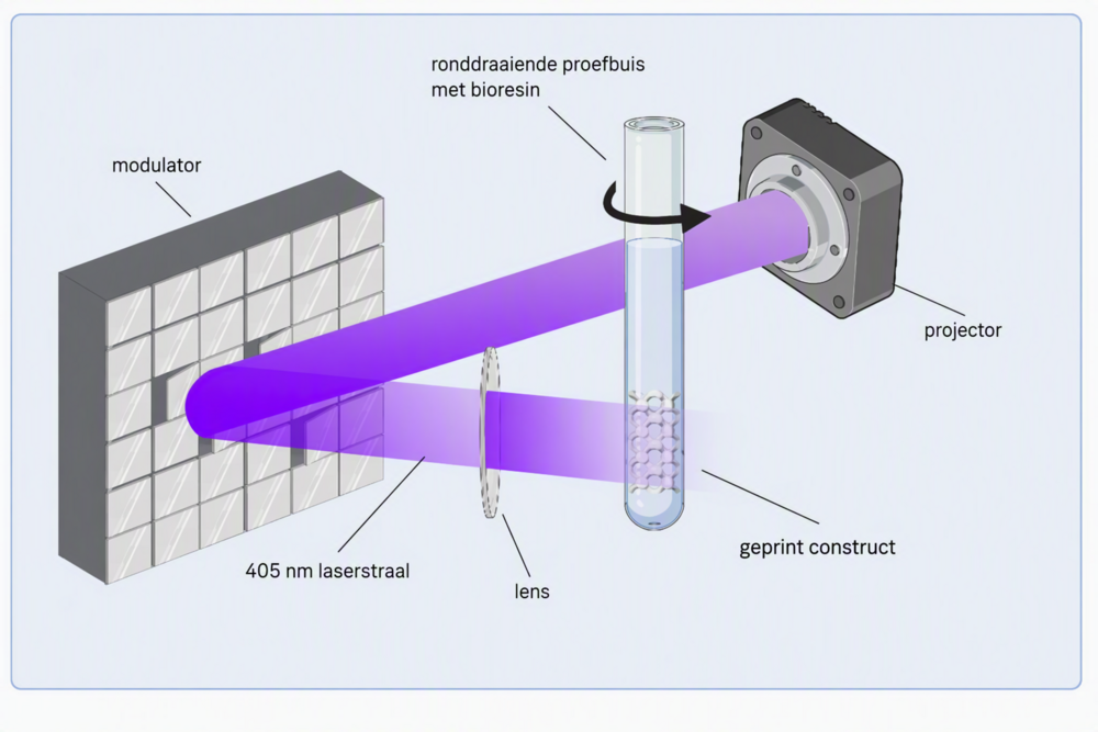
Schematische voorstelling van het werkingsprincipe van de VAM-printer.
De echte uitdaging zit in het materiaal
Wanneer mensen aan “bioprinting” denken, denken ze meestal aan de printer. Voor onderzoekers is het materiaal echter minstens even belangrijk.
Een long is niet zomaar een verzameling cellen. De cellen bevinden zich in een complexe omgeving (= extracellulaire matrix) van eiwitten en andere moleculen die hen voortdurend signalen geven. Wanneer die omgeving ontbreekt, gedragen cellen zich vaak anders dan in het menselijk lichaam.
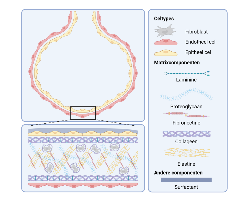
Componenten die te vinden zijn in de extracellulaire matrix van de long.
Daarom ontwikkelde ik een nieuwe bioink: GelMANB. Een bioink is een soort inkt die bestaat uit water en matrixcomponenten, kortom, een inkt waarin levende cellen zich goed thuis voelen. De basis van dit GelMANB bioink bestaat uit gelatine. De meeste mensen kennen gelatine uit snoepgoed, maar in de biomedische wereld vormt het een belangrijk bouwmateriaal omdat het eigenschappen bezit die lijken op die van menselijke weefsels.
Het bijzondere aan GelMANB is dat het twee verschillende chemische mechanismen combineert waarmee het materiaal kan vernet worden. Daardoor kunnen onderzoekers veel preciezer bepalen hoe stevig, elastisch of biologisch actief het uiteindelijke materiaal wordt. Uit mijn resultaten bleek dat die eigenschappen over een brede range konden variëren, waardoor het materiaal geschikt is voor verschillende toepassingen binnen de weefseltechnologie.
Waarom een long veerkracht nodig heeft
Een van de meest opmerkelijke onderdelen van het onderzoek draaide rond elastine.
Elastine is het eiwit dat onze longen hun veerkracht geeft. Telkens wanneer we in- en uitademen, wordt longweefsel uitgerekt en ontspant het opnieuw. Zonder elastine zouden longen veel moeilijker functioneren. Opvallend genoeg wordt elastine nauwelijks nog aangemaakt in volwassen longen, terwijl het tijdens de ontwikkeling juist een cruciale rol speelt.
Daarom ontwikkelde ik ook een nieuwe elastinegebaseerde component die aan het bioink kon worden toegevoegd. Het doel was om het materiaal niet alleen op een long te laten lijken, maar zich ook meer als een long te laten gedragen.
Blijven de cellen leven?
Een mooi geprint model heeft weinig waarde als de cellen erin niet overleven. Daarom onderzocht ik hoe menselijke longfibroblasten zich gedroegen in de verschillende materialen. Fibroblasten zijn belangrijke ondersteunende cellen die meehelpen bij de opbouw van longweefsel.
De resultaten waren veelbelovend. GelMANB bleek niet alleen goed printbaar, maar bood ook een geschikte omgeving voor de ingekapselde cellen. Vooral formuleringen met de elastinecomponent vertoonden een hoge celviabiliteit (=hoeveel cellen er nog in leven zijn) en zorgden ervoor dat de cellen een natuurlijkere vorm aannamen. Dat suggereert dat deze materialen de omstandigheden van echt longweefsel beter benaderen dan sommige bestaande alternatieven.
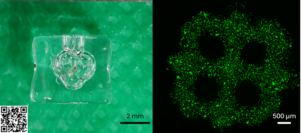
Geprinte alveoli gemaakt van GelMANB (links) en een ander geprint design waarin levende cellen te zien zijn dankzij een specifieke kleurstof die ze groen aankleurt (rechts). Scan de QR-code om het model te zien “ademen”.
Zal een geprinte long ooit ademen
Een volledig functionele, transplanteerbare long uit een printer blijft voorlopig toekomstmuziek. De menselijke long bevat een ingewikkeld netwerk van luchtwegen, bloedvaten en gespecialiseerde cellen dat vandaag nog niet volledig kan worden nagebootst.
Toch brengen nieuwe materialen zoals GelMANB en bioprinttechnieken zoals VAM onderzoekers een stap dichter bij realistische longmodellen. Zulke modellen kunnen helpen om longontwikkeling beter te begrijpen, behandelingen voor premature baby's te verbeteren en de afhankelijkheid van dierproeven te verminderen.
Een volledig geprinte long zal er morgen niet zijn. Maar de materialen en modellen die vandaag worden ontwikkeld, kunnen wel bijdragen aan betere behandelingen voor premature baby's die vandaag nog vechten voor elke ademhaling.
Bibliografie
1. Ohuma, E. O. et al. National, regional, and global estimates of preterm birth in 2020, with trends from 2010: a systematic analysis. Lancet 402, 1261–1271 (2023).
2. Zivaljevic, J., Jovandaric, M. Z., Babic, S. & Raus, M. Complications of Preterm Birth—The Importance of Care for the Outcome: A Narrative Review. Medicina (Kaunas) 60, 1014 (2024).
3. Chen, I.-L. & Chen, H.-L. New developments in neonatal respiratory management. Pediatr Neonatol63, 341–347 (2022).
4. Morrisey, E. E. & Hogan, B. L. M. Preparing for the First Breath: Genetic and Cellular Mechanisms in Lung Development. Developmental Cell 18, 8–23 (2010).
5. Hooper, S. B., Polglase, G. R. & Roehr, C. C. Cardiopulmonary changes with aeration of the newborn lung. Paediatric Respiratory Reviews 16, 147–150 (2015).
6. Yadav, S. & Lee, B. Neonatal Respiratory Distress Syndrome. in StatPearls (StatPearls Publishing, Treasure Island (FL), 2025).
7. Dehaene, I. PRETURN : ‘PREdiction in preTerm birth meets caUsal infeReNce’. (Ghent University, 2023).
8. Zelenina, M., Zelenin, S. & Aperia, A. Water Channels (Aquaporins) and Their Role for Postnatal Adaptation. Pediatr Res 57, 47–53 (2005).
9. Roberts, D., Brown, J., Medley, N. & Dalziel, S. R. Antenatal corticosteroids for accelerating fetal lung maturation for women at risk of preterm birth. Cochrane Database Syst Rev 2017, CD004454 (2017).
10. Herriges, M. & Morrisey, E. E. Lung development: orchestrating the generation and regeneration of a complex organ. Development 141, 502–513 (2014).
11. Rehman, S. & Bacha, D. Embryology, Pulmonary. in StatPearls (StatPearls Publishing, Treasure Island (FL), 2025).
12. He, P. et al. A human fetal lung cell atlas uncovers proximal-distal gradients of differentiation and key regulators of epithelial fates. Cell 185, 4841-4860.e25 (2022).
13. Sun, X. et al. A census of the lung: CellCards from LungMAP. Developmental Cell 57, 112-145.e2 (2022).
14. Zhang, X. et al. Dissecting pulmonary fibroblasts heterogeneity in lung development, health and diseases. Heliyon 9, e19428 (2023).
15. Min, H. et al. Fgf-10 is required for both limb and lung development and exhibits striking functional similarity to Drosophila branchless. Genes Dev 12, 3156–3161 (1998).
16. Chen, L., Acciani, T., Le Cras, T., Lutzko, C. & Perl, A.-K. T. Dynamic regulation of platelet-derived growth factor receptor α expression in alveolar fibroblasts during realveolarization. Am J Respir Cell Mol Biol 47, 517–527 (2012).
17. Nabhan, A. N., Brownfield, D. G., Harbury, P. B., Krasnow, M. A. & Desai, T. J. Single-cell Wnt signaling niches maintain stemness of alveolar type 2 cells. Science 359, 1118–1123 (2018).
18. Sawhney, A. S. et al. A molecular circuit regulates fate plasticity in emerging and adult AT2 cells. Nat Commun 16, 8924 (2025).
19. Weibel, E. R. On the Tricks Alveolar Epithelial Cells Play to Make a Good Lung. Am J Respir Crit Care Med 191, 504–513 (2015).
20. Dushianthan, A., Grocott, M. P. W., Murugan, G. S., Wilkinson, T. M. A. & Postle, A. D. Pulmonary Surfactant in Adult ARDS: Current Perspectives and Future Directions. Diagnostics (Basel) 13, 2964 (2023).
21. Holm, B. A., Wang, Z., Egan, E. A. & Notter, R. H. Content of Dipalmitoyl Phosphatidylcholine in Lung Surfactant: Ramifications for Surface Activity. Pediatr Res 39, 805–811 (1996).
22. Agassandian, M. & Mallampalli, R. K. Surfactant phospholipid metabolism. Biochim Biophys Acta 1831, 612–625 (2013).
23. Tang, Z. et al. Mechanical Forces Program the Orientation of Cell Division during Airway Tube Morphogenesis. Developmental Cell 44, 313-325.e5 (2018).
24. Li, J. et al. The Strength of Mechanical Forces Determines the Differentiation of Alveolar Epithelial Cells. Developmental Cell 44, 297-312.e5 (2018).
25. Hackett, T.-L. et al. The Role of the Dynamic Lung Extracellular Matrix Environment on Fibroblast Morphology and Inflammation. Cells 11, 185 (2022).
26. Bayne, E. F. et al. High-Throughput Extracellular Matrix Proteomics of Human Lungs Enabled by Photocleavable Surfactant and diaPASEF. J. Proteome Res. 23, 2908–2918 (2024).
27. de Hilster, R. H. J. et al. Human lung extracellular matrix hydrogels resemble the stiffness and viscoelasticity of native lung tissue. American Journal of Physiology-Lung Cellular and Molecular Physiology 318, L698–L704 (2020).
28. Kirkness, M. W., Lehmann, K. & Forde, N. R. Mechanics and structural stability of the collagen triple helix. Curr Opin Chem Biol 53, 98–105 (2019).
29. Ito, J. T. et al. Extracellular Matrix Component Remodeling in Respiratory Diseases: What Has Been Found in Clinical and Experimental Studies? Cells 8, 342 (2019).
30. Mereness, J. A. et al. Collagen VI Deficiency Results in Structural Abnormalities in the Mouse Lung. The American Journal of Pathology 190, 426–441 (2020).
31. Heino, J. The collagen family members as cell adhesion proteins. BioEssays 29, 1001–1010 (2007).
32. Emsley, J., Knight, C. G., Farndale, R. W., Barnes, M. J. & Liddington, R. C. Structural basis of collagen recognition by integrin alpha2beta1. Cell 101, 47–56 (2000).
33. Gullberg, D. et al. Analysis of alpha 1 beta 1, alpha 2 beta 1 and alpha 3 beta 1 integrins in cell--collagen interactions: identification of conformation dependent alpha 1 beta 1 binding sites in collagen type I. EMBO J 11, 3865–3873 (1992).
34. Sudhakar, A. et al. Human tumstatin and human endostatin exhibit distinct antiangiogenic activities mediated by alpha v beta 3 and alpha 5 beta 1 integrins. Proc Natl Acad Sci U S A 100, 4766–4771 (2003).
35. Mecham, R. P. Elastin in lung development and disease pathogenesis. Matrix Biology 73, 6–20 (2018).
36. McGowan, S. E. Extracellular matrix and the regulation of lung development and repair. FASEB J 6, 2895–2904 (1992).
37. Ozsvar, J. et al. Tropoelastin and Elastin Assembly. Front. Bioeng. Biotechnol. 9, (2021).
38. Rauscher, S., Baud, S., Miao, M., Keeley, F. W. & Pomès, R. Proline and Glycine Control Protein Self-Organization into Elastomeric or Amyloid Fibrils. Structure 14, 1667–1676 (2006).
39. Rauscher, S. & Pomès, R. The liquid structure of elastin. eLife 6, e26526 (2017).
40. Wendel, D. P., Taylor, D. G., Albertine, K. H., Keating, M. T. & Li, D. Y. Impaired Distal Airway Development in Mice Lacking Elastin. Am J Respir Cell Mol Biol 23, 320–326 (2000).
41. Lindahl, P. et al. Alveogenesis failure in PDGF-A-deficient mice is coupled to lack of distal spreading of alveolar smooth muscle cell progenitors during lung development. Development 124, 3943–3953 (1997).
42. Ruhrberg, C. et al. Spatially restricted patterning cues provided by heparin-binding VEGF-A control blood vessel branching morphogenesis. Genes Dev 16, 2684–2698 (2002).
43. Zhao, J., Sime, P. J., Bringas, P., Gauldie, J. & Warburton, D. Adenovirus-mediated decorin gene transfer preventsTGF-β-induced inhibition of lung morphogenesis. American Journal of Physiology-Lung Cellular and Molecular Physiology 277, L412–L422 (1999).
44. Thuringer, M., Zent, R., Lennon, R. & Plosa, E. J. Basement membranes in lung development, disease, and repair. Matrix Biology 140, 123–132 (2025).
45. Giard, D. J. et al. In Vitro Cultivation of Human Tumors: Establishment of Cell Lines Derived From a Series of Solid Tumors2. J Natl Cancer Inst 51, 1417–1423 (1973).
46. Petpiroon, N. et al. Development of lung tissue models and their applications. Life Sciences 334, 122208 (2023).
47. Murkar, R. S., Wiese-Rischke, C., Weigel, T., Kopp, S. & Walles, H. Developing human upper, lower, and deep lung airway models: Combining different scaffolds and developing complex co-cultures. J Tissue Eng 16, 20417314241299076 (2025).
48. Hajari, M. A. et al. A novel alveoli-on-chip platform for modeling cyclic stretch in patient-derived alveolar epithelial cells cultured from organoids. Lab Chip https://doi.org/10.1039/D5LC00473J (2025) doi:10.1039/D5LC00473J.
49. Doryab, A. & Groll, J. Biomimetic In Vitro Lung Models: Current Challenges and Future Perspective. Advanced Materials 35, 2210519 (2023).
50. Yang, S. et al. Organoids: The current status and biomedical applications. MedComm (2020) 4, e274 (2023).
51. Vazquez-Armendariz, A. I. & Tata, P. R. Recent advances in lung organoid development and applications in disease modeling. J Clin Invest 133, e170500.
52. Li, L., Feng, J., Zhao, S., Rong, Z. & Lin, Y. SOX9 inactivation affects the proliferation and differentiation of human lung organoids. Stem Cell Research & Therapy 12, 343 (2021).
53. Lim, M. J., Kim, S. J., Jo, A. & Kim, S. W. Enhanced application potential of alveolar organoids through epithelial and niche cell interactions. Sci Rep 15, 17538 (2025).
54. Zhou, T. et al. Generation of Induced Pluripotent Stem Cells from Urine. J Am Soc Nephrol 22, 1221–1228 (2011).
55. Alsobaie, S., Alsobaie, T., Alshammary, A. & Mantalaris, S. Differentiation of human induced pluripotent stem cells into functional lung alveolar epithelial cells in 3D dynamic culture. Front Bioeng Biotechnol 11, 1173149 (2023).
56. Lim, M. J., Jo, A. & Kim, S.-W. A novel method for generating induced pluripotent stem cell (iPSC)-derived alveolar organoids: a comparison of their ability depending on iPSC origin. Organoid 3, (2023).
57. Buckberry, S. et al. Transient naive reprogramming corrects hiPS cells functionally and epigenetically. Nature 620, 863–872 (2023).
58. Lo, B. & Parham, L. Ethical Issues in Stem Cell Research. Endocr Rev 30, 204–213 (2009).
59. Wang, Q., Yuan, F., Zuo, X. & Li, M. Breakthroughs and challenges of organoid models for assessing cancer immunotherapy: a cutting-edge tool for advancing personalised treatments. Cell Death Discov. 11, 222 (2025).
60. Yu, B. et al. Organoids for tissue repair and regeneration. Materials Today Bio 33, 102013 (2025).
61. Martinovich, K. M. et al. Conditionally reprogrammed primary airway epithelial cells maintain morphology, lineage and disease specific functional characteristics. Sci Rep 7, 17971 (2017).
62. Liu, C. et al. Bridging the gap: how patient-derived lung cancer organoids are transforming personalized medicine. Front Cell Dev Biol 13, 1554268 (2025).
63. Ringquist, R. et al. An immune-competent lung-on-a-chip for modelling the human severe influenza infection response. Nat. Biomed. Eng 1–23 (2025) doi:10.1038/s41551-025-01491-9.
64. Stucki, J. D. et al. Medium throughput breathing human primary cell alveolus-on-chip model. Sci Rep 8, 14359 (2018).
65. Kohon, A. I. et al. Targeting nanoparticles to lung cancer-derived A549 cells based on changes on interstitial stiffness in biomimetic models. iScience 27, 111015 (2024).
66. Huh, D. et al. Reconstituting Organ-Level Lung Functions on a Chip. Science 328, 1662–1668 (2010).
67. Stucki, A. O. et al. A lung-on-a-chip array with an integrated bio-inspired respiration mechanism. Lab Chip 15, 1302–1310 (2015).
68. Luk, C. H. et al. Autologous human iPSC–derived alveolus-on-chip reveals early pathological events of Mycobacterium tuberculosis infection. Science Advances 12, eaea9874 (2026).
69. Dabaghi, M. et al. Surface Modification of PDMS-Based Microfluidic Devices with Collagen Using Polydopamine as a Spacer to Enhance Primary Human Bronchial Epithelial Cell Adhesion. Micromachines (Basel) 12, 132 (2021).
70. Zamprogno, P. et al. Second-generation lung-on-a-chip with an array of stretchable alveoli made with a biological membrane. Commun Biol 4, 1–10 (2021).
71. Waters, C. M., Roan, E. & Navajas, D. Mechanobiology in Lung Epithelial Cells: Measurements, Perturbations, and Responses. Compr Physiol 2, 1–29 (2012).
72. Grigoryan, B. et al. Multivascular networks and functional intravascular topologies within biocompatible hydrogels. Science 364, 458–464 (2019).
73. Phogat, S., Guo, T. J. F., Thiam, F. & Osei, E. T. Establishing a 3D Vascularized Tri-Culture Model of the Human Airways via a Digital Light Processing Bioprinter. Biotechnology and Bioengineering 122, 2273–2280 (2025).
74. Kunwar, P. et al. Shaping hydrogel bioinks into 3D, multiscale, perfusable models using multimodal printing. 2026.01.29.702588 Preprint at https://doi.org/10.64898/2026.01.29.702588 (2026).
75. Li, W. et al. Stereolithography apparatus and digital light processing-based 3D bioprinting for tissue fabrication. iScience 26, 106039 (2023).
76. Maes, L. et al. Digital light processing of photo-crosslinkable gelatin to create biomimetic 3D constructs serving small intestinal tissue regeneration. Biomaterials Advances 171, 214232 (2025).
77. Leung, C. M. et al. A guide to the organ-on-a-chip. Nat Rev Methods Primers 2, 33 (2022).
78. Shusteff, M. et al. One-step volumetric additive manufacturing of complex polymer structures. Science Advances 3, eaao5496 (2017).
79. Loterie, D., Delrot, P. & Moser, C. High-resolution tomographic volumetric additive manufacturing. Nat Commun 11, 852 (2020).
80. Dhand, A. P., Davidson, M. D. & Burdick, J. A. Lithography-based 3D printing of hydrogels. Nat Rev Bioeng 3, 108–125 (2025).
81. Whyte, D. J., Doeven, E. H., Sutti, A., Kouzani, A. Z. & Adams, S. D. Volumetric additive manufacturing: A new frontier in layer-less 3D printing. Additive Manufacturing 84, 104094 (2024).
82. Fairbanks, B. D., Schwartz, M. P., Bowman, C. N. & Anseth, K. S. Photoinitiated polymerization of PEG-diacrylate with lithium phenyl-2,4,6-trimethylbenzoylphosphinate: polymerization rate and cytocompatibility. Biomaterials 30, 6702–6707 (2009).
83. Kelly, B. E. et al. Volumetric additive manufacturing via tomographic reconstruction. Science 363, 1075–1079 (2019).
84. Van Hoorick, J. et al. (Photo-)crosslinkable gelatin derivatives for biofabrication applications. Acta Biomaterialia 97, 46–73 (2019).
85. Rizzo, R., Wechsler, F., Zhang, Q. & Moser, C. Too Big, Too Small, Too $O_2$: The Pandoro Effect from Oxygen Gradients in Tomographic Volumetric Additive Manufacturing. Preprint at https://doi.org/10.48550/ARXIV.2604.06313 (2026).
86. Cramer, N. B. & Bowman, C. N. Thiol‐ene and Thiol‐yne Chemistry in Ideal Network Synthesis. https://doi.org/10.1039/9781849736961-00001 (2013) doi:10.1039/9781849736961-00001.
87. Thijssen, Q., Toombs, J., Li, C. C., Taylor, H. & Van Vlierberghe, S. From pixels to voxels: A mechanistic perspective on volumetric 3D-printing. Progress in Polymer Science147, 101755 (2023).
88. Stiles, A., Tison, T.-A., Pruitt, L. & Vaidya, U. Photoinitiator Selection and Concentration in Photopolymer Formulations towards Large-Format Additive Manufacturing. Polymers14, 2708 (2022).
89. Yue, K. et al. Synthesis, properties, and biomedical applications of gelatin methacryloyl (GelMA) hydrogels. Biomaterials 73, 254–271 (2015).
90. Chansoria, P. et al. Light from Afield: Fast, High-Resolution, and Layer-Free Deep Vat 3D Printing. Chem. Rev. 124, 8787–8822 (2024).
91. Duymaz, D., Karaoğlu, İ. C. & Kizilel, S. Effect of Photoinitiation Process on Photo-Crosslinking of Gelatin Methacryloyl Hydrogel Networks. Macromolecular Rapid Communications 46, e00376 (2025).
92. Van Hoorick, J. et al. Thiol-norbornene gelatin hydrogels: influence of thiolated crosslinker on network properties and high definition 3D printing. Biofabrication 13, (2020).
93. Malafaia, A. P., Sobreiro-Almeida, R., Rodrigues, J. M. M. & Mano, J. F. Thiol-ene click chemistry: Enabling 3D printing of natural-based inks for biomedical applications. Biomaterials Advances 167, 214105 (2025).
94. Van Hoorick, J. et al. Highly Reactive Thiol-Norbornene Photo-Click Hydrogels: Toward Improved Processability. Macromol Rapid Commun39, e1800181 (2018).
95. Northrop, B. H. & Coffey, R. N. Thiol–Ene Click Chemistry: Computational and Kinetic Analysis of the Influence of Alkene Functionality. J. Am. Chem. Soc. 134, 13804–13817 (2012).
96. Tanneberger, A. E. et al. Biomaterial-based 3D human lung models replicate pathological characteristics of early pulmonary fibrosis. Acta Biomaterialia 204, 277–292 (2025).
97. POLYMÈRES À BASE D’UN BIOPOLYMÈRE MODIFIÉ BIFONCTIONNEL ET HYDROGELS POUVANT ÊTRE OBTENUS À PARTIR DE CES POLYMÈRES À BASE D’UN BIOPOLYMÈRE MODIFIÉ BIFONCTIONNEL (Patents) - Data INPI. https://data.inpi.fr/brevets/WO2019229140?q=#WO2019229140.
98. Pien, N. et al. Development of photo-crosslinkable collagen hydrogel building blocks for vascular tissue engineering applications: A superior alternative to methacrylated gelatin? Materials Science and Engineering: C 130, 112460 (2021).
99. Parmentier, L. et al. 2D fibrillar osteoid niche mimicry through inclusion of visco-elastic and topographical cues in gelatin-based networks. Int J Biol Macromol 254, 127619 (2024).
100. Meeremans, M., Damme, L. V., Spiegelaere, W. D., Vlierberghe, S. V. & Schauwer, C. D. Equine Tenocyte Seeding on Gelatin Hydrogels Improves Elongated Morphology. Polymers13, (2021).
101. Burns, J. A., Butler, J. C., Moran, J. & Whitesides, G. M. Selective reduction of disulfides by tris(2-carboxyethyl)phosphine. J. Org. Chem. 56, 2648–2650 (1991).
102. Almalla, A. et al. Chemistry matters: A side-by-side comparison of two chemically distinct methacryloylated dECM bioresins for vat photopolymerization. Biomaterials Advances 160, 213850 (2024).
103. Parmentier, L., D’Haese, S., Carpentier, N., Dmitriev, R. I. & Van Vlierberghe, S. Bottom-Up Extrusion-Based Biofabrication of the Osteoid Niche. Macromol Biosci 24, e2300395 (2024).
104. Pien, N. et al. Exploring the impact of volumetric additive manufacturing of photo-crosslinkable gelatin on mesenchymal stromal cell behavior and differentiation. Additive Manufacturing109, 104850 (2025).
105. Bernal, P. N. et al. Volumetric Bioprinting of Organoids and Optically Tuned Hydrogels to Build Liver-Like Metabolic Biofactories. Advanced Materials 34, 2110054 (2022).
106. Duquesne, J. et al. Volumetric bioprinting of the osteoid niche. Biofabrication 17, 025002 (2025).
107. Tytgat, L. et al. Additive manufacturing of photo-crosslinked gelatin scaffolds for adipose tissue engineering. Acta Biomater 94, 340–350 (2019).
108. Parmentier, L. et al. Structure-function relationship of methacryloyl-modified gelatin revealed by proteomics and 3D modeling. Applied Materials Today 48, 103072 (2026).
109. de Farias, B. S. et al. Influence of gelatin type on physicochemical properties of electrospun nanofibers. Sci Rep 13, 15195 (2023).
110. Tanaka, N., Suyama, K., Mai, E. & Nose, T. Stepwise LCST-Type Phase Separation in Mixtures of Short-Chain Elastin-Like Peptides With Minimal Structural Differences. Biopolymers117, e70084 (2026).
111. Tanaka, N., Suyama, K., Tomohara, K. & Nose, T. Exploring LCST- and UCST-like Behavior of Branched Molecules Bearing Repeat Units of Elastin-like Peptides as Side Components. Biomacromolecules25, 7156–7166 (2024).
112. Asai, D. et al. Injectable Polypeptide Hydrogel Depot System for Assessment of the Immune Response–Inducing Efficacy of Sustained Antigen Release Alone. Macromolecular Bioscience 19, 1900167 (2019).
113. Asai, D. et al. Protein polymer hydrogels by in situ, rapid and reversible self-gelation. Biomaterials33, 5451–5458 (2012).
114. Polio, S. R. et al. Cross-platform mechanical characterization of lung tissue. PLoS One 13, e0204765 (2018).
115. Trębacz, H. & Barzycka, A. Mechanical Properties and Functions of Elastin: An Overview. Biomolecules13, 574 (2023).
116. Bou Jawde, S., Takahashi, A., Bates, J. H. T. & Suki, B. An Analytical Model for Estimating Alveolar Wall Elastic Moduli From Lung Tissue Uniaxial Stress-Strain Curves. Front. Physiol. 11, (2020).
117. Jandl, K. & Kwapiszewska, G. Stiffness of the Extracellular Matrix: A Regulator of Prostaglandins in Pulmonary Fibrosis? Am J Respir Cell Mol Biol 63, 721–722 (2020).
118. Meyvis, T. K. L. et al. A comparison between the use of dynamic mechanical analysis and oscillatory shear rheometry for the characterisation of hydrogels. International Journal of Pharmaceutics 244, 163–168 (2002).
119. Morley, C. J., Thornton, A. J., Fowler, M. A., Cole, T. J. & Hewson, P. H. Respiratory rate and severity of illness in babies under 6 months old. Arch Dis Child 65, 834–837 (1990).
120. Gehlen, J., Qiu, W., Schädli, G. N., Müller, R. & Qin, X.-H. Tomographic volumetric bioprinting of heterocellular bone-like tissues in seconds. Acta Biomaterialia 156, 49–60 (2023).
121. Ribezzi, D. et al. Multi-material Volumetric Bioprinting and Plug-and-play Suspension Bath Biofabrication via Bioresin Molecular Weight Tuning and via Multiwavelength Alignment Optics. Advanced Materials 37, 2409355 (2025).
122. Duquesne, J. Engineering vascularisatie in botweefsel scaffolds via volumetrisch bioprinting. (Master of Science in Biomedical Engineering, 2023).
123. Tavares-Negrete, J. A., Babayigit, C., Boyraz, O. & Esfandyarpour, R. Impact of exposure time and power intensity on cell viability in 3D bioprinting. in Laser 3D Manufacturing XII vol. 13354 1335402 (SPIE, 2025).
124. Tissue Engineering of Recellularized Small-Diameter Vascular Grafts. https://journals.sagepub.com/doi/epdf/10.1089/ten.2005.11.778?src=getft… doi:10.1089/ten.2005.11.778.
125. High cell density and high-resolution 3D bioprinting for fabricating vascularized tissues. https://www.science.org/doi/10.1126/sciadv.ade7923 doi:10.1126/sciadv.ade7923.
126. Ng, W. L. et al. Fabrication and Characterization of 3D Bioprinted Triple-layered Human Alveolar Lung Models. Int J Bioprint 7, 332 (2021).
127. Firsova, A. B. et al. Spatial single-cell atlas reveals regional variations in healthy and diseased human lung. Nat Commun 16, 9745 (2025).
128. Falandt, M. et al. Spatial-Selective Volumetric 4D Printing and Single-Photon Grafting of Biomolecules within Centimeter-Scale Hydrogels via Tomographic Manufacturing. Advanced Materials Technologies 8, 2300026 (2023).
129. Bagheri, A., Zakerzadeh, M. R., Sadigh, M. J. & Salehabadi, R. Volumetric additive manufacturing of complex geometries around complex inserts. Sci Rep 16, 6522 (2026).
130. Regehly, M. et al. Xolography for linear volumetric 3D printing. Nature 588, 620–624 (2020).