
Nieuwe melkparameter voor een verbeterde opvolging van uiergezondheid bij melkkoeien - differentiële somatische celtelling
Mastitis, oftewel uierontsteking, is nog altijd een zeer groot probleem op de almaar groter wordende Vlaamse melkveebedrijven. Mastitis zorgt niet alleen voor een lagere melkproductie, maar ook voor een negatief effect op de melksamenstelling: enerzijds kan de concentratie van nuttige componenten zoals lactose, caseïne en botervet tot wel 20 procent dalen en anderzijds kunnen ongewenste elementen in de melk toenemen. Er kunnen ook problemen optreden met de houdbaarheid, de verwerking van de melk voor de kaasmakerij wordt bemoeilijkt en de pH van de melk neemt toe als gevolg van uierinfecties. Er is nood aan een nieuwe parameter om mastitis beter te kunnen beheersen. Mastitis vroeg detecteren is cruciaal om economische verliezen te beperken en de kans op genezing te verhogen.
Een uierontsteking wordt meestal veroorzaakt door pathogene bacteriën die het tepelkanaal hebben weten binnen te dringen. Mastitis kan een klinische, subklinische en een chronische vorm aannemen. Klinische mastitis is de zichtbare vorm die behoorlijke schade aanricht en een uitgebreide behandeling vraagt. De onzichtbare, subklinische vorm is een uierontsteking zonder klinische symptomen en waarbij de melkproductie stapsgewijs vermindert. Als de ziekte zich blijft handhaven bij eenzelfde dier spreken we van chronische mastitis. Mastitis oefent een aanzienlijk effect uit op de bedrijfseconomische resultaten. De totale kost van mastitis loopt gemiddeld op tot € 240 per melkgevende koe per jaar en per jaar wordt een derde van de melkkoeien getroffen door mastitis. Nieuwe technieken zijn dus nodig om mastitis onder controle te houden.
Defensiemechanismen
Witte bloedcellen, met name de macrofagen en neutrofielen, vervullen een cruciale rol in de aangeboren defensie tegen mastitis. Ze herkennen indringers en bewaken als het ware het niet-geïnfecteerde weefsel. Een ontsteking wordt over het algemeen gedefinieerd als een toename van de witte bloedcellen. De witte bloedcellen worden aangetrokken naar de plaats van infectie en komen zo in de melk terecht. Ze proberen de infectie op te lossen door de schadelijke micro-organismen te "fagocyteren" oftewel op te slokken. Macrofagen zijn het overheersende celtype in gezond uierweefsel. Zij ruimen alles in de herstellende fase van een infectie op. Neutrofielen zijn het overheersende celtype tijdens de beginstadia van een infectie.
Detectie mastitis
Tot op vandaag wordt de somatische celtelling (“het celgetal”) gebruikt om de uiergezondheid te beoordelen. Het somatisch celgetal geeft de totale hoeveelheid witte bloedcellen in de melk weer. Een somatische celtelling van 200 000 (voor vaarzen 150 000) cellen per milliliter wordt gebruikt als "magische grenswaarde". Lager dan 200 000 cellen per milliliter neemt men aan dat de uier gezond is en hoger dan 200 000 cellen per milliliter wordt aangenomen dat de melkklieren geïnfecteerd zijn. Het totale celgetal geeft echter niet weer in welke fase van infectie de ontsteking zich bevindt, omdat het de immuuncellen niet onderverdeeld. De nieuwe parameter, het gedifferentieerd somatisch celgetal oftewel de DSCC, geeft de verhouding weer tussen het aantal macrofagen en neutrofielen in de melk. Deze nieuwe parameter verschaft aldus meer gedetailleerd inzicht in de actuele gezondheidstoestand van de uier. De differentiële celtelling detecteert veranderingen in relatieve celpopulaties al vóórdat het totaal aantal cellen toeneemt. Het gedifferentieerde celgetal is een percentage dat het aandeel neutrofielen in de melk weergeeft. Het percentage macrofagen kan worden berekend door DSCC van 100% af te trekken. Gezond uierweefsel bevat hoofdzakelijk macrofagen: dit betekent dus een lage DSCC. Wanneer er een ontstekingsreactie plaatsvindt, zal het aantal neutrofielen toenemen en bijgevolg zal ook de DSCC stijgen. De DSCC geeft dus snel en accuraat aan of het uierweefsel ontstoken is en in welke fase die ontsteking zich dan wel bevindt. Een hoge DSCC doet zich voor tijdens de beginstadia van een infectie (ook als het celgetal nog laag is), een dalende DSCC geeft de herstellende fase van de infectie weer.
Resultaten
Allereerst werden van alle beschikbare data statistische kengetallen berekend om de betrouwbaarheid ervan na te gaan. De gemiddelde DSCC was 73 %, dit wil zeggen 73 % neutrofielen en 27 % macrofagen. De helft van de DSCC-waarden lagen tussen 66 en 83 %. Deze resultaten liggen in lijn met wat eerder gevonden is in ander onderzoek.
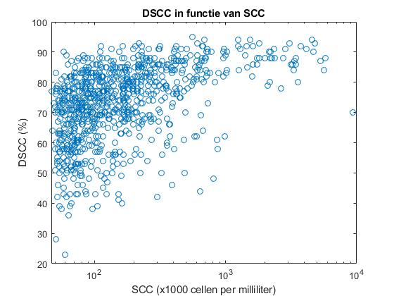
Brede ranges van DSCC zijn beschreven bij lage celgetalwaarden, maar als het celgetal (SCC) stijgt, zal ook de DSCC stijgen en zich in een minder groot bereik bevinden. Dat er zowel hoge als lage DSCC-waarden horen bij lage celgetalwaarden, wil zeggen dat er meerdere fasen van een infectie vertegenwoordigd worden. Hoge celgetalwaarden (meer dan 200 000 cellen per milliliter) in combinatie met hoge DSCC-waarden (meer dan 80 procent) zijn indicators voor vroege infectiefasen van mastitis. Er worden namelijk veel neutrofielen aangetrokken om een immuunreactie te induceren. Aan de andere kant worden hoge celgetalwaarden in combinatie met lage DSCC-waarden gerelateerd aan de herstellende fase van de infectie, omdat lage DSCC-waarden overeenkomen met een hoog percentage macrofagen, die een goede uiergezondheidstoestand weerspiegelen. Het celgetal en de DSCC zijn positief gecorreleerd en er kan een lineair verband opgesteld worden tussen de DSCC en de logaritme van het celgetal. Om het onderscheid te maken tussen gezond en geïnfecteerd uierweefsel, kan er een drempel op DSCC van 80 % gezet worden. Met andere woorden: stijgt de DSCC boven de 80 %, dan kan dit gebruikt worden als een vroege indicator voor mastitis. Het voordeel van de DSCC ten opzichte van het totaal celgetal is dat hij mogelijk sneller reageert dan het totaal celgetal. Hierdoor kan er sneller ingegrepen worden en zal de infectie efficiënter bestreden kunnen worden. De DSCC van eerstekalfskoeien verschilt niet significant van deze van meerderekalfskoeien. Het aantal dagen in lactatie beïnvloedt de DSCC wel: de DSCC volgt namelijk het patroon van een lactatiecurve. Ook de historiek van eerdere infecties speelt een rol: het gemiddelde gedifferentieerd celgetal is hoger naarmate dieren al vijf of meer keer eerder leden aan een uierinfectie.
Besluit
Er moet nog veel onderzoek gebeuren naar het gedrag van de DSCC, onder andere naar de drempelwaarde van 80 %, hoe snel hij verandert bij een uierinfectie, de correlatie met andere parameters enzovoort. Maar deze parameter zal vast en zeker bijdragen tot het verduurzamen van de melkproductie en de verbetering van de uiergezondheid.
Bibliografie
AAD. (2015). What is subklinical mastitis? Geraadpleegd van https://www.qscoutlab.com/what-is-subclinical-mastitis AB Register. (2019). Producenten. Geraadpleegd van https://www.abregister.be/PRODUCENTEN.php Alnakip, M., Quintela-Baluja, M., Böhme, K., Fernández-No, I., Caamaño-Antelo, S., CaloMata, P., & Barros-Velázquez J. (2014). The Immunology of Mammary Gland of Dairy Ruminants between Healthy and Inflammatory Conditions. Journal of Veterinary Medicine, 2014(31). Aref, N., Sayed, A., Zahran, A., Abdelaal, G., & Nasser, E. (2018). Flow cytometric analysis of somatic cells and oxidant/antioxidant profile in dairy cows with subclinical mastitis. Bulgarian Journal of Veterinary Medicine, 21(3), 347–357. Bobbo, T., Cipolat-Gotet, C., Bittante, G., & Cecchinato, A. (2016). The nonlinear effect of somatic cell count on milk composition, coagulation properties, curd firmness modeling, cheese yield, and curd nutrient recovery. Journal of Dairy Science, 99(7), 5104–5119. Bobbo, T., Ruegg, P.L., Stocco, G., Fiore, E., Gianesella, M., Morgante, M., Pasotto, D., Bittante, G., & Cecchinato, A. (2017). Associations between pathogen-specific cases of subclinical mastitis and milk yield, quality, protein composition, and cheese-making traits in dairy cows. Journal of Dairy Science, 100(6), 4868–4883. Bobbo, T., Penasa, M., & Cassandro, M. (2019). Short communication: Genetic aspects of milk differential somatic cell count in Holstein cows: A preliminary analysis. Journal of Dairy Science, 102(5), 4275–4279. Bradley, A. & Green, M. (2005). Use and interpretation of somatic cell count data in dairy cows. In Practice, 27(6), 310–315. Biebaut, E. (2018). Toepassingen en interpretatie van de california mastitis test als diagnostische tool voor subklinische mastitis bij melkvee [masterthesis]. Gent: Universiteit Gent, Faculteit Diergeneeskunde. Breen, J.E., Green, M.J., & Bradley, A.J. (2009). Quarter and cow risk factors associated with the occurrence of clinical mastitis in dairy cows in the United Kingdom. Journal of Dairy Science, 92(2009), 2551–2561. Blowey, R. & Edmondson, P. (2010). Mastitis Control in Dairy Herds. Ipswich: Farming press. Clark, G., Stickinger, H., Balderas, R., Val Zelm, M., Zola, H., Hart, D., & Engel, P. (2016). Nomenclature of CD molecules from the Tenth Human Leucocyte Differentiation Antigen Workshop. Clinical & Translational Immunology, 5(1). Damm, M., Holm, C., Blaabjerg, M., Bro, M.N., & Schwarz, D. (2017). Differential somatic cell count—A novel method for routine mastitis screening in the frame of Dairy Herd Improvement testing programs. Journal of Dairy Science, 100(6), 4926–4940. De Hertogh, M. (2014). Klinische mastitis, een veel voorkomend economisch probleem in de melkveeindustrie [masterthesis]. Gent: Universiteit Gent, Faculteit Diergeneeskunde. De Schutter, H. (2015) Droogzetten met beperkt gebruik van antimicrobiële middelen: Toepassing in de Vlaamse melkveehouderij [masterthesis]. Geel: Katholieke Universiteit Leuven, Faculteit Industriële Ingenieurswetenschappen. De Vliegher, S., Fox, L.K., Piepers, S., McDougall, S., & Barkema, H.W. (2012). Invited review: Mastitis in dairy heifers: Nature of the disease, potential impact, prevention, and control. Journal of Dairy Science, 95(3), 1025–1040. 65 Denis, M., Parlane, N., Lacy-Hulbert, J., Summers, E., Buddle, B., & Wedlock, N. (2006). Bactericidal activity of macrophages against Streptococcus uberis is different in mammary gland secretions of lactating and drying off cows. Veterinary Immunology and Immunopathology, 114(1), 111–120. Dickrell, J. (z.d.). QScout Farm Lab Diagnoses Subclinical Mastitis. Geraadpleegd van https://www.agweb.com/article/qscout-farm-lab-diagnoses-subclinical-mas… Dosogne, H., Vangroenweghe, F., Mehrzad, J., Massart-Leën, A.M., & Burvenich, C. (2003). Differential Leukocyte Count Method for Bovine Low Somatic Cell Count Milk. Journal of Dairy Science, 86(3), 828–834. GD (z.d.). Slotgat en tepelkanaal. Geraadpleegd op 24 april 2019 via https://www.gddiergezondheid.nl/diergezondheid/management/uiergezondhei… tand/slotgat-en-tepelkanaal Gomes, F. & Henriques, M. (2015). Control of Bovine Mastitis: Old and Recent Therapeutic Approaches. Current Microbiology, 72(4), 377–382. Goldsby, R., Kindt, T., & Osborne, B. (2007). Immunology (6e editie). New York, NY: W.H. Freeman and Co. Gonçalves, J.L., Lyman, R.L., Hockett, M., Rodriguez, R., Dos Santos, M.V., & Anderson, K.L. (2017). Using milk leukocyte differentials for diagnosis of subclinical bovine mastitis. Journal of Dairy Research, 84(3), 309–317. Halasa, T. (2012). Bioeconomic modeling of intervention against clinical mastitis caused by contagious pathogens. Journal of Dairy Science, 95(10), 5740–5749. Huijps, K., De Vliegher, S., Lam, T., & Hogeveen, H. (2009). Cost estimation of heifer mastitis in early lactation by stochastic modelling. Veterinary Microbiology, 134(1), 121–127. Hurley, W.L. (1989). Symposium: Mammary Gland Function During Involution and the Declining Phase of Lactation. Journal of Dairy Science, 72, 1637–1646. Hockett, M., Payne, M., & Rodriguez, R., (2014a). Evaluation of cow-level selective dry cow therapy based on diagnosis by milk leukocyte differential. National Mastitis Council Regional Meeting Proceedings, Gent: National Mastitis Council. Hockett, M., Payne, M., & Rodriguez, R., (2014b). Milk Leukocyte Differential as a Tool to Guide Quarter-Level, Selective Dry Cow Therapy. Gent: National Mastitis Council. Hogan, J. & Smith, K. L. (2003). Coliform mastitis. Journal of Veterinary Research, 34(2003), 507–519. Hogeveen, H., & Lam, T. (2012). Udder Health and Communication. Wageningen: Wageningen Academic Publishers. Holm, C. (2010). Nummer 2630487B1. Hillerød, Denemarken: Octrooibureau Denemarken. IKM Vlaanderen. (2018). Registratie antimicrobiële diergeneesmiddelen. Geraadpleegd van http://www.ikm.be/news/files/Infobrief_verschaffers_sept2018.pdf Invitrogen. (z.d.). Acridine orange, geraadpleegd van https://www.thermofisher.com/order/catalog/product/A1301?SID=srch-srp-A… Kirkeby, C., Toft, N., Schwarz, D., Farre, M., Nielsen, S., Zervers, L., Hechinger, S., & Halasa, T. (2020). Differential somatic cell count as an additional indicator for intramammary infections in dairy cows. Journal of Dairy Science, 103(2), 1759-1775. Kirkpatrick, M.A., & Olson, J.D. (2015). Somatic cell counts at first test: More than a number. National Mastitis Council 54th Annual Meeting: Proceedings. Memphis, TN: National Mastitis Council. 66 Kroll, W., Rieke, E., & Wüzburg, U. (1988). Nummer 0342501A2. Darmstadt, Duitsland: Octrooibureau Duitsland. M-team. (2019). Aandacht voor uiergezondheid in Vlaanderen blijft nodig. [Nieuwsbrief] MCC Vlaanderen. (2017). Jaarverslag 2017. Geraadpleegd van https://www.mccvlaanderen.be/sites/default/files/publicatiofiles/MCC_20… Murphy, J.M., Pfau, K.O., Lepard, O.L., & Bartlett, J.W. (1944). Comparison of the incidence of udder infection and mastitis in two cow families. Cornell Veterinary, 34(1944), 185– 192 Østerås, O. & Sølverød, L. (2009). Norwegian mastitis control programme. Irish Veterinary Journal, 62(4), 26–33. Pankey, J.W., Eberhart, R.J., Cuming, A.L., Daggett, R.D., Farnsworth, R.J., & Mcduff, C.K. (1984). Uptake on postmilking teat antisepsis. Journal of Dairy Science, 67(6), 1336– 1353. Pellegrino, M., Giraudo, J., Raspanti, C., Odierno, L., & Bogni, C. (2010). Efficacy of immunization against bovine mastitis using a Staphylococcus aureus avirulent mutant vaccine. Vaccine, 28(28), 4523–4528. Petzer, I.-M., Karzis, J., Meyer, I.A., & van der Schans, T.J. (2013). A cost-benefit model comparing the California Milk Cell Test and Milk Electrical Resistance Test. Onderstepoort Journal of Veterinary Research, 80(1), 538. Piepers, S., De Meulemeester, L., de Kruif, A., Opsomer, G., Barkema, H., & De Vliegher, S., (2007). Prevalence and distribution of mastitis pathogens in subclinically infected dairy cows in Flanders, Belgium. Journal of Dairy Research, 74(4), 478–483. Pilla, R., Schwarz, D., König, S., & Piccinini, R. (2012). Microscopic differential cell counting to identify inflammatory reactions in dairy cow quarter milk samples. Journal of Dairy Science, 95(8), 4410–4420. Pillai, S.R., Kunze, E., Sordillo, L.M., & Jayarao, B.M. (2001). Application of differential inflammatory cell count as a tool to monitor udder health. Journal of Dairy Science, 84(6),1413-1420. Polk, B. & Frey, M. (2012). Physiology of the Gastrointestinal Tract (5e editie). Amsterdam: Elsevier. PR Newswire. (z.d.). Advanced Animal Diagnostics Raises $6 Million for On-farm Diagnostics. Geraadpleegd van https://search-proquestcom.kuleuven.ezproxy.kuleuven.be/docview/1324975…% 3Aprimo#center Rees, A., Fischer-Tenhagen, C., & Heuwieser, W. (2017). Udder firmness as a possible indicator for clinical mastitis. Journal of Dairy Science, 100(3), 2170–2183. Reneau, J. (1986). Effective Use of Dairy Herd Improvement Somatic Cell Counts in Mastitis Control. Journal of Dairy Science, 69(6), 1708–1720. Reyher, K. K., Dohoo, I. R., Scholl, D. T., & Keefe, G. P. (2012). Evaluation of minor pathogen intramammary infection, susceptibility parameters, and somatic cell counts on the development of new intramammary infections with major mastitis pathogens. Journal of Dairy Science, 95(7), 3766–3780. Rivas, A.L., Quimby, F.W., Blue, J., & Coksaygan, O. (2001). Longitudinal evaluation of bovine mammary gland health status by somatic cell counting, flow cytometry, and cytology. Journal of Veterinairy Diagnostic Investigation, 13(5), 399–407. Rodriguez, R.R., & Galanaugh, C.F. (2007). Nummer 2007112332A2. Darmstadt, Duitsland: Octrooibureau Duitsland. 67 Roos, D. (1995). In en om de neutrofiel: neutrofielen en ontstekingen. NTKC, 20, 76–81. Rossi, R.S., Amarante, A.F., Correia, L.B.N., Guerra, S.T., Nobrega, D.B., Latosinski, G.S., Rossi, B.F., Rall, V.L.M., & Pantoja, J.C.F. (2018). Diagnostic accuracy of Somaticell, California Mastitis Test, and microbiological examination of composite milk to detect Streptococcus agalactiae intramammary infections. Journal of Dairy Science, 101(11), 10220–10229. Ruegg, P.L. (2017). A 100-Year Review: Mastitis detection, management, and prevention. Journal of Dairy Science, 100(12), 10381–10397. Shook, G.E. & Schutz, M.M. (1994). Selection on somatic cell score to improve resistance to mastitis in the United States. Journal of Dairy Science, 77(1994), 648–658. Schalm, O. & Noorlander, D. (1957). Experiments and observations leading to the development of California mastitis test. Journal of American Veterinary Medical Association, 130, 199–204. Schreiner, D.A., & Ruegg, P.L. (2003). Relationship between udder and leg hygiene scores and subclinical mastitis. Journal of Dairy Science, 86(2003), 3460–3465. Schwarz, D. (z.d.) Differential Somatic Cell Count with the Fossomatic 7 DC - a novel parameter. Denemarken: Foss. Schwarz, D., Diesterbeck, U.S., Failing, K., König, S., Brügemann, K., Zschöck, M., Wolter, W., & Czerny, C.-P. (2010). Somatic cell counts and bacteriological status in quarter foremilk samples of cows in Hesse, Germany—A longitudinal study. Journal of Dairy Science, 93(12), 5716–5728. Schwarz, D., Diesterbeck, K., König, S., Brügemann, K., Schlez, K., Zschöck, M., Wolter, W., & Czerny, C.-P. (2011a). Flow cytometric differential cell counts in milk for the evaluation of inflammatory reactions in clinically healthy and subclinically infected bovine mammary glands. Journal of Dairy Science, 94(10), 5033–5044. Schwarz, D., Diesterbeck, K., König, S., Brügemann, K., Schlez, K., Zschöck, M., Wolter, W., & Czerny, C.-P. (2011b). Microscopic differential cell counts in milk for the evaluation of inflammatory reactions in clinically healthy and subclinically infected bovine mammary glands. Journal of Dairy Research, 78(4), 448–455. Schwarz, D., Lipkens, Z., Piepers, S., & De Vliegher, S. (2019). Investigation of differential somatic cell count as a potential new supplementary indicator to somatic cell count for identification of intramammary infection in dairy cows at the end of the lactation period. Preventive Veterinary Medicine 172(2019). Sordillo, L.M., Shafer-Weaver, K., & Derosa, D. (1997). Immunobiology of the Mammary Gland. Journal of Dairy Science, 80(8), 1851–1865. Supré, K., Lommelen, K., & De Meulemeester, L. (2014). Antimicrobial susceptibility and distribution of inhibition zone diameters of bovine mastitis pathogens in Flanders, Belgium, Veterinary Microbiology, 171(3-4), 374–381. Van Soest, F., Santman-Berends, I., Lam, T., & Hogeveen, H. (2016). Failure and preventive costs of mastitis on Dutch dairy farms. Journal of Dairy Science, 99(10), 8365–8374. Vanden Bosch, M. (2014). Gestructureerde aanpak van uiergezondheidsproblemen op een melkveebedrijf – focus op Staphylococcus aureus [masterthesis]. Gent: Universiteit Gent, Faculteit Diergeneeskunde. Verbeke, J., Piepers, S., Supré, K., & De Vliegher, S. (2014). Pathogen-specific incidence rate of clinical mastitis in Flemish dairy herds, severity, and association with herd hygiene. Journal of Dairy Science, 97(11), 6926–6934. 68 Verschelde, J. (2013). Lokale afweer van de uier: wordt het celgetal van een gezond kwartier beïnvloed door een infectie in naburige kwartieren? [masterthesis]. Gent: Universiteit Gent, Faculteit Diergeneeskunde. Wall, S., Wellnitz, O., Bruckmaier, R., & Schwarz, D. (2018). Differential somatic cell count in milk before, during, and after lipopolysaccharide- and lipoteichoic-acid-induced mastitis in dairy cows. Journal of Dairy Science, 101(6), 5362–5373. Wickström, E., Persson-Waller, K., Lindmark-Månsson, H., Östensson, K., & Sternesjö, Å. (2009). Relationship between somatic cell count, polymorphonuclear leucocyte count and quality parameters in bovine bulk tank milk. Journal of Dairy Research, 76(2), 195– 201. Wilkinson, P. (1998). Encyclopedia of Immunology (2e editie). Amsterdam: Elsevier. Zecconi, A., Vairani, D., Cipolla, M., Rizzi, N., & Zanini, L. (2018). Assessment of subclinical mastitis diagnostic accuracy by differential cell count in individual cow milk. Italian Journal of Animal Science, 18(1), 460–465.


